Next Issue
Volume 15, August
Previous Issue
Volume 15, June


Mar. Drugs, Volume 15, Issue 7 (July 2017) – 42 articles

Cover Story (view full-size image): 
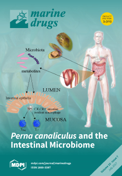
Treatment options for managing chronic conditions is an ever evolving and continuous process. Green lipped mussel extracts from marine molluscs can influence inflammatory symptomology. Two genera are discussed in this review for their potential therapeutic use, Perna canaliculus and Mytilus. Saltzman et al. explore P. canaliculus, positing prebiotic benefits for the intestinal microbiome that improve pro-inflammatory symptoms in patients diagnosed with osteoarthritis of the knee. Saltzman et al. outline how P. canaliculus influences the intestinal microbiome, suppressing exogenous and endogenous intestinal activities and promoting intestinal epithelial homeostasis. They also discuss how marine-derived omega-3 fatty acid supplementation could be utilized in the management of metabolic syndrome, associated complications and chemotherapy-induced mucositis. View Paper here
  • Issues are regarded as officially published after their release is announced to the table of contents alert mailing list.
  • You may sign up for e-mail alerts to receive table of contents of newly released issues.
  • PDF is the official format for papers published in both, html and pdf forms. To view the papers in pdf format, click on the "PDF Full-text" link, and use the free Adobe Reader to open them.
Order results
Result details
Section
Select all
Export citation of selected articles as:
827 KiB  
Article
Functional Comparison for Lipid Metabolism and Intestinal and Fecal Microflora Enzyme Activities between Low Molecular Weight Chitosan and Chitosan Oligosaccharide in High-Fat-Diet-Fed Rats
by Chen-Yuan Chiu, Shih-An Feng, Shing-Hwa Liu and Meng-Tsan Chiang
Mar. Drugs 2017, 15(7), 234; https://doi.org/10.3390/md15070234 - 24 Jul 2017
Cited by 31 | Viewed by 6082
Abstract
The present study investigated and compared the regulatory effects on the lipid-related metabolism and intestinal disaccharidase/fecal bacterial enzyme activities between low molecular weight chitosan and chitosan oligosaccharide in high-fat-diet-fed rats. Diet supplementation of low molecular weight chitosan showed greater efficiency than chitosan oligosaccharide [...] Read more.
The present study investigated and compared the regulatory effects on the lipid-related metabolism and intestinal disaccharidase/fecal bacterial enzyme activities between low molecular weight chitosan and chitosan oligosaccharide in high-fat-diet-fed rats. Diet supplementation of low molecular weight chitosan showed greater efficiency than chitosan oligosaccharide in suppressing the increased weights in body and in liver and adipose tissues of high-fat-diet-fed rats. Supplementation of low molecular weight chitosan also showed a greater improvement than chitosan oligosaccharide in imbalance of plasma, hepatic, and fecal lipid profiles, and intestinal disaccharidase activities in high-fat-diet-fed rats. Moreover, both low molecular weight chitosan and chitosan oligosaccharide significantly decreased the fecal microflora mucinase and β-glucuronidase activities in high-fat-diet-fed rats. These results suggest that low molecular weight chitosan exerts a greater positive improvement than chitosan oligosaccharide in lipid metabolism and intestinal disaccharidase activity in high-fat-diet-induced obese rats. Full article
Show Figures

Figure 1

318 KiB  
Review
Toxicity at the Edge of Life: A Review on Cyanobacterial Toxins from Extreme Environments
by Samuel Cirés, María Cristina Casero and Antonio Quesada
Mar. Drugs 2017, 15(7), 233; https://doi.org/10.3390/md15070233 - 24 Jul 2017
Cited by 61 | Viewed by 6499
Abstract
Cyanotoxins are secondary metabolites produced by cyanobacteria, of varied chemical nature and toxic effects. Although cyanobacteria thrive in all kinds of ecosystems on Earth even under very harsh conditions, current knowledge on cyanotoxin distribution is almost restricted to freshwaters from temperate latitudes. In [...] Read more.
Cyanotoxins are secondary metabolites produced by cyanobacteria, of varied chemical nature and toxic effects. Although cyanobacteria thrive in all kinds of ecosystems on Earth even under very harsh conditions, current knowledge on cyanotoxin distribution is almost restricted to freshwaters from temperate latitudes. In this review, we bring to the forefront the presence of cyanotoxins in extreme environments. Cyanotoxins have been reported especially in polar deserts (both from the Arctic and Antarctica) and alkaline lakes, but also in hot deserts, hypersaline environments, and hot springs. Cyanotoxins detected in these ecosystems include neurotoxins—anatoxin-a, anatoxin-a (S), paralytic shellfish toxins, β-methylaminopropionic acid, N-(2-aminoethyl) glycine and 2,4-diaminobutyric acid- and hepatotoxins –cylindrospermopsins, microcystins and nodularins—with microcystins being the most frequently reported. Toxin production there has been linked to at least eleven cyanobacterial genera yet only three of these (Arthrospira, Synechococcus and Oscillatoria) have been confirmed as producers in culture. Beyond a comprehensive analysis of cyanotoxin presence in each of the extreme environments, this review also identifies the main knowledge gaps to overcome (e.g., scarcity of isolates and –omics data, among others) toward an initial assessment of ecological and human health risks in these amazing ecosystems developing at the very edge of life. Full article
(This article belongs to the Special Issue Algal Toxins II, 2017)
3478 KiB  
Review
Ladder-Shaped Ion Channel Ligands: Current State of Knowledge
by Yuri B. Shmukler and Denis A. Nikishin
Mar. Drugs 2017, 15(7), 232; https://doi.org/10.3390/md15070232 - 20 Jul 2017
Cited by 20 | Viewed by 5320
Abstract
Ciguatoxins (CTX) and brevetoxins (BTX) are polycyclic ethereal compounds biosynthesized by the worldwide distributed planktonic and epibenthic dinoflagellates of Gambierdiscus and Karenia genera, correspondingly. Ciguatera, evoked by CTXs, is a type of ichthyosarcotoxism, which involves a variety of gastrointestinal and neurological symptoms, while [...] Read more.
Ciguatoxins (CTX) and brevetoxins (BTX) are polycyclic ethereal compounds biosynthesized by the worldwide distributed planktonic and epibenthic dinoflagellates of Gambierdiscus and Karenia genera, correspondingly. Ciguatera, evoked by CTXs, is a type of ichthyosarcotoxism, which involves a variety of gastrointestinal and neurological symptoms, while BTXs cause so-called neurotoxic shellfish poisoning. Both types of toxins are reviewed together because of similar mechanisms of their action. These are the only molecules known to activate voltage-sensitive Na+-channels in mammals through a specific interaction with site 5 of its α-subunit and may compete for it, which results in an increase in neuronal excitability, neurotransmitter release and impairment of synaptic vesicle recycling. Most marine ciguatoxins potentiate Nav channels, but a considerable number of them, such as gambierol and maitotoxin, have been shown to affect another ion channel. Although the extrinsic function of these toxins is probably associated with the function of a feeding deterrent, it was suggested that their intrinsic function is coupled with the regulation of photosynthesis via light-harvesting complex II and thioredoxin. Antagonistic effects of BTXs and brevenal may provide evidence of their participation as positive and negative regulators of this mechanism. Full article
(This article belongs to the Special Issue Marine Drugs and Ion Currents)
Show Figures

Graphical abstract

2526 KiB  
Article
Mertensene, a Halogenated Monoterpene, Induces G2/M Cell Cycle Arrest and Caspase Dependent Apoptosis of Human Colon Adenocarcinoma HT29 Cell Line through the Modulation of ERK-1/-2, AKT and NF-κB Signaling
by Safa Tarhouni-Jabberi, Ons Zakraoui, Efstathia Ioannou, Ichrak Riahi-Chebbi, Meriam Haoues, Vassilios Roussis, Riadh Kharrat and Khadija Essafi-Benkhadir
Mar. Drugs 2017, 15(7), 221; https://doi.org/10.3390/md15070221 - 20 Jul 2017
Cited by 37 | Viewed by 4207
Abstract
Conventional treatment of advanced colorectal cancer is associated with tumor resistance and toxicity towards normal tissues. Therefore, development of effective anticancer therapeutic alternatives is still urgently required. Nowadays, marine secondary metabolites have been extensively investigated due to the fact that they frequently exhibit [...] Read more.
Conventional treatment of advanced colorectal cancer is associated with tumor resistance and toxicity towards normal tissues. Therefore, development of effective anticancer therapeutic alternatives is still urgently required. Nowadays, marine secondary metabolites have been extensively investigated due to the fact that they frequently exhibit anti-tumor properties. However, little attention has been given to terpenoids isolated from seaweeds. In this study, we isolated the halogenated monoterpene mertensene from the red alga Pterocladiella capillacea (S.G. Gmelin) Santelices and Hommersand and we highlight its inhibitory effect on the viability of two human colorectal adenocarcinoma cell lines HT29 and LS174. Interestingly, exposure of HT29 cells to different concentrations of mertensene correlated with the activation of MAPK ERK-1/-2, Akt and NF-κB pathways. Moreover, mertensene-induced G2/M cell cycle arrest was associated with a decrease in the phosphorylated forms of the anti-tumor transcription factor p53, retinoblastoma protein (Rb), cdc2 and chkp2. Indeed, a reduction of the cellular level of cyclin-dependent kinases CDK2 and CDK4 was observed in mertensene-treated cells. We also demonstrated that mertensene triggers a caspase-dependent apoptosis in HT29 cancer cells characterized by the activation of caspase-3 and the cleavage of poly (ADP-ribose) polymerase (PARP). Besides, the level of death receptor-associated protein TRADD increased significantly in a concentration-dependent manner. Taken together, these results demonstrate the potential of mertensene as a drug candidate for the treatment of colon cancer. Full article
Show Figures

Figure 1

3184 KiB  
Article
Rapid Estimation of Astaxanthin and the Carotenoid-to-Chlorophyll Ratio in the Green Microalga Chromochloris zofingiensis Using Flow Cytometry
by Junhui Chen, Dong Wei and Georg Pohnert
Mar. Drugs 2017, 15(7), 231; https://doi.org/10.3390/md15070231 - 19 Jul 2017
Cited by 45 | Viewed by 8342
Abstract
The green microalga Chromochloris zofingiensis can accumulate significant amounts of valuable carotenoids, mainly natural astaxanthin, a product with applications in functional food, cosmetics, nutraceuticals, and with potential therapeutic value in cardiovascular and neurological diseases. To optimize the production of astaxanthin, it is essential [...] Read more.
The green microalga Chromochloris zofingiensis can accumulate significant amounts of valuable carotenoids, mainly natural astaxanthin, a product with applications in functional food, cosmetics, nutraceuticals, and with potential therapeutic value in cardiovascular and neurological diseases. To optimize the production of astaxanthin, it is essential to monitor the content of astaxanthin in algal cells during cultivation. The widely used HPLC (high-performance liquid chromatography) method for quantitative astaxanthin determination is time-consuming and laborious. In the present work, we present a method using flow cytometry (FCM) for in vivo determination of the astaxanthin content and the carotenoid-to-chlorophyll ratio (Car/Chl) in mixotrophic C. zofingiensis. The method is based on the assessment of fluorescent characteristics of cellular pigments. The mean fluorescence intensity (MFI) of living cells was determined by FCM to monitor pigment formation based on the correlation between MFI detected in particular channels (FL1: 533 ± 15 nm; FL2: 585 ± 20 nm; FL3: >670 nm) and pigment content in algal cells. Through correlation and regression analysis, a linear relationship was observed between MFI in FL2 (band-pass filter, emission at 585 nm in FCM) and astaxanthin content (in HPLC) and applied for predicting astaxanthin content. With similar procedures, the relationships between MFI in different channels and Car/Chl ratio in mixotrophic C. zofingiensis were also determined. Car/Chl ratios could be estimated by the ratios of MFI (FL1/FL3, FL2/FL3). FCM is thus a highly efficient and feasible method for rapid estimation of astaxanthin content in the green microalga C. zofingiensis. The rapid FCM method is complementary to the current HPLC method, especially for rapid evaluation and prediction of astaxanthin formation as it is required during the high-throughput culture in the laboratory and mass cultivation in industry. Full article
(This article belongs to the Collection Bioactive Compounds from Marine Plankton)
Show Figures

Figure 1

881 KiB  
Communication
Microindolinone A, a Novel 4,5,6,7-Tetrahydroindole, from the Deep-Sea-Derived Actinomycete Microbacterium sp. MCCC 1A11207
by Siwen Niu, Ting-Ting Zhou, Chun-Lan Xie, Gai-Yun Zhang and Xian-Wen Yang
Mar. Drugs 2017, 15(7), 230; https://doi.org/10.3390/md15070230 - 19 Jul 2017
Cited by 28 | Viewed by 4986
Abstract
A novel indole, microindolinone A (1), was isolated from a deep-sea-derived actinomycete Microbacterium sp. MCCC 1A11207, together with 18 known compounds (219). By detailed analysis of the 1H, 13C, HSQC, COSY, HMBC, high resolution electron [...] Read more.
A novel indole, microindolinone A (1), was isolated from a deep-sea-derived actinomycete Microbacterium sp. MCCC 1A11207, together with 18 known compounds (219). By detailed analysis of the 1H, 13C, HSQC, COSY, HMBC, high resolution electron spray ionization mass spectrum (HRESIMS), and circular dichroism (CD) data, the absolute configuration of 1 was elucidated as 5R-hydroxy-4,5,6,7-tetrahydroindole-4-one. It is noteworthy that 1 is the second example of a saturated indole isolated from nature. Full article
(This article belongs to the Special Issue Natural Products from Marine Actinomycetes)
Show Figures

Graphical abstract

657 KiB  
Review
The Biological Activities of Sesterterpenoid-Type Ophiobolins
by Wei Tian, Zixin Deng and Kui Hong
Mar. Drugs 2017, 15(7), 229; https://doi.org/10.3390/md15070229 - 18 Jul 2017
Cited by 47 | Viewed by 5396
Abstract
Ophiobolins (Ophs) are a group of tricarbocyclic sesterterpenoids whose structures contain a tricyclic 5-8-5 carbotricyclic skeleton. Thus far, 49 natural Ophs have been reported and assigned into A–W subgroups in order of discovery. While these sesterterpenoids were first characterized as highly effective phytotoxins, [...] Read more.
Ophiobolins (Ophs) are a group of tricarbocyclic sesterterpenoids whose structures contain a tricyclic 5-8-5 carbotricyclic skeleton. Thus far, 49 natural Ophs have been reported and assigned into A–W subgroups in order of discovery. While these sesterterpenoids were first characterized as highly effective phytotoxins, later investigations demonstrated that they display a broad spectrum of biological and pharmacological characteristics such as phytotoxic, antimicrobial, nematocidal, cytotoxic, anti-influenza and inflammation-promoting activities. These bioactive molecules are promising drug candidates due to the developments of their anti-proliferative activities against a vast number of cancer cell lines, multidrug resistance (MDR) cells and cancer stem cells (CSCs). Despite numerous studies on the biological functions of Ophs, their pharmacological mechanism still requires further research. This review summarizes the chemical structures, sources, and biological activities of the oph family and discusses its mechanisms and structure–activity relationship to lay the foundation for the future developments and applications of these promising molecules. Full article
Show Figures

Graphical abstract

3110 KiB  
Article
A Transcriptomic Survey of Ion Channel-Based Conotoxins in the Chinese Tubular Cone Snail (Conus betulinus)
by Yu Huang, Chao Peng, Yunhai Yi, Bingmiao Gao and Qiong Shi
Mar. Drugs 2017, 15(7), 228; https://doi.org/10.3390/md15070228 - 18 Jul 2017
Cited by 5 | Viewed by 4038
Abstract
Conotoxins in the venom of cone snails (Conus spp.) are a mixture of active peptides that work as blockers, agonists, antagonists, or inactivators of various ion channels. Recently we reported a high-throughput method to identify 215 conotoxin transcripts from the Chinese tubular [...] Read more.
Conotoxins in the venom of cone snails (Conus spp.) are a mixture of active peptides that work as blockers, agonists, antagonists, or inactivators of various ion channels. Recently we reported a high-throughput method to identify 215 conotoxin transcripts from the Chinese tubular cone snail, C. betulinus. Here, based on the previous datasets of four transcriptomes from three venom ducts and one venom bulb, we explored ion channel-based conotoxins and predicted their related ion channel receptors. Homologous analysis was also performed for the most abundant ion channel protein, voltage-gated potassium (Kv; with Kv1.1 as the representative), and the most studied ion channel receptor, nicotinic acetylcholine receptor (nAChR; with α2-nAChR as the representative), in different animals. Our transcriptomic survey demonstrated that ion channel-based conotoxins and related ion channel proteins/receptors transcribe differentially between the venom duct and the venom bulb. In addition, we observed that putative κ-conotoxins were the most common conotoxins with the highest transcription levels in the examined C. betulinus. Furthermore, Kv1.1 and α2-nAChR were conserved in their functional domains of deduced protein sequences, suggesting similar effects of conotoxins via the ion channels in various species, including human beings. In a word, our present work suggests a high-throughput way to develop conotoxins as potential drugs for treatment of ion channel-associated human diseases. Full article
(This article belongs to the Special Issue Marine Drugs and Ion Currents)
Show Figures

Figure 1

5613 KiB  
Article
The Inhibitory Activity of Luzonicosides from the Starfish Echinaster luzonicus against Human Melanoma Cells
by Olesya S. Malyarenko, Sergey A. Dyshlovoy, Alla A. Kicha, Natalia V. Ivanchina, Timofey V. Malyarenko, Bokemeyer Carsten, Von Amsberg Gunhild, Valentin A. Stonik and Svetlana P. Ermakova
Mar. Drugs 2017, 15(7), 227; https://doi.org/10.3390/md15070227 - 18 Jul 2017
Cited by 23 | Viewed by 4027
Abstract
Malignant melanoma is the most dangerous form of skin cancer, with a rapidly increasing incidence rate. Despite recent advances in melanoma research following the approval of several novel targeted and immuno-therapies, the majority of oncological patients will ultimately perish from the disease. Thus, [...] Read more.
Malignant melanoma is the most dangerous form of skin cancer, with a rapidly increasing incidence rate. Despite recent advances in melanoma research following the approval of several novel targeted and immuno-therapies, the majority of oncological patients will ultimately perish from the disease. Thus, new effective drugs are still required. Starfish steroid glycosides possess different biological activities, including antitumor activity. The current study focused on the determination of the in vitro inhibitory activity and the mechanism of action of cyclic steroid glycosides isolated from the starfish Echinaster luzonicus—luzonicoside A (LuzA) and luzonicoside D (LuzD)—in human melanoma RPMI-7951 and SK-Mel-28 cell lines. LuzA inhibited proliferation, the formation of colonies, and the migration of SK-Mel-28 cells significantly more than LuzD. Anti-cancer activity has been ascribed to cell cycle regulation and apoptosis induction. The molecular mechanism of action appears to be related to the regulation of the activity of cleaved caspase-3 and poly(ADP-ribose) polymerase (PARP), along with Survivin, Bcl-2, p21 and cyclin D1 level. Overall, our findings support a potential anti-cancer efficacy of luzonicosides A and D on human melanoma cells. Full article
Show Figures

Figure 1

3258 KiB  
Article
Accumulation of Microcystin (LR, RR and YR) in Three Freshwater Bivalves in Microcystis aeruginosa Bloom Using Dual Isotope Tracer
by Min-Seob Kim, Yeon-Jung Lee, Sun-Yong Ha, Baik-Ho Kim, Soon-Jin Hwang, Jung-Taek Kwon, Jong-Woo Choi and Kyung-Hoon Shin
Mar. Drugs 2017, 15(7), 226; https://doi.org/10.3390/md15070226 - 17 Jul 2017
Cited by 19 | Viewed by 4345
Abstract
Abstract: Stable isotope tracers were first applied to evaluate the Microcystis cell assimilation efficiency of Sinanodonta bivalves, since the past identification method has been limited to tracking the changes of each chl-a, clearity, and nutrient. The toxicity profile and accumulation [...] Read more.
Abstract: Stable isotope tracers were first applied to evaluate the Microcystis cell assimilation efficiency of Sinanodonta bivalves, since the past identification method has been limited to tracking the changes of each chl-a, clearity, and nutrient. The toxicity profile and accumulation of MC-LR, -RR and -YR in different organs (foot and digestive organs) from the three filter-feeders (Sinanodonta woodina, Sinanodonta arcaeformis, and Unio douglasiae) were assessed under the condition of toxigenic cyanobacteria (Microcystis aeruginosa) blooms through an in situ pond experiment using 13C and 15N dual isotope tracers. Chl-a concentration in the manipulated pond was dramatically decreased after the beginning of the second day, ranging from 217.5 to 15.6 μg·L−1. The highest amount of MCs was incorporated into muscle and gland tissues in U. douglasiae during the study period, at nearly 2 or 3 times higher than in S.woodiana and S. arcaeformis. In addition, the incorporated 13C and 15N atom % in the U. douglasiae bivalve showed lower values than in other bivalves. The results demonstrate that U. douglasiae has less capacity to assimilate toxic cyanobacteria derived from diet. However, the incorporated 13C and 15N atom % of S. arcaeformis showed a larger feeding capacity than U. douglasiae and S. wodiana. Our results therefore also indicate that S. arcaeformis can eliminate the toxin more rapidly than U. douglasiae, having a larger detoxification capacity. Full article
(This article belongs to the Special Issue Algal Toxins II, 2017)
Show Figures

Figure 1

1286 KiB  
Article
Urinary Excretion of Tetrodotoxin Modeled in a Porcine Renal Proximal Tubule Epithelial Cell Line, LLC-PK1
by Takuya Matsumoto, Yui Ishizaki, Keika Mochizuki, Mitsuru Aoyagi, Yoshiharu Mitoma, Shoichiro Ishizaki and Yuji Nagashima
Mar. Drugs 2017, 15(7), 225; https://doi.org/10.3390/md15070225 - 17 Jul 2017
Cited by 3 | Viewed by 4085
Abstract
This study examined the urinary excretion of tetrodotoxin (TTX) modeled in a porcine renal proximal tubule epithelial cell line, LLC-PK1. Time course profiles of TTX excretion and reabsorption across the cell monolayers at 37 °C showed that the amount of TTX [...] Read more.
This study examined the urinary excretion of tetrodotoxin (TTX) modeled in a porcine renal proximal tubule epithelial cell line, LLC-PK1. Time course profiles of TTX excretion and reabsorption across the cell monolayers at 37 °C showed that the amount of TTX transported increased linearly for 60 min. However, at 4 °C, the amount of TTX transported was approximately 20% of the value at 37 °C. These results indicate that TTX transport is both a transcellular and carrier-mediated process. Using a transport inhibition assay in which cell monolayers were incubated with 50 µM TTX and 5 mM of a transport inhibitor at 37 °C for 30 min, urinary excretion was significantly reduced by probenecid, tetraethylammonium (TEA), l-carnitine, and cimetidine, slightly reduced by p-aminohippuric acid (PAH), and unaffected by 1-methyl-4-phenylpyridinium (MPP+), oxaliplatin, and cefalexin. Renal reabsorption was significantly reduced by PAH, but was unaffected by probenecid, TEA and l-carnitine. These findings indicate that TTX is primarily excreted by organic cation transporters (OCTs) and organic cation/carnitine transporters (OCTNs), partially transported by organic anion transporters (OATs) and multidrug resistance-associated proteins (MRPs), and negligibly transported by multidrug and toxic compound extrusion transporters (MATEs). Full article
(This article belongs to the Special Issue Tetrodotoxin)
Show Figures

Figure 1

3772 KiB  
Article
Selenium-Containing Polysaccharide-Protein Complex in Se-Enriched Ulva fasciata Induces Mitochondria-Mediated Apoptosis in A549 Human Lung Cancer Cells
by Xian Sun, Yu Zhong, Hongtian Luo and Yufeng Yang
Mar. Drugs 2017, 15(7), 215; https://doi.org/10.3390/md15070215 - 16 Jul 2017
Cited by 34 | Viewed by 4464
Abstract
The role of selenium (Se) and Ulva fasciata as potent cancer chemopreventive and chemotherapeutic agents has been supported by epidemiological, preclinical, and clinical studies. In this study, Se-containing polysaccharide-protein complex (Se-PPC), a novel organoselenium compound, a Se-containing polysaccharide-protein complex in Se-enriched Ulva fasciata [...] Read more.
The role of selenium (Se) and Ulva fasciata as potent cancer chemopreventive and chemotherapeutic agents has been supported by epidemiological, preclinical, and clinical studies. In this study, Se-containing polysaccharide-protein complex (Se-PPC), a novel organoselenium compound, a Se-containing polysaccharide-protein complex in Se-enriched Ulva fasciata, is a potent anti-proliferative agent against human lung cancer A549 cells. Se-PPC markedly inhibited the growth of cancer cells via induction of apoptosis which was accompanied by the formation of apoptotic bodies, an increase in the population of apoptotic sub-G1 phase cells, upregulation of p53, and activation of caspase-3 in A549 cells. Further investigation on intracellular mechanisms indicated that cytochrome C was released from mitochondria into cytosol in A549 cells after Se-PPC treatment. Se-PPC induced depletion of mitochondrial membrane potential (ΔΨm) in A549 cells through regulating the expression of anti-apoptotic (Bcl-2, Bcl-XL) and pro-apoptotic (Bax, Bid) proteins, resulting in disruption of the activation of caspase-9. This is the first report to demonstrate the cytotoxic effect of Se-PPC on human cancer cells and to provide a possible mechanism for this activity. Thus, Se-PPC is a promising novel organoselenium compound with potential to treat human cancers. Full article
(This article belongs to the Collection Marine Compounds and Cancer)
Show Figures

Figure 1

3465 KiB  
Article
Inhibitory Growth of Oral Squamous Cell Carcinoma Cancer via Bacterial Prodigiosin
by Ming-Fang Cheng, Chun-Shu Lin, Yu-Hsin Chen, Ping-Jyun Sung, Shian-Ren Lin, Yi-Wen Tong and Ching-Feng Weng
Mar. Drugs 2017, 15(7), 224; https://doi.org/10.3390/md15070224 - 15 Jul 2017
Cited by 35 | Viewed by 4982
Abstract
Chemotherapy drugs for oral cancers always cause side effects and adverse effects. Currently natural sources and herbs are being searched for treated human oral squamous carcinoma cells (OSCC) in an effort to alleviate the causations of agents in oral cancers chemotherapy. This study [...] Read more.
Chemotherapy drugs for oral cancers always cause side effects and adverse effects. Currently natural sources and herbs are being searched for treated human oral squamous carcinoma cells (OSCC) in an effort to alleviate the causations of agents in oral cancers chemotherapy. This study investigates the effect of prodigiosin (PG), an alkaloid and natural red pigment as a secondary metabolite of Serratia marcescens, to inhibit human oral squamous carcinoma cell growth; thereby, developing a new drug for the treatment of oral cancer. In vitro cultured human OSCC models (OECM1 and SAS cell lines) were used to test the inhibitory growth of PG via cell cytotoxic effects (MTT assay), cell cycle analysis, and Western blotting. PG under various concentrations and time courses were shown to effectively cause cell death and cell-cycle arrest in OECM1 and SAS cells. Additionally, PG induced autophagic cell death in OECM1 and SAS cells by LC3-mediated P62/LC3-I/LC3-II pathway at the in vitro level. These findings elucidate the role of PG, which may target the autophagic cell death pathways as a potential agent in cancer therapeutics. Full article
(This article belongs to the Special Issue Development and Application of Herbal Medicine from Marine Origin)
Show Figures

Graphical abstract

1757 KiB  
Article
Synthesis of Poly(norbornene-methylamine), a Biomimetic of Chitosan, by Ring-Opening Metathesis Polymerization (ROMP)
by Na Li, Huanhuan Wang, Xiaosai Qu and Yu Chen
Mar. Drugs 2017, 15(7), 223; https://doi.org/10.3390/md15070223 - 14 Jul 2017
Cited by 9 | Viewed by 6011
Abstract
ROMP is an effective method for preparing functional polymers due to its having characteristics of “living” polymerization and rapid development of catalysts. In the present work, poly(norbornene-methylamine), a mimic of chitosan, was synthesized via ROMP reaction. The amino-protected product, 5-norbornene-2-(N-methyl)-phthalimide, was [...] Read more.
ROMP is an effective method for preparing functional polymers due to its having characteristics of “living” polymerization and rapid development of catalysts. In the present work, poly(norbornene-methylamine), a mimic of chitosan, was synthesized via ROMP reaction. The amino-protected product, 5-norbornene-2-(N-methyl)-phthalimide, was prepared by a reaction of 5-norbornene-2-methylamine with phthalic anhydride, which was then subjected to the ROMP reaction in the presence of Hoveyda-Grubbs 2nd catalyst to afford poly(norbornene-(N-methyl)-phthalimide). The target product, poly(norbornene-methylamine), was obtained by deprotection reaction of poly(norbornene-(N-methyl)-phthalimide). The products in each step were characterized by FTIR and 1H-NMR, and their thermal stabilities were determined by TG analysis. The effects of molar ratio between monomer ([M]/[I]) and catalyst on the average relative molecular weight ( M n ¯ ) and molecular weight distribution of the produced polymer products were determined by gel permeation chromatography (GPC). It was found that the M n ¯ of poly(norbornene-(N-methyl)-phthalimide) was controllable and exhibited a narrow polydispersity index (PDI) (~1.10). The synthesis condition of 5-norbornene-2-(N-methyl)-phthalimide was optimized by determining the yields at different reaction temperatures and reaction times. The highest yield was obtained at a reaction temperature of 130 °C and a reaction time of 20 min. Our work provides a new strategy to synthesize polymers with controllable structures and free –NH2 groups via ROMP. Full article
(This article belongs to the Special Issue Marine Biomimetics)
Show Figures

Graphical abstract

3923 KiB  
Article
Sponge-Inspired Dibromohemibastadin Prevents and Disrupts Bacterial Biofilms without Toxicity
by Tiffany Le Norcy, Hendrik Niemann, Peter Proksch, Karen Tait, Isabelle Linossier, Karine Réhel, Claire Hellio and Fabienne Faÿ
Mar. Drugs 2017, 15(7), 222; https://doi.org/10.3390/md15070222 - 12 Jul 2017
Cited by 10 | Viewed by 3840
Abstract
Since the banning of several families of compounds in antifouling (AF) coatings, the search for environmentally friendly AF compounds has intensified. Natural sources of AF compounds have been identified in marine organisms and can be used to create analogues in laboratory. In a [...] Read more.
Since the banning of several families of compounds in antifouling (AF) coatings, the search for environmentally friendly AF compounds has intensified. Natural sources of AF compounds have been identified in marine organisms and can be used to create analogues in laboratory. In a previous study, we identified that dibromohemibastadin-1 (DBHB) is a promising AF molecule, leading to the inhibition of the activity of phenoloxidase, an enzyme involved in the attachment of mussels to surfaces. This paper describes the activity of the DBHB on biofilm formation and its detachment and on bacterial adhesion and communication: quorum sensing. DBHB has an anti-biofilm activity without affecting adhesion of marine and terrestrial bacteria at a dose of 10 µM. Moreover, DBHB activity on quorum sensing (QS) is demonstrated at doses of 8 and 16 µM. The activity of DBHB on QS is compared to kojic acid, a quorum sensing inhibitor already described. This compound is a promising environmentally friendly molecule potentially useful for the inhibition of microfouling. Full article
Show Figures

Figure 1

2611 KiB  
Article
Maitotoxin-4, a Novel MTX Analog Produced by Gambierdiscus excentricus
by Francesco Pisapia, Manoëlla Sibat, Christine Herrenknecht, Korian Lhaute, Greta Gaiani, Pierre-Jean Ferron, Valérie Fessard, Santiago Fraga, Silvia M. Nascimento, R. Wayne Litaker, William C. Holland, Catherine Roullier and Philipp Hess
Mar. Drugs 2017, 15(7), 220; https://doi.org/10.3390/md15070220 - 11 Jul 2017
Cited by 56 | Viewed by 8166
Abstract
Maitotoxins (MTXs) are among the most potent toxins known. These toxins are produced by epi-benthic dinoflagellates of the genera Gambierdiscus and Fukuyoa and may play a role in causing the symptoms associated with Ciguatera Fish Poisoning. A recent survey revealed that, of the [...] Read more.
Maitotoxins (MTXs) are among the most potent toxins known. These toxins are produced by epi-benthic dinoflagellates of the genera Gambierdiscus and Fukuyoa and may play a role in causing the symptoms associated with Ciguatera Fish Poisoning. A recent survey revealed that, of the species tested, the newly described species from the Canary Islands, G. excentricus, is one of the most maitotoxic. The goal of the present study was to characterize MTX-related compounds produced by this species. Initially, lysates of cells from two Canary Island G. excentricus strains VGO791 and VGO792 were partially purified by (i) liquid-liquid partitioning between dichloromethane and aqueous methanol followed by (ii) size-exclusion chromatography. Fractions from chromatographic separation were screened for MTX toxicity using both the neuroblastoma neuro-2a (N2a) cytotoxicity and Ca2+ flux functional assays. Fractions containing MTX activity were analyzed using liquid chromatography coupled to high-resolution mass spectrometry (LC-HRMS) to pinpoint potential MTX analogs. Subsequent non-targeted HRMS analysis permitted the identification of a novel MTX analog, maitotoxin-4 (MTX4, accurate mono-isotopic mass of 3292.4860 Da, as free acid form) in the most toxic fractions. HRMS/MS spectra of MTX4 as well as of MTX are presented. In addition, crude methanolic extracts of five other strains of G. excentricus and 37 other strains representing one Fukuyoa species and ten species, one ribotype and one undetermined strain/species of Gambierdiscus were screened for the presence of MTXs using low resolution tandem mass spectrometry (LRMS/MS). This targeted analysis indicated the original maitotoxin (MTX) was only present in one strain (G. australes S080911_1). Putative maitotoxin-2 (p-MTX2) and maitotoxin-3 (p-MTX3) were identified in several other species, but confirmation was not possible because of the lack of reference material. Maitotoxin-4 was detected in all seven strains of G. excentricus examined, independently of their origin (Brazil, Canary Islands and Caribbean), and not detected in any other species. MTX4 may therefore serve as a biomarker for the highly toxic G. excentricus in the Atlantic area. Full article
Show Figures

Figure 1

2339 KiB  
Article
The Epiphytic Genus Gambierdiscus (Dinophyceae) in the Kermadec Islands and Zealandia Regions of the Southwestern Pacific and the Associated Risk of Ciguatera Fish Poisoning
by Lesley L. Rhodes, Kirsty F. Smith, Sam Murray, D. Tim Harwood, Tom Trnski and Rex Munday
Mar. Drugs 2017, 15(7), 219; https://doi.org/10.3390/md15070219 - 11 Jul 2017
Cited by 32 | Viewed by 5448
Abstract
Species in the genus Gambierdiscus produce ciguatoxins (CTXs) and/or maitotoxins (MTXs), which may cause ciguatera fish poisoning (CFP) in humans if contaminated fish are consumed. Species of Gambierdiscus have previously been isolated from macroalgae at Rangitahua (Raoul Island and North Meyer Islands, northern [...] Read more.
Species in the genus Gambierdiscus produce ciguatoxins (CTXs) and/or maitotoxins (MTXs), which may cause ciguatera fish poisoning (CFP) in humans if contaminated fish are consumed. Species of Gambierdiscus have previously been isolated from macroalgae at Rangitahua (Raoul Island and North Meyer Islands, northern Kermadec Islands), and the opportunity was taken to sample for Gambierdiscus at the more southerly Macauley Island during an expedition in 2016. Gambierdiscus cells were isolated, cultured, and DNA extracted and sequenced to determine the species present. Bulk cultures were tested for CTXs and MTXs by liquid chromatography-mass spectrometry (LC-MS/MS). The species isolated were G. australes, which produced MTX-1 (ranging from 3 to 36 pg/cell), and G. polynesiensis, which produced neither MTX-1 nor, unusually, any known CTXs. Isolates of both species produced putative MTX-3. The risk of fish, particularly herbivorous fish, causing CFP in the Zealandia and Kermadec Islands region is real, although in mainland New Zealand the risk is currently low. Both Gambierdiscus and Fukuyoa have been recorded in the sub-tropical northern region of New Zealand, and so the risk may increase with warming seas and shift in the distribution of Gambierdiscus species. Full article
(This article belongs to the Special Issue Harmful Marine Phytoplankton)
Show Figures

Figure 1

4385 KiB  
Article
Genome Sequence of Pseudomonas stutzeri 273 and Identification of the Exopolysaccharide EPS273 Biosynthesis Locus
by Shimei Wu, Rikuan Zheng, Zhenxia Sha and Chaomin Sun
Mar. Drugs 2017, 15(7), 218; https://doi.org/10.3390/md15070218 - 10 Jul 2017
Cited by 13 | Viewed by 5871
Abstract
Pseudomonas stutzeri 273 is a marine bacterium producing exopolysaccharide 273 (EPS273) with high anti-biofilm activity against P. aeruginosa PAO1. Here, the complete genome of P. stutzeri 273 was sequenced and the genome contained a circular 5.03 Mb chromosome. With extensive analysis of [...] Read more.
Pseudomonas stutzeri 273 is a marine bacterium producing exopolysaccharide 273 (EPS273) with high anti-biofilm activity against P. aeruginosa PAO1. Here, the complete genome of P. stutzeri 273 was sequenced and the genome contained a circular 5.03 Mb chromosome. With extensive analysis of the genome, a genetic locus containing 18 genes was predicted to be involved in the biosynthesis of EPS273. In order to confirm this prediction, two adjacent genes (eps273-H and eps273-I) encoding glycosyltransferases and one gene (eps273-O) encoding tyrosine protein kinase within the genetic locus were deleted and biosynthesis of EPS273 was checked in parallel. The molecular weight profile of EPS purified from the mutant Δeps273-HI was obviously different from that purified from wild-type P. stutzeri 273, while the corresponding EPS was hardly detected from the mutant Δeps273-O, which indicated the involvement of the proposed 18-gene cluster in the biosynthesis of EPS273. Moreover, the mutant Δeps273-HI had the biofilm formed earlier compared with the wild type, and the mutant Δeps273-O almost completely lost the ability of biofilm formation. Therefore, EPS273 might facilitate the biofilm formation for its producing strain P. stutzeri 273 while inhibiting the biofilm formation of P. aeruginosa PAO1. This study can contribute to better understanding of the biosynthesis of EPS273 and disclose the biological function of EPS273 for its producing strain P. stutzeri 273. Full article
(This article belongs to the Special Issue Marine Products for Health and Beauty)
Show Figures

Figure 1

3181 KiB  
Article
Discovery of DNA Topoisomerase I Inhibitors with Low-Cytotoxicity Based on Virtual Screening from Natural Products
by Lan-Ting Xin, Lu Liu, Chang-Lun Shao, Ri-Lei Yu, Fang-Ling Chen, Shi-Jun Yue, Mei Wang, Zhong-Long Guo, Ya-Chu Fan, Hua-Shi Guan and Chang-Yun Wang
Mar. Drugs 2017, 15(7), 217; https://doi.org/10.3390/md15070217 - 09 Jul 2017
Cited by 26 | Viewed by 4633
Abstract
Currently, DNA topoisomerase I (Topo I) inhibitors constitute a family of antitumor agents with demonstrated clinical effects on human malignancies. However, the clinical uses of these agents have been greatly limited due to their severe toxic effects. Therefore, it is urgent to find [...] Read more.
Currently, DNA topoisomerase I (Topo I) inhibitors constitute a family of antitumor agents with demonstrated clinical effects on human malignancies. However, the clinical uses of these agents have been greatly limited due to their severe toxic effects. Therefore, it is urgent to find and develop novel low toxic Topo I inhibitors. In recent years, during our ongoing research on natural antitumor products, a collection of low cytotoxic or non-cytotoxic compounds with various structures were identified from marine invertebrates, plants, and their symbiotic microorganisms. In the present study, new Topo I inhibitors were discovered from low cytotoxic and non-cytotoxic natural products by virtual screening with docking simulations in combination with bioassay test. In total, eight potent Topo I inhibitors were found from 138 low cytotoxic or non-cytotoxic compounds from coral-derived fungi and plants. All of these Topo I inhibitors demonstrated activities against Topo I-mediated relaxation of supercoiled DNA at the concentrations of 5–100 µM. Notably, the flavonoids showed higher Topo I inhibitory activities than other compounds. These newly discovered Topo I inhibitors exhibited structurally diverse and could be considered as a good starting point for the development of new antitumor lead compounds. Full article
(This article belongs to the Collection Marine Compounds and Cancer)
Show Figures

Graphical abstract

1903 KiB  
Article
Phytosterols from Dunaliella tertiolecta Reduce Cell Proliferation in Sheep Fed Flaxseed during Post Partum
by Maria Giovanna Ciliberti, Matteo Francavilla, Simona Intini, Marzia Albenzio, Rosaria Marino, Antonella Santillo and Mariangela Caroprese
Mar. Drugs 2017, 15(7), 216; https://doi.org/10.3390/md15070216 - 06 Jul 2017
Cited by 9 | Viewed by 3687
Abstract
The post partum period is characterized by immunosuppression and increased disease susceptibility. Both phytosterols from microalga Dunaniella tertiolecta and dietary supplementation with n-3 polyunsaturated fatty acids (PUFA) influence cell proliferation and cytokine release during inflammation. The objective of this paper was the evaluation [...] Read more.
The post partum period is characterized by immunosuppression and increased disease susceptibility. Both phytosterols from microalga Dunaniella tertiolecta and dietary supplementation with n-3 polyunsaturated fatty acids (PUFA) influence cell proliferation and cytokine release during inflammation. The objective of this paper was the evaluation of the effects of physterols, extracted and purified from D. tertiolecta, on the in vitro immune responses of ewes supplemented with flaxseed during post partum. Twenty Comisana parturient ewes were divided in two balanced groups, and supplemented with flaxseed (FS, 250 g/day) or fed with a conventional diet (CON). Blood samples (15 mL) were collected for five weeks, starting from lambing, in order to isolate peripheral blood mononuclear cells (PBMC). Stimulated PBMC were treated with a total sterols fraction from D. tertiolecta (TS), a mix of ergosterol and 7-dehydroporiferasterol (purified extract, PE), and a mix of acetylated ergosterol and 7-dehydroporiferasterol (acetylated purified extract, AcPE), extracted and purified from D. tertiolecta at two concentrations (0.4 and 0.8 mg/mL). Results of the experiment demonstrated that n-3 PUFA from flaxseed induced an anti-inflammatory cytokine profile, with an increase of both IL-10, IL-6 and a decrease of IL-1β. TS, PE, and AcPE purified from D. tertiolecta showed an anti-proliferative effect on sheep PBMC regardless their chemical composition and concentration. Full article
(This article belongs to the Special Issue Marine Compounds and Inflammation II, 2017)
Show Figures

Figure 1

1712 KiB  
Article
Producing Novel Fibrinolytic Isoindolinone Derivatives in Marine Fungus Stachybotrys longispora FG216 by the Rational Supply of Amino Compounds According to Its Biosynthesis Pathway
by Ying Yin, Qiang Fu, Wenhui Wu, Menghao Cai, Xiangshan Zhou and Yuanxing Zhang
Mar. Drugs 2017, 15(7), 214; https://doi.org/10.3390/md15070214 - 05 Jul 2017
Cited by 19 | Viewed by 4773
Abstract
Many fungi in the Stachybotrys genus can produce various isoindolinone derivatives. These compounds are formed by a spontaneous reaction between a phthalic aldehyde precursor and an ammonium ion or amino compounds. In this study, we suggested the isoindolinone biosynthetic gene cluster in Stachybotrys [...] Read more.
Many fungi in the Stachybotrys genus can produce various isoindolinone derivatives. These compounds are formed by a spontaneous reaction between a phthalic aldehyde precursor and an ammonium ion or amino compounds. In this study, we suggested the isoindolinone biosynthetic gene cluster in Stachybotrys by genome mining based on three reported core genes. Remarkably, there is an additional nitrate reductase (NR) gene copy in the proposed cluster. NR is the rate-limiting enzyme of nitrate reduction. Accordingly, this cluster was speculated to play a role in the balance of ammonium ion concentration in Stachybotrys. Ammonium ions can be replaced by different amino compounds to create structural diversity in the biosynthetic process of isoindolinone. We tested a rational supply of amino compounds ((±)-3-amino-2-piperidinone, glycine, and l-threonine) in the culture of an isoindolinone high-producing marine fungus, Stachybotrys longispora FG216. As a result, we obtained four new kinds of isoindolinone derivatives (FGFC4–GFC7) by this method. Furthermore, high yields of FGFC4–FGFC7 confirmed the outstanding production capacity of FG216. Among the four new isoindolinone derivatives, FGFC6 and FGFC7 showed promising fibrinolytic activities. The knowledge of biosynthesis pathways may be an important attribute for the discovery of novel bioactive marine natural products. Full article
(This article belongs to the Special Issue Genome Mining and Marine Microbial Natural Products)
Show Figures

Graphical abstract

3369 KiB  
Article
Genome Sequences of Marine Shrimp Exopalaemon carinicauda Holthuis Provide Insights into Genome Size Evolution of Caridea
by Jianbo Yuan, Yi Gao, Xiaojun Zhang, Jiankai Wei, Chengzhang Liu, Fuhua Li and Jianhai Xiang
Mar. Drugs 2017, 15(7), 213; https://doi.org/10.3390/md15070213 - 05 Jul 2017
Cited by 49 | Viewed by 5739
Abstract
Crustacea, particularly Decapoda, contains many economically important species, such as shrimps and crabs. Crustaceans exhibit enormous (nearly 500-fold) variability in genome size. However, limited genome resources are available for investigating these species. Exopalaemon carinicauda Holthuis, an economical caridean shrimp, is a potential ideal [...] Read more.
Crustacea, particularly Decapoda, contains many economically important species, such as shrimps and crabs. Crustaceans exhibit enormous (nearly 500-fold) variability in genome size. However, limited genome resources are available for investigating these species. Exopalaemon carinicauda Holthuis, an economical caridean shrimp, is a potential ideal experimental animal for research on crustaceans. In this study, we performed low-coverage sequencing and de novo assembly of the E. carinicauda genome. The assembly covers more than 95% of coding regions. E. carinicauda possesses a large complex genome (5.73 Gb), with size twice higher than those of many decapod shrimps. As such, comparative genomic analyses were implied to investigate factors affecting genome size evolution of decapods. However, clues associated with genome duplication were not identified, and few horizontally transferred sequences were detected. Ultimately, the burst of transposable elements, especially retrotransposons, was determined as the major factor influencing genome expansion. A total of 2 Gb repeats were identified, and RTE-BovB, Jockey, Gypsy, and DIRS were the four major retrotransposons that significantly expanded. Both recent (Jockey and Gypsy) and ancestral (DIRS) originated retrotransposons responsible for the genome evolution. The E. carinicauda genome also exhibited potential for the genomic and experimental research of shrimps. Full article
Show Figures

Figure 1

1262 KiB  
Article
Sterols from the Octocoral Nephthea columnaris
by Ta-Yuan Whuang, Hong-Chieh Tsai, Yin-Di Su, Tsong-Long Hwang and Ping-Jyun Sung
Mar. Drugs 2017, 15(7), 212; https://doi.org/10.3390/md15070212 - 04 Jul 2017
Cited by 8 | Viewed by 3353
Abstract
Two new sterols, columnaristerols B (1) and C (2), along with two known analogues, 5,6-epoxylitosterol (3) and litosterol (4), were obtained from the octocoral Nephthea columnaris. The structures of new sterols 1 and 2 [...] Read more.
Two new sterols, columnaristerols B (1) and C (2), along with two known analogues, 5,6-epoxylitosterol (3) and litosterol (4), were obtained from the octocoral Nephthea columnaris. The structures of new sterols 1 and 2 were elucidated by using spectroscopic methods and comparing the spectroscopic data with those of known related metabolites. Sterol 3 was found to suppress superoxide anion production and elastase secretion by human neutrophils. Full article
(This article belongs to the Special Issue Natural Products from Coral Reef Organisms)
Show Figures

Graphical abstract

1289 KiB  
Article
Cytotoxic Effects of Sarcophyton sp. Soft Corals—Is There a Correlation to Their NMR Fingerprints?
by Mohamed A. Farag, Mostafa I. Fekry, Montasser A. Al-Hammady, Mohamed N. Khalil, Hesham R. El-Seedi, Achim Meyer, Andrea Porzel, Hildegard Westphal and Ludger A. Wessjohann
Mar. Drugs 2017, 15(7), 211; https://doi.org/10.3390/md15070211 - 04 Jul 2017
Cited by 23 | Viewed by 5419
Abstract
Sarcophyton sp. soft corals are rich in cembranoid diterpenes, which represent the main chemical defense of corals against their natural predators in addition to their myriad biological effects in humans. Quantitative NMR (qNMR) was applied for assessing the diterpene variation in 16 soft [...] Read more.
Sarcophyton sp. soft corals are rich in cembranoid diterpenes, which represent the main chemical defense of corals against their natural predators in addition to their myriad biological effects in humans. Quantitative NMR (qNMR) was applied for assessing the diterpene variation in 16 soft coral specimens in the context of their genotype, origin, and growing habitat. qNMR revealed high diterpene levels in Sarcophyton sp. compared to Sinularia and Lobophyton, with (ent)sarcophines as major components (17–100 µg/mg) of the coral tissues. Multivariate data analysis was employed to classify samples based on the quantified level of diterpenes, and compared to the untargeted NMR approach. Results revealed that qNMR provided a stronger classification model of Sarcophyton sp. than untargeted NMR fingerprinting. Additionally, cytotoxicity of soft coral crude extracts was assessed against androgen-dependent prostate cancer cell lines (PC3) and androgen-independent colon cancer cell lines (HT-29), with IC50 values ranging from 10–60 µg/mL. No obvious correlation between the extracts’ IC50 values and their diterpene levels was found using either Spearman or Pearson correlations. This suggests that this type of bioactivity may not be easily predicted by NMR metabolomics in soft corals, or is not strongly correlated to measured diterpene levels. Full article
Show Figures

Figure 1

2516 KiB  
Article
24-Methyl-Cholesta-5,24(28)-Diene-3β,19-diol-7β-Monoacetate Inhibits Human Small Cell Lung Cancer Growth In Vitro and In Vivo via Apoptosis Induction
by Ting-Wen Chung, Jui-Hsin Su, Chi-Chen Lin, Yi-Rong Li, Ya-Hsuan Chao, Sheng-Hao Lin and Hong-Lin Chan
Mar. Drugs 2017, 15(7), 210; https://doi.org/10.3390/md15070210 - 01 Jul 2017
Cited by 10 | Viewed by 4009
Abstract
24-methyl-cholesta-5,24(28)-diene-3β,19-diol-7β-monoacetate (MeCDDA) is a natural steroid compound isolated from a wild-type soft coral (Nephthea erecta). The present study aimed to investigate the anti-small cell lung cancer (SCLC) effects of MeCDDA in vitro and in vivo, as well as to elucidate its [...] Read more.
24-methyl-cholesta-5,24(28)-diene-3β,19-diol-7β-monoacetate (MeCDDA) is a natural steroid compound isolated from a wild-type soft coral (Nephthea erecta). The present study aimed to investigate the anti-small cell lung cancer (SCLC) effects of MeCDDA in vitro and in vivo, as well as to elucidate its underlying mechanism. Our results indicated that H1688 and H146 cells show relevant sensitivity to MeCDDA, and the exposure to MeCDDA in SCLC cells caused dose-dependent growth inhibitory responses. In addition, MeCDDA treatment promoted cell apoptosis and increased the activities of caspases in H1688 cells, reducing the mitochondrial membrane potential and stimulating the release of cytochrome c into the cytosol. Along with the increase in Bax expression and reduction in Bcl-2, the MeCDDA treatment also significantly decreased Akt and mTOR phosphorylation. Finally, MeCDDA treatment in the mouse xenograft model of H1688 cells exhibited significant inhibition of tumor growth, corroborating MeCDDA as a potential pre-clinical candidate for the treatment of SCLC. Overall, our results demonstrate that the cytotoxic effects of MeCDDA towards H1688 and H146 cells, possibly through the activation of the mitochondrial apoptotic pathway and inhibition of the PI3K/Akt/mTOR pathway, merit further studies for its possible clinical application in chemotherapy. Full article
(This article belongs to the Special Issue Natural Products from Coral Reef Organisms)
Show Figures

Figure 1

1231 KiB  
Article
Bistratamides M and N, Oxazole-Thiazole Containing Cyclic Hexapeptides Isolated from Lissoclinum bistratum Interaction of Zinc (II) with Bistratamide K
by Carlos Urda, Rogelio Fernández, Jaime Rodríguez, Marta Pérez, Carlos Jiménez and Carmen Cuevas
Mar. Drugs 2017, 15(7), 209; https://doi.org/10.3390/md15070209 - 01 Jul 2017
Cited by 18 | Viewed by 5193
Abstract
Two novel oxazole-thiazole containing cyclic hexapeptides, bistratamides M (1) and N (2) have been isolated from the marine ascidian Lissoclinum bistratum (L. bistratum) collected in Raja Ampat (Papua Bar, Indonesia). The planar structure of 1 and 2 was assigned [...] Read more.
Two novel oxazole-thiazole containing cyclic hexapeptides, bistratamides M (1) and N (2) have been isolated from the marine ascidian Lissoclinum bistratum (L. bistratum) collected in Raja Ampat (Papua Bar, Indonesia). The planar structure of 1 and 2 was assigned on the basis of extensive 1D and 2D NMR spectroscopy and mass spectrometry. The absolute configuration of the amino acid residues in 1 and 2 was determined by the application of the Marfey’s and advanced Marfey’s methods after ozonolysis followed by acid-catalyzed hydrolysis. The interaction between zinc (II) and the naturally known bistratamide K (3), a cyclic hexapeptide isolated from a different specimen of Lissoclinum bistratum, was monitored by 1H and 13C NMR. The results obtained are consistent with the proposal that these peptides are biosynthesized for binding to metal ions. Compounds 1 and 2 display moderate cytotoxicity against four human tumor cell lines with GI50 values in the micromolar range. Full article
Show Figures

Graphical abstract

223 KiB  
Article
Ciguatoxins and Maitotoxins in Extracts of Sixteen Gambierdiscus Isolates and One Fukuyoa Isolate from the South Pacific and Their Toxicity to Mice by Intraperitoneal and Oral Administration
by Rex Munday, Sam Murray, Lesley L. Rhodes, Michaela E. Larsson and D. Tim Harwood
Mar. Drugs 2017, 15(7), 208; https://doi.org/10.3390/md15070208 - 30 Jun 2017
Cited by 47 | Viewed by 4621
Abstract
Ciguatoxins (CTXs), and possibly maitotoxins (MTXs), are responsible for Ciguatera Fish Poisoning, an important health problem for consumers of reef fish (such as inhabitants of islands in the South Pacific Ocean). The habitational range of the Gambierdiscus species is expanding, and new species [...] Read more.
Ciguatoxins (CTXs), and possibly maitotoxins (MTXs), are responsible for Ciguatera Fish Poisoning, an important health problem for consumers of reef fish (such as inhabitants of islands in the South Pacific Ocean). The habitational range of the Gambierdiscus species is expanding, and new species are being discovered. In order to provide information on the potential health risk of the Gambierdiscus species, and one Fukuyoa species (found in the Cook Islands, the Kermadec Islands, mainland New Zealand, and New South Wales, Australia), 17 microalgae isolates were collected from these areas. Unialgal cultures were grown and extracts of the culture isolates were analysed for CTXs and MTXs by liquid chromatography tandem mass spectrometry (LC-MS/MS), and their toxicity to mice was determined by intraperitoneal and oral administration. An isolate of G. carpenteri contained neither CTXs nor MTXs, while 15 other isolates (including G. australes, G. cheloniae, G. pacificus, G. honu, and F. paulensis) contained only MTX-1 and/or MTX-3. An isolate of G. polynesiensis contained both CTXs and MTX-3. All the extracts were toxic to mice by intraperitoneal injection, but those containing only MTX-1 and/or -3 were much less toxic by oral administration. The extract of G. polynesiensis was highly toxic by both routes of administration. Full article
(This article belongs to the Special Issue Harmful Marine Phytoplankton)
982 KiB  
Review
Perna canaliculus and the Intestinal Microbiome
by Emma Tali Saltzman, Michael Thomsen, Sean Hall and Luis Vitetta
Mar. Drugs 2017, 15(7), 207; https://doi.org/10.3390/md15070207 - 30 Jun 2017
Cited by 6 | Viewed by 8175
Abstract
Natural medicines are often an attractive option for patients diagnosed with chronic conditions. Three main classes of bioactives that have been reported from marine mussel extracts include proteins, lipids and carbohydrates. Commercially, the most relevant species of marine mollusks belong to two genera, [...] Read more.
Natural medicines are often an attractive option for patients diagnosed with chronic conditions. Three main classes of bioactives that have been reported from marine mussel extracts include proteins, lipids and carbohydrates. Commercially, the most relevant species of marine mollusks belong to two genera, Perna and Mytilus. Specifically, the Perna canaliculus species has been repeatedly demonstrated to harbor anti-inflammatory compounds such as omega-3 polyunsaturated fatty acids (ω-3 PUFAs) that can ameliorate pro-inflammatory conditions, or proteins that can promote thrombin inhibitory activity. Recent clinical studies have posited that extracts from green-lipped mussels may lead to prebiotic activity in the intestinal microbiome that in turn has been reported to improve symptoms of osteoarthritis of the knee. Prebiotics have been reported to favorably interact with the intestinal microbiome through the proliferation of beneficial bacteria in the gut, suppressing exogenous and endogenous intestinal infections and promoting homeostasis by balancing local pro- and anti-inflammatory actions. Bioactive compounds from Perna canaliculus are functional foods and, in this regard, may positively interact with the intestinal microbiome and provide novel therapeutic solutions for intra-intestinal and extra-intestinal inflammatory conditions. Full article
(This article belongs to the Special Issue Nutraceuticals and Functional Foods)
Show Figures

Figure 1

828 KiB  
Article
Tricholides A and B and Unnarmicin D: New Hybrid PKS-NRPS Macrocycles Isolated from an Environmental Collection of Trichodesmium thiebautii
by Matthew J. Bertin, Alexandre F. Roduit, Jiadong Sun, Gabriella E. Alves, Christopher W. Via, Miguel A. Gonzalez, Paul V. Zimba and Peter D. R. Moeller
Mar. Drugs 2017, 15(7), 206; https://doi.org/10.3390/md15070206 - 30 Jun 2017
Cited by 11 | Viewed by 5397
Abstract
Bioassay-guided isolation of the lipophilic extract of Trichodesmium thiebautii bloom material led to the purification and structure characterization of two new hybrid polyketide-non-ribosomal peptide (PKS-NRPS) macrocyclic compounds, tricholides A and B (1 and 2). A third macrocyclic compound, unnarmicin D ( [...] Read more.
Bioassay-guided isolation of the lipophilic extract of Trichodesmium thiebautii bloom material led to the purification and structure characterization of two new hybrid polyketide-non-ribosomal peptide (PKS-NRPS) macrocyclic compounds, tricholides A and B (1 and 2). A third macrocyclic compound, unnarmicin D (3), was identified as a new depsipeptide in the unnarmicin family, given its structural similarity to the existing compounds in this group. The planar structures of 13 were determined using 1D and 2D NMR spectra and complementary spectroscopic and spectrometric procedures. The absolute configurations of the amino acid components of 13 were determined via acid hydrolysis, derivitization with Marfey’s reagent and HPLC-UV comparison to authentic amino acid standards. The absolute configuration of the 3-hydroxydodecanoic acid moiety in 3 was determined using a modified Mosher’s esterification procedure on a linear derivative of tricharmicin (4) and additionally by a comparison of 13C NMR shifts of 3 to known depsipeptides with β-hydroxy acid subunits. Tricholide B (2) showed moderate cytotoxicity to Neuro-2A murine neuroblastoma cells (EC50: 14.5 ± 6.2 μM). Full article
(This article belongs to the Special Issue Compounds from Cyanobacteria II)
Show Figures

Graphical abstract

2172 KiB  
Article
Antibacterial Activity of AI-Hemocidin 2, a Novel N-Terminal Peptide of Hemoglobin Purified from Arca inflata
by Chunlei Li, Jianhua Zhu, Yanqing Wang, Yuyan Chen, Liyan Song, Weiming Zheng, Jingjing Li and Rongmin Yu
Mar. Drugs 2017, 15(7), 205; https://doi.org/10.3390/md15070205 - 29 Jun 2017
Cited by 20 | Viewed by 4459
Abstract
The continued emergence of antibiotic resistant bacteria in recent years is of great concern. The search for new classes of antibacterial agents has expanded to non-traditional sources such as shellfish. An antibacterial subunit of hemoglobin (Hb-I) was purified from the mantle of Arca [...] Read more.
The continued emergence of antibiotic resistant bacteria in recent years is of great concern. The search for new classes of antibacterial agents has expanded to non-traditional sources such as shellfish. An antibacterial subunit of hemoglobin (Hb-I) was purified from the mantle of Arca inflata by phosphate extraction and ion exchange chromatography. A novel antibacterial peptide, AI-hemocidin 2, derived from Hb-I, was discovered using bioinformatics analysis. It displayed antibacterial activity across a broad spectrum of microorganisms, including several Gram-positive and Gram-negative bacteria, with minimal inhibitory concentration (MIC) values ranging from 37.5 to 300 μg/mL, and it exhibited minimal hemolytic or cytotoxic activities. The antibacterial activity of AI-hemocidin 2 was thermostable (25–100 °C) and pH resistant (pH 3–10). The cellular integrity was determined by flow cytometry. AI-hemocidin 2 was capable of permeating the cellular membrane. Changes in the cell morphology were observed with a scanning electron microscope. Circular dichroism spectra suggested that AI-hemocidin 2 formed an α-helix structure in the membrane mimetic environment. The results indicated that the anti-bacterial mechanism for AI-hemocidin 2 occurred through disrupting the cell membrane. AI-hemocidin 2 might be a potential candidate for tackling antibiotic resistant bacteria. Full article
(This article belongs to the Collection Bioactive Compounds from Marine Invertebrates)
Show Figures

Graphical abstract

Previous Issue
Next Issue
Back to TopTop