Next Issue
Volume 10, November
Previous Issue
Volume 10, September


Bioengineering, Volume 10, Issue 10 (October 2023) – 129 articles

Cover Story (view full-size image): 
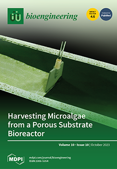
Ensuring the quantity and quality of freshwater has become a principal current environmental challenges. In many parts of the world, domestically and industrially polluted water is essentially untreated and released into the environment. Among the methods developed to treat polluted freshwater and to recycle nutrients, bioremediation using microalgae has gained increased attention in recent years. A technical innovation, the Porous Substrate Bioreactor (PSBR) showed promising results in the removal of pollutants from various types of wastewater (domestic, agricultural, and industrial). Prior studies using PSBRs were conducted at the laboratory and pilot scales and revealed some advantages over other microalgae-based, suspended cultivation technologies, particularly linked to cost-effectiveness, low-level water consumption, and high biomass productivity. View this paper
  • Issues are regarded as officially published after their release is announced to the table of contents alert mailing list.
  • You may sign up for e-mail alerts to receive table of contents of newly released issues.
  • PDF is the official format for papers published in both, html and pdf forms. To view the papers in pdf format, click on the "PDF Full-text" link, and use the free Adobe Reader to open them.
Order results
Result details
Section
Select all
Export citation of selected articles as:
14 pages, 3464 KiB  
Article
Machine Learning-Based Measurement of Regional and Global Spinal Parameters Using the Concept of Incidence Angle of Inflection Points
by Thong Phi Nguyen, Ji-Hwan Kim, Seong-Ha Kim, Jonghun Yoon and Sung-Hoon Choi
Bioengineering 2023, 10(10), 1236; https://doi.org/10.3390/bioengineering10101236 - 23 Oct 2023
Viewed by 1227
Abstract
This study delves into the application of convolutional neural networks (CNNs) in evaluating spinal sagittal alignment, introducing the innovative concept of incidence angles of inflection points (IAIPs) as intuitive parameters to capture the interplay between pelvic and spinal alignment. Pioneering the fusion of [...] Read more.
This study delves into the application of convolutional neural networks (CNNs) in evaluating spinal sagittal alignment, introducing the innovative concept of incidence angles of inflection points (IAIPs) as intuitive parameters to capture the interplay between pelvic and spinal alignment. Pioneering the fusion of IAIPs with machine learning for sagittal alignment analysis, this research scrutinized whole-spine lateral radiographs from hundreds of patients who visited a single institution, utilizing high-quality images for parameter assessments. Noteworthy findings revealed robust success rates for certain parameters, including pelvic and C2 incidence angles, but comparatively lower rates for sacral slope and L1 incidence. The proposed CNN-based machine learning method demonstrated remarkable efficiency, achieving an impressive 80 percent detection rate for various spinal angles, such as lumbar lordosis and thoracic kyphosis, with a precise error threshold of 3.5°. Further bolstering the study’s credibility, measurements derived from the novel formula closely aligned with those directly extracted from the CNN model. In conclusion, this research underscores the utility of the CNN-based deep learning algorithm in delivering precise measurements of spinal sagittal parameters, and highlights the potential for integrating machine learning with the IAIP concept for comprehensive data accumulation in the domain of sagittal spinal alignment analysis, thus advancing our understanding of spinal health. Full article
(This article belongs to the Section Biosignal Processing)
Show Figures

Figure 1

12 pages, 5011 KiB  
Article
Biocompatibility and Transplantation Efficacy of the C-Clear Artificial Cornea in a Rabbit Chemical Burn Model
by Ho-Seok Chung, Sanghyu Nam, Ko-Eun Lee, Do-Sun Jeong, Seheon Oh, Jeong-Hye Sunwoo, Hun Lee, Jae-Yong Kim and Hungwon Tchah
Bioengineering 2023, 10(10), 1235; https://doi.org/10.3390/bioengineering10101235 - 21 Oct 2023
Viewed by 1264
Abstract
We investigated the bioavailability and stability of a C-Clear artificial cornea in a rabbit chemical burn model. Thirty-six rabbits were divided into a control group (n = 16) and a chemical burn group that used NaOH solution (n = 20). After [...] Read more.
We investigated the bioavailability and stability of a C-Clear artificial cornea in a rabbit chemical burn model. Thirty-six rabbits were divided into a control group (n = 16) and a chemical burn group that used NaOH solution (n = 20). After lamellar dissection, the central posterior lamella was excised using a 3 mm diameter trephine, and an artificial cornea was transplanted into the lamellar pocket. After 2 weeks, the central anterior lamella was excised using a 3 mm diameter trephine to secure a clean visual axis. We examined the anterior segment of the eyes weekly for 12 weeks after transplantation. Successful subjects whose artificial corneas were maintained stably for 12 weeks were euthanized and underwent histologic examinations. Artificial corneas remained stable for up to 12 weeks in 62.5 and 50% of rabbits in the control and chemical burn groups, respectively. Two rabbits in the chemical burn group showed the formation of a retroprosthetic membrane, and one rabbit with visual axis blockage underwent membrane removal using a Nd:YAG laser. In histologic examinations, adhesion between artificial cornea and peripheral corneal stoma was observed. In conclusion, we confirmed structural stability and biocompatibility of the C-Clear artificial cornea for up to 12 weeks after implantation in control and chemical burn groups. Full article
(This article belongs to the Special Issue Bioengineering and the Eye, Volume II)
Show Figures

Figure 1

11 pages, 2341 KiB  
Article
An Integrated Multi-Channel Deep Neural Network for Mesial Temporal Lobe Epilepsy Identification Using Multi-Modal Medical Data
by Ruowei Qu, Xuan Ji, Shifeng Wang, Zhaonan Wang, Le Wang, Xinsheng Yang, Shaoya Yin, Junhua Gu, Alan Wang and Guizhi Xu
Bioengineering 2023, 10(10), 1234; https://doi.org/10.3390/bioengineering10101234 - 21 Oct 2023
Viewed by 1284
Abstract
Epilepsy is a chronic brain disease with recurrent seizures. Mesial temporal lobe epilepsy (MTLE) is the most common pathological cause of epilepsy. With the development of computer-aided diagnosis technology, there are many auxiliary diagnostic approaches based on deep learning algorithms. However, the causes [...] Read more.
Epilepsy is a chronic brain disease with recurrent seizures. Mesial temporal lobe epilepsy (MTLE) is the most common pathological cause of epilepsy. With the development of computer-aided diagnosis technology, there are many auxiliary diagnostic approaches based on deep learning algorithms. However, the causes of epilepsy are complex, and distinguishing different types of epilepsy accurately is challenging with a single mode of examination. In this study, our aim is to assess the combination of multi-modal epilepsy medical information from structural MRI, PET image, typical clinical symptoms and personal demographic and cognitive data (PDC) by adopting a multi-channel 3D deep convolutional neural network and pre-training PET images. The results show better diagnosis accuracy than using one single type of medical data alone. These findings reveal the potential of a deep neural network in multi-modal medical data fusion. Full article
(This article belongs to the Special Issue Artificial Intelligence in Biomedical Imaging)
Show Figures

Graphical abstract

18 pages, 5693 KiB  
Article
A Comparison of In Vivo Bone Tissue Generation Using Calcium Phosphate Bone Substitutes in a Novel 3D Printed Four-Chamber Periosteal Bioreactor
by D. S. Abdullah Al Maruf, Kai Cheng, Hai Xin, Veronica K. Y. Cheung, Matthew Foley, Innes K. Wise, Will Lewin, Catriona Froggatt, James Wykes, Krishnan Parthasarathi, David Leinkram, Dale Howes, Natalka Suchowerska, David R. McKenzie, Ruta Gupta, Jeremy M. Crook and Jonathan R. Clark
Bioengineering 2023, 10(10), 1233; https://doi.org/10.3390/bioengineering10101233 - 21 Oct 2023
Cited by 1 | Viewed by 1577
Abstract
Autologous bone replacement remains the preferred treatment for segmental defects of the mandible; however, it cannot replicate complex facial geometry and causes donor site morbidity. Bone tissue engineering has the potential to overcome these limitations. Various commercially available calcium phosphate-based bone substitutes (Novabone [...] Read more.
Autologous bone replacement remains the preferred treatment for segmental defects of the mandible; however, it cannot replicate complex facial geometry and causes donor site morbidity. Bone tissue engineering has the potential to overcome these limitations. Various commercially available calcium phosphate-based bone substitutes (Novabone®, BioOss®, and Zengro®) are commonly used in dentistry for small bone defects around teeth and implants. However, their role in ectopic bone formation, which can later be applied as vascularized graft in a bone defect, is yet to be explored. Here, we compare the above-mentioned bone substitutes with autologous bone with the aim of selecting one for future studies of segmental mandibular repair. Six female sheep, aged 7–8 years, were implanted with 40 mm long four-chambered polyether ether ketone (PEEK) bioreactors prepared using additive manufacturing followed by plasma immersion ion implantation (PIII) to improve hydrophilicity and bioactivity. Each bioreactor was wrapped with vascularized scapular periosteum and the chambers were filled with autologous bone graft, Novabone®, BioOss®, and Zengro®, respectively. The bioreactors were implanted within a subscapular muscle pocket for either 8 weeks (two sheep), 10 weeks (two sheep), or 12 weeks (two sheep), after which they were removed and assessed by microCT and routine histology. Moderate bone formation was observed in autologous bone grafts, while low bone formation was observed in the BioOss® and Zengro® chambers. No bone formation was observed in the Novabone® chambers. Although the BioOss® and Zengro® chambers contained relatively small amounts of bone, endochondral ossification and retained hydroxyapatite suggest their potential in new bone formation in an ectopic site if a consistent supply of progenitor cells and/or growth factors can be ensured over a longer duration. Full article
(This article belongs to the Section Regenerative Engineering)
Show Figures

Graphical abstract

18 pages, 1008 KiB  
Review
Three-Dimensional Bioprinting in Soft Tissue Engineering for Plastic and Reconstructive Surgery
by Astrid Bülow, Benedikt Schäfer and Justus P. Beier
Bioengineering 2023, 10(10), 1232; https://doi.org/10.3390/bioengineering10101232 - 21 Oct 2023
Cited by 2 | Viewed by 1652
Abstract
Skeletal muscle tissue engineering (TE) and adipose tissue engineering have undergone significant progress in recent years. This review focuses on the key findings in these areas, particularly highlighting the integration of 3D bioprinting techniques to overcome challenges and enhance tissue regeneration. In skeletal [...] Read more.
Skeletal muscle tissue engineering (TE) and adipose tissue engineering have undergone significant progress in recent years. This review focuses on the key findings in these areas, particularly highlighting the integration of 3D bioprinting techniques to overcome challenges and enhance tissue regeneration. In skeletal muscle TE, 3D bioprinting enables the precise replication of muscle architecture. This addresses the need for the parallel alignment of cells and proper innervation. Satellite cells (SCs) and mesenchymal stem cells (MSCs) have been utilized, along with co-cultivation strategies for vascularization and innervation. Therefore, various printing methods and materials, including decellularized extracellular matrix (dECM), have been explored. Similarly, in adipose tissue engineering, 3D bioprinting has been employed to overcome the challenge of vascularization; addressing this challenge is vital for graft survival. Decellularized adipose tissue and biomimetic scaffolds have been used as biological inks, along with adipose-derived stem cells (ADSCs), to enhance graft survival. The integration of dECM and alginate bioinks has demonstrated improved adipocyte maturation and differentiation. These findings highlight the potential of 3D bioprinting techniques in skeletal muscle and adipose tissue engineering. By integrating specific cell types, biomaterials, and printing methods, significant progress has been made in tissue regeneration. However, challenges such as fabricating larger constructs, translating findings to human models, and obtaining regulatory approvals for cellular therapies remain to be addressed. Nonetheless, these advancements underscore the transformative impact of 3D bioprinting in tissue engineering research and its potential for future clinical applications. Full article
(This article belongs to the Special Issue 3D-Bioprinting in Bioengineering)
Show Figures

Figure 1

16 pages, 6355 KiB  
Article
Aneurysm Rupture Prediction Based on Strain Energy-CFD Modelling
by Ahmed M. Al-Jumaily, Abd Halim Bin Embong, Mohammad AL-Rawi, Giri Mahadevan and Shukei Sugita
Bioengineering 2023, 10(10), 1231; https://doi.org/10.3390/bioengineering10101231 - 21 Oct 2023
Cited by 1 | Viewed by 1174
Abstract
This paper presents a Patient-Specific Aneurysm Model (PSAM) analyzed using Computational Fluid Dynamics (CFD). The PSAM combines the energy strain function and stress–strain relationship of the dilated vessel wall to predict the rupture of aneurysms. This predictive model is developed by analyzing ultrasound [...] Read more.
This paper presents a Patient-Specific Aneurysm Model (PSAM) analyzed using Computational Fluid Dynamics (CFD). The PSAM combines the energy strain function and stress–strain relationship of the dilated vessel wall to predict the rupture of aneurysms. This predictive model is developed by analyzing ultrasound images acquired with a 6–9 MHz Doppler transducer, which provides real-time data on the arterial deformations. The patient-specific cyclic loading on the PSAM is extrapolated from the strain energy function developed using historical stress–strain relationships. Multivariant factors are proposed to locate points of arterial weakening that precede rupture. Biaxial tensile tests are used to calculate the material properties of the artery wall, enabling the observation of the time-dependent material response in wall rupture formation. In this way, correlations between the wall deformation and tissue failure mode can predict the aneurysm’s propensity to rupture. This method can be embedded within the ultrasound measures used to diagnose potential AAA ruptures. Full article
(This article belongs to the Special Issue Computational Models in Cardiovascular Medicine)
Show Figures

Figure 1

15 pages, 2672 KiB  
Article
Identification and Characterization of a Novel Mannanase from Klebsiella grimontii
by Changzheng Chen, Kuikui Li, Tang Li, Junyan Li, Qishun Liu and Heng Yin
Bioengineering 2023, 10(10), 1230; https://doi.org/10.3390/bioengineering10101230 - 21 Oct 2023
Cited by 2 | Viewed by 1215
Abstract
Konjac glucomannan (KGM) is a natural polysaccharide derived from konjac, which has been widely used in various fields due to its numerous beneficial properties. However, the high viscosity and water absorption of KGM limit its application. Compared with KGM, Konjac glucomannan oligosaccharides (KGMOS) [...] Read more.
Konjac glucomannan (KGM) is a natural polysaccharide derived from konjac, which has been widely used in various fields due to its numerous beneficial properties. However, the high viscosity and water absorption of KGM limit its application. Compared with KGM, Konjac glucomannan oligosaccharides (KGMOS) have higher water solubility and stronger application value. In this paper, a novel mannanase KgManA was cloned from Klebsiella grimontii to develop a new KGMOS-producing enzyme. Bioinformatic analysis shows that the structural similarity between KgManA and other enzymes was less than 18.33%. Phylogenetic analysis shows that KgManA shares different branches with the traditional mannanases containing the CMB35 domain, indicating that it is a novel mannanase. Then, the enzymatic properties were determined and substrate specificity was characterized. Surprisingly, KgManA is stable in a very wide pH range of 3.0 to 10.0; it has a special substrate specificity and seems to be active only for mannans without galactose in the side chain. Additionally, the three-dimensional structure of the enzyme was simulated and molecular docking of the mannotetraose substrate was performed. As far as we know, this is the first report to characterize the enzymatic properties and to simulate the structure of mannanase from K. grimontii. This work will contribute to the development and characterization of novel K. grimontii-derived mannanases. The above results indicate that KgManA is a promising tool for the production of KGMOS. Full article
Show Figures

Figure 1

17 pages, 18439 KiB  
Article
AI-Driven Segmentation and Automated Analysis of the Whole Sagittal Spine from X-ray Images for Spinopelvic Parameter Evaluation
by Sang-Youn Song, Min-Seok Seo, Chang-Won Kim, Yun-Heung Kim, Byeong-Cheol Yoo, Hyun-Ju Choi, Sung-Hyo Seo, Sung-Wook Kang, Myung-Geun Song, Dae-Cheol Nam and Dong-Hee Kim
Bioengineering 2023, 10(10), 1229; https://doi.org/10.3390/bioengineering10101229 - 20 Oct 2023
Cited by 3 | Viewed by 1279
Abstract
Spinal–pelvic parameters are utilized in orthopedics for assessing patients’ curvature and body alignment in diagnosing, treating, and planning surgeries for spinal and pelvic disorders. Segmenting and autodetecting the whole spine from lateral radiographs is challenging. Recent efforts have employed deep learning techniques to [...] Read more.
Spinal–pelvic parameters are utilized in orthopedics for assessing patients’ curvature and body alignment in diagnosing, treating, and planning surgeries for spinal and pelvic disorders. Segmenting and autodetecting the whole spine from lateral radiographs is challenging. Recent efforts have employed deep learning techniques to automate the segmentation and analysis of whole-spine lateral radiographs. This study aims to develop an artificial intelligence (AI)-based deep learning approach for the automated segmentation, alignment, and measurement of spinal–pelvic parameters through whole-spine lateral radiographs. We conducted the study on 932 annotated images from various spinal pathologies. Using a deep learning (DL) model, anatomical landmarks of the cervical, thoracic, lumbar vertebrae, sacrum, and femoral head were automatically distinguished. The algorithm was designed to measure 13 radiographic alignment and spinal–pelvic parameters from the whole-spine lateral radiographs. Training data comprised 748 digital radiographic (DR) X-ray images, while 90 X-ray images were used for validation. Another set of 90 X-ray images served as the test set. Inter-rater reliability between orthopedic spine specialists, orthopedic residents, and the DL model was evaluated using the intraclass correlation coefficient (ICC). The segmentation accuracy for anatomical landmarks was within an acceptable range (median error: 1.7–4.1 mm). The inter-rater reliability between the proposed DL model and individual experts was fair to good for measurements of spinal curvature characteristics (all ICC values > 0.62). The developed DL model in this study demonstrated good levels of inter-rater reliability for predicting anatomical landmark positions and measuring radiographic alignment and spinal–pelvic parameters. Automated segmentation and analysis of whole-spine lateral radiographs using deep learning offers a promising tool to enhance accuracy and efficiency in orthopedic diagnostics and treatments. Full article
(This article belongs to the Section Biosignal Processing)
Show Figures

Figure 1

22 pages, 911 KiB  
Review
Immunotherapy in Acute Myeloid Leukemia: A Literature Review of Emerging Strategies
by Luca Guarnera, Carlos Bravo-Perez and Valeria Visconte
Bioengineering 2023, 10(10), 1228; https://doi.org/10.3390/bioengineering10101228 - 20 Oct 2023
Cited by 1 | Viewed by 1739
Abstract
In the last twenty years, we have witnessed a paradigm shift in the treatment and prognosis of acute myeloid leukemia (AML), thanks to the introduction of new efficient drugs or approaches to refine old therapies, such as Gemtuzumab Ozogamicin, CPX 3-5-1, hypomethylating agents, [...] Read more.
In the last twenty years, we have witnessed a paradigm shift in the treatment and prognosis of acute myeloid leukemia (AML), thanks to the introduction of new efficient drugs or approaches to refine old therapies, such as Gemtuzumab Ozogamicin, CPX 3-5-1, hypomethylating agents, and Venetoclax, the optimization of conditioning regimens in allogeneic hematopoietic stem cell transplantation and the improvement of supportive care. However, the long-term survival of non-M3 and non-core binding factor-AML is still dismal. For this reason, the expectations for the recently developed immunotherapies, such as antibody-based therapy, checkpoint inhibitors, and chimeric antigen receptor strategies, successfully tested in other hematologic malignancies, were very high. The inherent characteristics of AML blasts hampered the development of these treatments, and the path of immunotherapy in AML has been bumpy. Herein, we provide a detailed review of potential antigenic targets, available data from pre-clinical and clinical trials, and future directions of immunotherapies in AML. Full article
(This article belongs to the Special Issue Advances in Treatment of Leukemia)
Show Figures

Graphical abstract

14 pages, 5812 KiB  
Article
The Paracrine Effect of Hyaluronic Acid-Treated Endothelial Cells Promotes BMP-2-Mediated Osteogenesis
by Xiaojie Tong, Jin Chen, Renqin Wang, Dan Hou, Gang Wu, Chang Liu and Janak Lal Pathak
Bioengineering 2023, 10(10), 1227; https://doi.org/10.3390/bioengineering10101227 - 20 Oct 2023
Cited by 1 | Viewed by 1160
Abstract
The combination of hyaluronic acid (HA) and BMP-2 has been reported to promote bone regeneration. However, the interaction of endothelial cells and bone marrow mesenchymal stem cells (BMSCs) during HA + BMP-2 treatment is not fully understood. This study aimed to analyze the [...] Read more.
The combination of hyaluronic acid (HA) and BMP-2 has been reported to promote bone regeneration. However, the interaction of endothelial cells and bone marrow mesenchymal stem cells (BMSCs) during HA + BMP-2 treatment is not fully understood. This study aimed to analyze the direct effect of HA, as well as the paracrine effect of HA-treated endothelial cells, on the BMP-2-mediated osteogenic differentiation of BMSCs. The angiogenic differentiation potential of HA at different molecular weights and different concentrations was tested. The direct effect of HA, as well as the indirect effect of HA-treated human umbilical cord endothelial cells (HUVECs, i.e., conditioned medium (CM)-based co-culture) on the BMP-2-mediated osteogenic differentiation of BMSCs was analyzed using alkaline phosphatase (ALP) staining and activity, alizarin red S (ARS) staining, and RT-qPCR of osteogenic markers. Angiogenic differentiation markers were also analyzed in HUVECs after treatment with HA + BMP-2. The bone regeneration potential of BMP-2 and HA + BMP-2 was analyzed in a rat ectopic model. We found that 1600 kDa HA at 300 µg/mL promoted tube formation by HUVECs in vitro and upregulated the mRNA expression of the angiogenic markers CD31, VEGF, and bFGF. HA inhibited, but conditioned medium from HA-treated HUVECs promoted, the BMP-2-mediated osteogenic differentiation of BMSCs, as indicated by the results of ALP staining and activity, ARS staining, and the mRNA expression of the osteogenic markers RUNX-2, ALP, COLI, and OPN. HA + BMP-2 (50 ng/mL) upregulated the expression of the angiogenesis-related genes VEGF and bFGF in HUVECs and bone regeneration in vivo compared to BMP-2 treatment. In conclusion, the paracrine effect of hyaluronic acid-treated endothelial cells promotes BMP-2-mediated osteogenesis, suggesting the application potential of HA + BMP-2 in bone tissue engineering. Full article
(This article belongs to the Special Issue Biomaterials for Bone Repair and Regeneration)
Show Figures

Graphical abstract

15 pages, 2473 KiB  
Article
Verification and Validation of Lower Body Negative Pressure as a Non-Invasive Bioengineering Tool for Testing Technologies for Monitoring Human Hemorrhage
by Victor A. Convertino, Eric J. Snider, Sofia I. Hernandez-Torres, James P. Collier, Samantha K. Eaton, David R. Holmes III, Clifton R. Haider and Jose Salinas
Bioengineering 2023, 10(10), 1226; https://doi.org/10.3390/bioengineering10101226 - 20 Oct 2023
Viewed by 920
Abstract
Since hemorrhage is a leading cause of preventable death in both civilian and military settings, the development of advanced decision support monitoring capabilities is necessary to promote improved clinical outcomes. The emergence of lower body negative pressure (LBNP) has provided a bioengineering technology [...] Read more.
Since hemorrhage is a leading cause of preventable death in both civilian and military settings, the development of advanced decision support monitoring capabilities is necessary to promote improved clinical outcomes. The emergence of lower body negative pressure (LBNP) has provided a bioengineering technology for inducing progressive reductions in central blood volume shown to be accurate as a model for the study of the early compensatory stages of hemorrhage. In this context, the specific aim of this study was to provide for the first time a systematic technical evaluation to meet a commonly accepted engineering standard based on the FDA-recognized Standard for Assessing Credibility of Modeling through Verification and Validation (V&V) for Medical Devices (ASME standard V&V 40) specifically highlighting LBNP as a valuable resource for the safe study of hemorrhage physiology in humans. As an experimental tool, evidence is presented that LBNP is credible, repeatable, and validated as an analog for the study of human hemorrhage physiology compared to actual blood loss. The LBNP tool can promote the testing and development of advanced monitoring algorithms and evaluating wearable sensors with the goal of improving clinical outcomes during use in emergency medical settings. Full article
Show Figures

Figure 1

20 pages, 3429 KiB  
Article
Explainable Vision Transformer with Self-Supervised Learning to Predict Alzheimer’s Disease Progression Using 18F-FDG PET
by Uttam Khatri and Goo-Rak Kwon
Bioengineering 2023, 10(10), 1225; https://doi.org/10.3390/bioengineering10101225 - 20 Oct 2023
Viewed by 1903
Abstract
Alzheimer’s disease (AD) is a progressive neurodegenerative disorder that affects millions of people worldwide. Early and accurate prediction of AD progression is crucial for early intervention and personalized treatment planning. Although AD does not yet have a reliable therapy, several medications help slow [...] Read more.
Alzheimer’s disease (AD) is a progressive neurodegenerative disorder that affects millions of people worldwide. Early and accurate prediction of AD progression is crucial for early intervention and personalized treatment planning. Although AD does not yet have a reliable therapy, several medications help slow down the disease’s progression. However, more study is still needed to develop reliable methods for detecting AD and its phases. In the recent past, biomarkers associated with AD have been identified using neuroimaging methods. To uncover biomarkers, deep learning techniques have quickly emerged as a crucial methodology. A functional molecular imaging technique known as fluorodeoxyglucose positron emission tomography (18F-FDG-PET) has been shown to be effective in assisting researchers in understanding the morphological and neurological alterations to the brain associated with AD. Convolutional neural networks (CNNs) have also long dominated the field of AD progression and have been the subject of substantial research, while more recent approaches like vision transformers (ViT) have not yet been fully investigated. In this paper, we present a self-supervised learning (SSL) method to automatically acquire meaningful AD characteristics using the ViT architecture by pretraining the feature extractor using the self-distillation with no labels (DINO) and extreme learning machine (ELM) as classifier models. In this work, we examined a technique for predicting mild cognitive impairment (MCI) to AD utilizing an SSL model which learns powerful representations from unlabeled 18F-FDG PET images, thus reducing the need for large-labeled datasets. In comparison to several earlier approaches, our strategy showed state-of-the-art classification performance in terms of accuracy (92.31%), specificity (90.21%), and sensitivity (95.50%). Then, to make the suggested model easier to understand, we highlighted the brain regions that significantly influence the prediction of MCI development. Our methods offer a precise and efficient strategy for predicting the transition from MCI to AD. In conclusion, this research presents a novel Explainable SSL-ViT model that can accurately predict AD progress based on 18F-FDG PET scans. SSL, attention, and ELM mechanisms are integrated into the model to make it more predictive and interpretable. Future research will enable the development of viable treatments for neurodegenerative disorders by combining brain areas contributing to projection with observed anatomical traits. Full article
Show Figures

Figure 1

12 pages, 4826 KiB  
Article
An Analysis of Trabecular Bone Structure Based on Principal Stress Trajectory
by Jiwu Zhang, Haoran Li, Yuqing Zhou, Songhao Chen and Qiguo Rong
Bioengineering 2023, 10(10), 1224; https://doi.org/10.3390/bioengineering10101224 - 20 Oct 2023
Cited by 1 | Viewed by 1212
Abstract
To understand the mechanism of Wolff’s law, a finite element analysis was performed for a human proximal femur, and the principal stress trajectories of the femur were extracted using the principal stress visualization method. The mechanism of Wolff’s law was evaluated theoretically based [...] Read more.
To understand the mechanism of Wolff’s law, a finite element analysis was performed for a human proximal femur, and the principal stress trajectories of the femur were extracted using the principal stress visualization method. The mechanism of Wolff’s law was evaluated theoretically based on the distribution of the principal stress trajectories. Due to the dynamics of the loads, there was no one-to-one correspondence between the stress trajectories of the fixed load and the trabeculae in the cancellous architecture of the real bone. The trabeculae in the cancellous bone were influenced by the magnitude of the principal stress trajectory. Equivalent principal stress trajectories suitable for different load changes were proposed through the change in load cycle and compared with the anatomical structure of the femur. In addition, the three-dimensional distribution of the femoral principal stress trajectory was established, and the adaptability potential of each load was discussed. The principal stress visualization method could also be applied to bionic structure design. Full article
(This article belongs to the Section Biomechanics and Sports Medicine)
Show Figures

Graphical abstract

17 pages, 10204 KiB  
Article
Proton Conduction in Gly-X (X = Ser, Ser-Gly-Ser) and GS50
by Hitoki Semizo, Ryusei Yabu, Yamato Ohgishi, Haruka Kai, Hitoshi Nishimura and Yasumitsu Matsuo
Bioengineering 2023, 10(10), 1223; https://doi.org/10.3390/bioengineering10101223 - 19 Oct 2023
Viewed by 970
Abstract
In recent years, the use of biomaterials has been required from the viewpoint of biocompatibility of electronic devices. In this study, the proton conductivity of Glycyl-L-serine (Gly-Ser) was investigated to clarify the relationship between hydration and proton conduction in peptides. From the crystal [...] Read more.
In recent years, the use of biomaterials has been required from the viewpoint of biocompatibility of electronic devices. In this study, the proton conductivity of Glycyl-L-serine (Gly-Ser) was investigated to clarify the relationship between hydration and proton conduction in peptides. From the crystal and conductivity data, it was inferred that the proton conductivity in hydrated Gly-Ser crystals is caused by the cleavage and rearrangement of hydrogen bonds between hydration shells formed by hydrogen bonds between amino acids and water molecules. Moreover, a staircase-like change in proton conduction with hydration was observed at n = 0.3 and 0.5. These results indicate that proton transport in Gly-Ser is realized by hydration water. In addition, we also found that hydration of GSGS and GS50 can achieve proton conduction of Gly-Ser tetrameric GSGS and GS50 containing repeating sequences. The proton conductivity at n = 0.3 is due to percolation by the formation of proton-conducting pathways. In addition to these results, we found that proton conductivity at GS50 is realized by the diffusion constant of 3.21 × 10⁻⁸ cm2/s at GS50. Full article
(This article belongs to the Section Biomedical Engineering and Biomaterials)
Show Figures

Graphical abstract

17 pages, 2302 KiB  
Article
Evaluation of the Photoplethysmogram-Based Deep Learning Model for Continuous Respiratory Rate Estimation in Surgical Intensive Care Unit
by Chi Shin Hwang, Yong Hwan Kim, Jung Kyun Hyun, Joon Hwang Kim, Seo Rak Lee, Choong Min Kim, Jung Woo Nam and Eun Young Kim
Bioengineering 2023, 10(10), 1222; https://doi.org/10.3390/bioengineering10101222 - 19 Oct 2023
Viewed by 1154
Abstract
The respiratory rate (RR) is a significant indicator to evaluate a patient’s prognosis and status; however, it requires specific instrumentation or estimates from other monitored signals. A photoplethysmogram (PPG) is extensively used in clinical environments as well as in intensive care units (ICUs) [...] Read more.
The respiratory rate (RR) is a significant indicator to evaluate a patient’s prognosis and status; however, it requires specific instrumentation or estimates from other monitored signals. A photoplethysmogram (PPG) is extensively used in clinical environments as well as in intensive care units (ICUs) to primarily monitor peripheral circulation while capturing indirect information about intrathoracic pressure changes. This study aims to apply and evaluate several deep learning models using a PPG for the continuous and accurate estimation of the RRs of patients. The dataset was collected twice for 2 min each in 100 patients aged 18 years and older from the surgical intensive care unit of a tertiary referral hospital. The BIDMC and CapnoBase public datasets were also analyzed. The collected dataset was preprocessed and split according to the 5-fold cross-validation. We used seven deep learning models, including our own Dilated Residual Neural Network, to check how accurately the RR estimates match the ground truth using the mean absolute error (MAE). As a result, when validated using the collected dataset, our model showed the best results with a 1.2628 ± 0.2697 MAE on BIDMC and RespNet and with a 3.1268 ± 0.6363 MAE on our dataset, respectively. In conclusion, RR estimation using PPG-derived models is still challenging and has many limitations. However, if there is an equal amount of data from various breathing groups to train, we expect that various models, including our Dilated ResNet model, which showed good results, can achieve better results than the current ones. Full article
(This article belongs to the Special Issue Artificial Intelligence for Biomedical Signal Processing)
Show Figures

Graphical abstract

11 pages, 1391 KiB  
Brief Report
The Three-Dimensional Body Center of Mass at the Workplace under Hypogravity
by Tatiana Maillard
Bioengineering 2023, 10(10), 1221; https://doi.org/10.3390/bioengineering10101221 - 19 Oct 2023
Viewed by 952
Abstract
The center of mass dynamics of the seated posture of humans in a work environment under hypogravity (0 < g < 1) have rarely been investigated, and such research is yet to be carried out. The present study determined the difference in the [...] Read more.
The center of mass dynamics of the seated posture of humans in a work environment under hypogravity (0 < g < 1) have rarely been investigated, and such research is yet to be carried out. The present study determined the difference in the body system of 32 participants working under simulated 1/6 g (Moon) and 1 g (Earth) for comparison using static and dynamic task measurements. This was based on a markerless motion capture method that analyzed participants’ center of mass at the start, middle and end of the task when they began to get fatigued. According to this analysis, there is a positive relationship (p < 0.01) with a positive coefficient of correlation between the downward center of mass body shift along the proximodistal axis and gravity level for males and females. At the same time, the same positive relationship (p < 0.01) between the tilt of the body backward along the anterior–posterior axis and the level of gravity was found only in females. This offers fresh perspectives for comprehending hypogravity in a broader framework regarding its impact on musculoskeletal disorders. It can also improve workplace ergonomics, body stability, equipment design, and biomechanics. Full article
(This article belongs to the Special Issue Human Movement and Ergonomics)
Show Figures

Figure 1

13 pages, 3557 KiB  
Article
Rapid Segmentation and Diagnosis of Breast Tumor Ultrasound Images at the Sonographer Level Using Deep Learning
by Lei Yang, Baichuan Zhang, Fei Ren, Jianwen Gu, Jiao Gao, Jihua Wu, Dan Li, Huaping Jia, Guangling Li, Jing Zong, Jing Zhang, Xiaoman Yang, Xueyuan Zhang, Baolin Du, Xiaowen Wang and Na Li
Bioengineering 2023, 10(10), 1220; https://doi.org/10.3390/bioengineering10101220 - 19 Oct 2023
Cited by 2 | Viewed by 1911
Abstract
Background: Breast cancer is one of the most common malignant tumors in women. A noninvasive ultrasound examination can identify mammary-gland-related diseases and is well tolerated by dense breast, making it a preferred method for breast cancer screening and of significant clinical value. However, [...] Read more.
Background: Breast cancer is one of the most common malignant tumors in women. A noninvasive ultrasound examination can identify mammary-gland-related diseases and is well tolerated by dense breast, making it a preferred method for breast cancer screening and of significant clinical value. However, the diagnosis of breast nodules or masses via ultrasound is performed by a doctor in real time, which is time-consuming and subjective. Junior doctors are prone to missed diagnoses, especially in remote areas or grass-roots hospitals, due to limited medical resources and other factors, which bring great risks to a patient’s health. Therefore, there is an urgent need to develop fast and accurate ultrasound image analysis algorithms to assist diagnoses. Methods: We propose a breast ultrasound image-based assisted-diagnosis method based on convolutional neural networks, which can effectively improve the diagnostic speed and the early screening rate of breast cancer. Our method consists of two stages: tumor recognition and tumor classification. (1) Attention-based semantic segmentation is used to identify the location and size of the tumor; (2) the identified nodules are cropped to construct a training dataset. Then, a convolutional neural network for the diagnosis of benign and malignant breast nodules is trained on this dataset. We collected 2057 images from 1131 patients as the training and validation dataset, and 100 images of the patients with accurate pathological criteria were used as the test dataset. Results: The experimental results based on this dataset show that the MIoU of tumor location recognition is 0.89 and the average accuracy of benign and malignant diagnoses is 97%. The diagnosis performance of the developed diagnostic system is basically consistent with that of senior doctors and is superior to that of junior doctors. In addition, we can provide the doctor with a preliminary diagnosis so that it can be diagnosed quickly. Conclusion: Our proposed method can effectively improve diagnostic speed and the early screening rate of breast cancer. The system provides a valuable aid for the ultrasonic diagnosis of breast cancer. Full article
(This article belongs to the Special Issue Artificial Intelligence in Biomedical Imaging)
Show Figures

Figure 1

14 pages, 24344 KiB  
Article
Impact of Variation in Commissural Angle between Fused Leaflets in the Functionally Bicuspid Aortic Valve on Hemodynamics and Tissue Biomechanics
by Elias Sundström and Justin T. Tretter
Bioengineering 2023, 10(10), 1219; https://doi.org/10.3390/bioengineering10101219 - 18 Oct 2023
Cited by 1 | Viewed by 1368
Abstract
In subjects with functionally bicuspid aortic valves (BAVs) with fusion between the coronary leaflets, there is a natural variation of the commissural angle. What is not fully understood is how this variation influences the hemodynamics and tissue biomechanics. These variables may influence valvar [...] Read more.
In subjects with functionally bicuspid aortic valves (BAVs) with fusion between the coronary leaflets, there is a natural variation of the commissural angle. What is not fully understood is how this variation influences the hemodynamics and tissue biomechanics. These variables may influence valvar durability and function, both in the native valve and following repair, and influence ongoing aortic dilation. A 3D aortic valvar model was reconstructed from a patient with a normal trileaflet aortic valve using cardiac magnetic resonance (CMR) imaging. Fluid–structure interaction (FSI) simulations were used to compare the effects of the varying commissural angles between the non-coronary with its adjacent coronary leaflet. The results showed that the BAV with very asymmetric commissures (120 degree commissural angle) reduces the aortic opening area during peak systole and with a jet that impacts on the right posterior wall proximally of the ascending aorta, giving rise to elevated wall shear stress. This manifests in a shear layer with a retrograde flow and strong swirling towards the fused leaflet side. In contrast, a more symmetrical commissural angle (180 degree commissural angle) reduces the jet impact on the posterior wall and leads to a linear decrease in stress and strain levels in the non-fused non-coronary leaflet. These findings highlight the importance of considering the commissural angle in the progression of aortic valvar stenosis, the regional distribution of stresses and strain levels experienced by the leaflets which may predispose to valvar deterioration, and progression in thoracic aortic dilation in patients with functionally bicuspid aortic valves. Understanding the hemodynamics and biomechanics of the functionally bicuspid aortic valve and its variation in structure may provide insight into predicting the risk of aortic valve dysfunction and thoracic aortic dilation, which could inform clinical decision making and potentially lead to improved aortic valvar surgical outcomes. Full article
Show Figures

Graphical abstract

14 pages, 3912 KiB  
Article
Investigation of the Mechanical Response of the Foot Structure Considering Push-Off Angles in Speed Skating
by Haichun Wang, Yusen Wu, Jingxi Liu and Xiaolan Zhu
Bioengineering 2023, 10(10), 1218; https://doi.org/10.3390/bioengineering10101218 - 18 Oct 2023
Viewed by 1106
Abstract
The push-off angle is an important factor affecting speed-skating performance. However, quantitative evidence for the relationship between the push-off angle and foot injury is incomplete. This study aimed to establish a three-dimensional (3D) finite element model (FEM) and investigate the mechanical responses of [...] Read more.
The push-off angle is an important factor affecting speed-skating performance. However, quantitative evidence for the relationship between the push-off angle and foot injury is incomplete. This study aimed to establish a three-dimensional (3D) finite element model (FEM) and investigate the mechanical responses of foot structures to stress and strain to explore the relationship between injury and movement. A 3D FEM was reconstructed using CT and 3D scan data and validated by comparing the FEM-predicted and in vivo measurement data in the balanced standing state. A push-off angle obtained from a video of a champion was loaded into the FEM. The error rates of validation were less than 10%. With a decrease in the push-off angle, the stress on the metatarsal increased; the stress on the talus, ankle joint cartilage and plantar fascia decreased, as did the strain on the ankle joint cartilage and plantar fascia. The FEM was considered reasonable. Not all foot structures had an increased risk of injury with a decrease in the push-off angle from 70° to 42°. The FEM established in this study provides a possibility for further determining and quantifying the relationship between foot injury and skating technique. Full article
Show Figures

Figure 1

21 pages, 3497 KiB  
Article
Development of a Three-Dimensional Carotid Ultrasound Image Segmentation Workflow for Improved Efficiency, Reproducibility and Accuracy in Measuring Vessel Wall and Plaque Volume and Thickness
by Yuan Zhao, Mingjie Jiang, Wai Sum Chan and Bernard Chiu
Bioengineering 2023, 10(10), 1217; https://doi.org/10.3390/bioengineering10101217 - 18 Oct 2023
Viewed by 1069
Abstract
Automated segmentation of carotid lumen-intima boundary (LIB) and media-adventitia boundary (MAB) by deep convolutional neural networks (CNN) from three-dimensional ultrasound (3DUS) images has made assessment and monitoring of carotid atherosclerosis more efficient than manual segmentation. However, training of CNN still requires manual segmentation [...] Read more.
Automated segmentation of carotid lumen-intima boundary (LIB) and media-adventitia boundary (MAB) by deep convolutional neural networks (CNN) from three-dimensional ultrasound (3DUS) images has made assessment and monitoring of carotid atherosclerosis more efficient than manual segmentation. However, training of CNN still requires manual segmentation of LIB and MAB. Therefore, there is a need to improve the efficiency of manual segmentation and develop strategies to improve segmentation accuracy by the CNN for serial monitoring of carotid atherosclerosis. One strategy to reduce segmentation time is to increase the interslice distance (ISD) between segmented axial slices of a 3DUS image while maintaining the segmentation reliability. We, for the first time, investigated the effect of ISD on the reproducibility of MAB and LIB segmentations. The intra-observer reproducibility of LIB and MAB segmentations at ISDs of 1 mm and 2 mm was not statistically significantly different, whereas the reproducibility at ISD = 3 mm was statistically lower. Therefore, we conclude that segmentation with an ISD of 2 mm provides sufficient reliability for CNN training. We further proposed training the CNN by the baseline images of the entire cohort of patients for automatic segmentation of the follow-up images acquired for the same cohort. We validated that segmentation with this time-based partitioning approach is more accurate than that produced by patient-based partitioning, especially at the carotid bifurcation. This study forms the basis for an efficient, reproducible, and accurate 3DUS workflow for serial monitoring of carotid atherosclerosis useful in risk stratification of cardiovascular events and in evaluating the efficacy of new treatments. Full article
Show Figures

Graphical abstract

15 pages, 10190 KiB  
Article
Machine Learning-Based Segmentation of the Thoracic Aorta with Congenital Valve Disease Using MRI
by Elias Sundström and Marco Laudato
Bioengineering 2023, 10(10), 1216; https://doi.org/10.3390/bioengineering10101216 - 18 Oct 2023
Cited by 3 | Viewed by 1652
Abstract
Subjects with bicuspid aortic valves (BAV) are at risk of developing valve dysfunction and need regular clinical imaging surveillance. Management of BAV involves manual and time-consuming segmentation of the aorta for assessing left ventricular function, jet velocity, gradient, shear stress, and valve area [...] Read more.
Subjects with bicuspid aortic valves (BAV) are at risk of developing valve dysfunction and need regular clinical imaging surveillance. Management of BAV involves manual and time-consuming segmentation of the aorta for assessing left ventricular function, jet velocity, gradient, shear stress, and valve area with aortic valve stenosis. This paper aims to employ machine learning-based (ML) segmentation as a potential for improved BAV assessment and reducing manual bias. The focus is on quantifying the relationship between valve morphology and vortical structures, and analyzing how valve morphology influences the aorta’s susceptibility to shear stress that may lead to valve incompetence. The ML-based segmentation that is employed is trained on whole-body Computed Tomography (CT). Magnetic Resonance Imaging (MRI) is acquired from six subjects, three with tricuspid aortic valves (TAV) and three functionally BAV, with right–left leaflet fusion. These are used for segmentation of the cardiovascular system and delineation of four-dimensional phase-contrast magnetic resonance imaging (4D-PCMRI) for quantification of vortical structures and wall shear stress. The ML-based segmentation model exhibits a high Dice score (0.86) for the heart organ, indicating a robust segmentation. However, the Dice score for the thoracic aorta is comparatively poor (0.72). It is found that wall shear stress is predominantly symmetric in TAVs. BAVs exhibit highly asymmetric wall shear stress, with the region opposite the fused coronary leaflets experiencing elevated tangential wall shear stress. This is due to the higher tangential velocity explained by helical flow, proximally of the sinutubal junction of the ascending aorta. ML-based segmentation not only reduces the runtime of assessing the hemodynamic effectiveness, but also identifies the significance of the tangential wall shear stress in addition to the axial wall shear stress that may lead to the progression of valve incompetence in BAVs, which could guide potential adjustments in surgical interventions. Full article
Show Figures

Graphical abstract

14 pages, 2665 KiB  
Article
The Effects of Negative Pressure Induced by Flow Separation Vortices on Vocal Fold Dynamics during Voice Production
by Weili Jiang, Xudong Zheng, Charles Farbos de Luzan, Liran Oren, Ephraim Gutmark and Qian Xue
Bioengineering 2023, 10(10), 1215; https://doi.org/10.3390/bioengineering10101215 - 18 Oct 2023
Cited by 1 | Viewed by 958
Abstract
This study used a two-dimensional flow-structure-interaction computer model to investigate the effects of flow-separation-vortex-induced negative pressure on vocal fold vibration and flow dynamics during vocal fold vibration. The study found that negative pressure induced by flow separation vortices enhances vocal fold vibration by [...] Read more.
This study used a two-dimensional flow-structure-interaction computer model to investigate the effects of flow-separation-vortex-induced negative pressure on vocal fold vibration and flow dynamics during vocal fold vibration. The study found that negative pressure induced by flow separation vortices enhances vocal fold vibration by increasing aeroelastic energy transfer during vibration. The result showed that the intraglottal pressure was predominantly negative after flow separation before gradually recovering to zero at the glottis exit. When the negative pressure was removed, the vibration amplitude and flow rate were reduced by up to 20%, and the closing speed, flow skewness quotient, and maximum flow declination rate were reduced by up to 40%. The study provides insights into the complex interactions between flow dynamics, vocal fold vibration, and energy transfer during voice production. Full article
Show Figures

Figure 1

11 pages, 787 KiB  
Article
Predictors of Changes in Pelvic Rotation after Surgery with or without Femoral Derotational Osteotomy in Ambulatory Children with Cerebral Palsy
by Reiko Hara, Susan A. Rethlefsen, Tishya A. L. Wren and Robert M. Kay
Bioengineering 2023, 10(10), 1214; https://doi.org/10.3390/bioengineering10101214 - 18 Oct 2023
Viewed by 1261
Abstract
Asymmetry of pelvic rotation affects function. However, predicting the post-operative changes in pelvic rotation is difficult as the root causes are complex and likely multifactorial. This retrospective study explored potential predictors of the changes in pelvic rotation after surgery with or without femoral [...] Read more.
Asymmetry of pelvic rotation affects function. However, predicting the post-operative changes in pelvic rotation is difficult as the root causes are complex and likely multifactorial. This retrospective study explored potential predictors of the changes in pelvic rotation after surgery with or without femoral derotational osteotomy (FDRO) in ambulatory children with cerebral palsy (CP). The change in the mean pelvic rotation angle during the gait cycle, pre- to post-operatively, was examined based on the type of surgery (with or without FDRO) and CP distribution (unilateral or bilateral involvement). In unilaterally involved patients, pelvic rotation changed towards normal with FDRO (p = 0.04), whereas patients who did not undergo FDRO showed a significant worsening of pelvic asymmetry (p = 0.02). In bilaterally involved patients, the changes in pelvic rotation did not differ based on FDRO (p = 0.84). Pelvic rotation corrected more with a greater pre-operative asymmetry (β = −0.21, SE = 0.10, p = 0.03). Sex, age at surgery, GMFCS level, and follow-up time did not impact the change in pelvic rotation. For children with hemiplegia, internal hip rotation might cause compensatory deviation in pelvic rotation, which could be improved with surgical correction of the hip. The predicted changes in pelvic rotation should be considered when planning surgery for children with CP. Full article
(This article belongs to the Special Issue Biomechanics of Human Movement and Its Clinical Applications)
Show Figures

Figure 1

19 pages, 5288 KiB  
Article
Mechanical Characterization of the Human Abdominal Wall Using Uniaxial Tensile Testing
by Kyleigh Kriener, Raushan Lala, Ryan Anthony Peter Homes, Hayley Finley, Kate Sinclair, Mason Kelley Williams and Mark John Midwinter
Bioengineering 2023, 10(10), 1213; https://doi.org/10.3390/bioengineering10101213 - 17 Oct 2023
Cited by 1 | Viewed by 2064
Abstract
It is generally accepted that the human abdominal wall comprises skin, subcutaneous tissues, muscles and their aponeuroses, and the parietal peritoneum. Understanding these layers and their mechanical properties provides valuable information to those designing procedural skills trainers, supporting surgical procedures (hernia repair), and [...] Read more.
It is generally accepted that the human abdominal wall comprises skin, subcutaneous tissues, muscles and their aponeuroses, and the parietal peritoneum. Understanding these layers and their mechanical properties provides valuable information to those designing procedural skills trainers, supporting surgical procedures (hernia repair), and engineering-based work (in silico simulation). However, there is little literature available on the mechanical properties of the abdominal wall in layers or as a composite in the context of designing a procedural skills trainer. This work characterizes the tensile properties of the human abdominal wall by layer and as a partial composite. Tissues were collected from fresh-never-frozen and fresh-frozen cadavers and tested in uniaxial tension at a rate of 5 mm/min until failure. Stress–strain curves were created for each sample, and the values for elastic moduli, ultimate tensile strength, and strain at failure were obtained. The experimental outcomes from this study demonstrated variations in tensile properties within and between tissues. The data also suggest that the tensile properties of composite abdominal walls are not additive. Ultimately, this body of work contributes to a deeper comprehension of these mechanical properties and will serve to enhance patient care, refine surgical interventions, and assist with more sophisticated engineering solutions. Full article
(This article belongs to the Special Issue Biomechanics Analysis in Tissue Engineering)
Show Figures

Figure 1

19 pages, 2271 KiB  
Article
Anthropomorphic Characterization of Ankle Joint
by Dinesh Gundapaneni, James T. Tsatalis, Richard T. Laughlin and Tarun Goswami
Bioengineering 2023, 10(10), 1212; https://doi.org/10.3390/bioengineering10101212 - 17 Oct 2023
Cited by 1 | Viewed by 1189
Abstract
Even though total ankle replacement has emerged as an alternative treatment to arthrodesis, the long-term clinical results are unsatisfactory. Proper design of the ankle device is required to achieve successful arthroplasty results. Therefore, a quantitative knowledge of the ankle joint is necessary. In [...] Read more.
Even though total ankle replacement has emerged as an alternative treatment to arthrodesis, the long-term clinical results are unsatisfactory. Proper design of the ankle device is required to achieve successful arthroplasty results. Therefore, a quantitative knowledge of the ankle joint is necessary. In this pilot study, imaging data of 22 subjects (with both females and males and across three age groups) was used to measure the morphological parameters of the ankle joint. A total of 40 measurements were collected by creating sections in the sagittal and coronal planes for the tibia and talus. Statistical analyses were performed to compare genders, age groups, and image acquisition techniques used to generate 3D models. About 13 measurements derived for parameters (TiAL, SRTi, TaAL, SRTa, TiW, TaW, and TTL) that are very critical for the implant design showed significant differences (p-value < 0.05) between males and females. Young adults showed a significant difference (p-value < 0.05) compared to adults for 15 measurements related to critical tibial and talus parameters (TiAL, TiW, TML, TaAL, SRTa, TaW, and TTL), but no significant differences were observed between young adults and older adults, and between adults and older adults for most of the parameters. A positive correlation (r > 0.70) was observed between tibial and talar width values and between the sagittal radius values. When compared with morphological parameters obtained in this study, the sizes of current total ankle replacement devices can only fit a very limited group of people in this study. This pilot study contributes to the comprehensive understanding of the effects of gender and age group on ankle joint morphology and the relationship between tibial and talus parameters that can be used to plan and design ankle devices. Full article
(This article belongs to the Special Issue Computational Biomechanics, Volume II)
Show Figures

Figure 1

13 pages, 2330 KiB  
Article
Enhancing the Differentiation between Intestinal Behçet’s Disease and Crohn’s Disease through Quantitative Computed Tomography Analysis
by Yuanqiu Li, Ziman Xiong, Yuchen Jiang, Yaqi Shen, Xuemei Hu, Daoyu Hu and Zhen Li
Bioengineering 2023, 10(10), 1211; https://doi.org/10.3390/bioengineering10101211 - 17 Oct 2023
Viewed by 985
Abstract
Behçet’s disease (BD) behaves similarly to Crohn’s disease (CD) when the bowel is involved. Computed tomography enterography (CTE) can accurately show intestinal involvement and obtain body composition data. The objective of this study was to evaluate whether CTE could improve the ability to [...] Read more.
Behçet’s disease (BD) behaves similarly to Crohn’s disease (CD) when the bowel is involved. Computed tomography enterography (CTE) can accurately show intestinal involvement and obtain body composition data. The objective of this study was to evaluate whether CTE could improve the ability to distinguish between intestinal BD and CD. This study evaluated clinical, laboratory, endoscopic, and CTE features on first admission. Body composition analysis was based on the CTE arterial phase. The middle layers of the L1–L5 vertebral body were selected. The indicators assessed included: the area ratio of visceral adipose tissue (VAT)/subcutaneous adipose tissue (SAT) (VSR) in each layer, the total volume ratio of VAT/SAT, the quartile of VAT attenuation in each layer and the coefficient of variation (CV) of the VAT area for each patient was also calculated. Two models were developed based on the above indicators: one was a traditional model (age, gender, ulcer distribution) and the other was a comprehensive model (age, gender, ulcer distribution, proximal ileum involvement, asymmetrical thickening of bowel wall, intestinal stenosis, VSRL4, and CV). The areas under the receiver operating characteristic (ROC) curve of the traditional (sensitivity: 80.0%, specificity: 81.0%) and comprehensive (sensitivity: 95.0%, specificity: 87.2%) models were 0.862 and 0.941, respectively (p = 0.005). Full article
(This article belongs to the Section Biosignal Processing)
Show Figures

Graphical abstract

15 pages, 23985 KiB  
Article
Model-Optimizing Radiofrequency Parameters of 3D Finite Element Analysis for Ablation of Benign Thyroid Nodules
by Fabiano Bini, Andrada Pica, Franco Marinozzi, Alessandro Giusti, Andrea Leoncini and Pierpaolo Trimboli
Bioengineering 2023, 10(10), 1210; https://doi.org/10.3390/bioengineering10101210 - 17 Oct 2023
Viewed by 1145
Abstract
Radiofrequency (RF) ablation represents an efficient strategy to reduce the volume of thyroid nodules. In this study, a finite element model was developed with the aim of optimizing RF parameters, e.g., input power and treatment duration, in order to achieve the target volume [...] Read more.
Radiofrequency (RF) ablation represents an efficient strategy to reduce the volume of thyroid nodules. In this study, a finite element model was developed with the aim of optimizing RF parameters, e.g., input power and treatment duration, in order to achieve the target volume reduction rate (VRR) for a thyroid nodule. RF ablation is modelled as a coupled electro-thermal problem wherein the electric field is applied to induce tissue heating. The electric problem is solved with the Laplace equation, the temperature distribution is estimated with the Pennes bioheat equation, and the thermal damage is evaluated using the Arrhenius equation. The optimization model is applied to RF electrode with different active tip lengths in the interval from 5 mm to 40 mm at the 5 mm step. For each case, we also explored the influence of tumour blood perfusion rate on RF ablation outcomes. The model highlights that longer active tips are more efficient as they require lesser power and shorter treatment time to reach the target VRR. Moreover, this condition is characterized by a reduced transversal ablation zone. In addition, a higher blood perfusion increases the heat dispersion, requiring a different combination of RF power and time treatment to achieve the target VRR. The model may contribute to an improvement in patient-specific RF ablation treatment. Full article
(This article belongs to the Special Issue Advances in Thermal Therapy)
Show Figures

Figure 1

19 pages, 7067 KiB  
Article
Conditional Variational Autoencoder for Functional Connectivity Analysis of Autism Spectrum Disorder Functional Magnetic Resonance Imaging Data: A Comparative Study
by Mariia Sidulova and Chung Hyuk Park
Bioengineering 2023, 10(10), 1209; https://doi.org/10.3390/bioengineering10101209 - 16 Oct 2023
Cited by 1 | Viewed by 1424
Abstract
Generative models, such as Variational Autoencoders (VAEs), are increasingly employed for atypical pattern detection in brain imaging. During training, these models learn to capture the underlying patterns within “normal” brain images and generate new samples from those patterns. Neurodivergent states can be observed [...] Read more.
Generative models, such as Variational Autoencoders (VAEs), are increasingly employed for atypical pattern detection in brain imaging. During training, these models learn to capture the underlying patterns within “normal” brain images and generate new samples from those patterns. Neurodivergent states can be observed by measuring the dissimilarity between the generated/reconstructed images and the input images. This paper leverages VAEs to conduct Functional Connectivity (FC) analysis from functional Magnetic Resonance Imaging (fMRI) scans of individuals with Autism Spectrum Disorder (ASD), aiming to uncover atypical interconnectivity between brain regions. In the first part of our study, we compare multiple VAE architectures—Conditional VAE, Recurrent VAE, and a hybrid of CNN parallel with RNN VAE—aiming to establish the effectiveness of VAEs in application FC analysis. Given the nature of the disorder, ASD exhibits a higher prevalence among males than females. Therefore, in the second part of this paper, we investigate if introducing phenotypic data could improve the performance of VAEs and, consequently, FC analysis. We compare our results with the findings from previous studies in the literature. The results showed that CNN-based VAE architecture is more effective for this application than the other models. Full article
(This article belongs to the Special Issue Artificial Intelligence in Biomedical Imaging)
Show Figures

Graphical abstract

19 pages, 2780 KiB  
Article
Artificial Vision: The High-Frequency Electrical Stimulation of the Blind Mouse Retina Decay Spike Generation and Electrogenically Clamped Intracellular Ca2+ at Elevated Levels
by Lucia Peiroten, Eberhart Zrenner and Wadood Haq
Bioengineering 2023, 10(10), 1208; https://doi.org/10.3390/bioengineering10101208 - 16 Oct 2023
Cited by 2 | Viewed by 1273
Abstract
Background: The electrical stimulation (stim) of retinal neurons enables blind patients to experience limited artificial vision. A rapid response outage of the stimulated ganglion cells (GCs) allows for a low visual sensation rate. Hence, to elucidate the underlying mechanism, we investigated different stim [...] Read more.
Background: The electrical stimulation (stim) of retinal neurons enables blind patients to experience limited artificial vision. A rapid response outage of the stimulated ganglion cells (GCs) allows for a low visual sensation rate. Hence, to elucidate the underlying mechanism, we investigated different stim parameters and the role of the neuromodulator calcium (Ca2+). Methods: Subretinal stim was applied on retinal explants (blind rd1 mouse) using multielectrode arrays (MEAs) or single metal electrodes, and the GC activity was recorded using Ca2+ imaging or MEA, respectively. Stim parameters, including voltage, phase polarity, and frequency, were investigated using specific blockers. Results: At lower stim frequencies (<5 Hz), GCs responded synaptically according to the stim pulses (stim: biphasic, cathodic-first, −1.6/+1.5 V). In contrast, higher stim frequencies (≥5 Hz) also activated GCs directly and induced a rapid GC spike response outage (<500 ms, MEA recordings), while in Ca2+ imaging at the same frequencies, increased intracellular Ca2+ levels were observed. Conclusions: Our study elucidated the mechanisms involved in stim-dependent GC spike response outage: sustained high-frequency stim-induced spike outage, accompanied by electrogenically clamped intracellular Ca2+ levels at elevated levels. These findings will guide future studies optimizing stim paradigms for electrical implant applications for interfacing neurons. Full article
(This article belongs to the Special Issue Pathophysiology and Translational Research of Retinal Diseases)
Show Figures

Figure 1

12 pages, 2368 KiB  
Article
Implicit HbA1c Achieving 87% Accuracy within 90 Days in Non-Invasive Fasting Blood Glucose Measurements Using Photoplethysmography
by Justin Chu, Yao-Ting Chang, Shien-Kuei Liaw and Fu-Liang Yang
Bioengineering 2023, 10(10), 1207; https://doi.org/10.3390/bioengineering10101207 - 16 Oct 2023
Viewed by 1095
Abstract
To reduce the error induced by overfitting or underfitting in predicting non-invasive fasting blood glucose (NIBG) levels using photoplethysmography (PPG) data alone, we previously demonstrated that incorporating HbA1c led to a notable 10% improvement in NIBG prediction accuracy (the ratio in zone A [...] Read more.
To reduce the error induced by overfitting or underfitting in predicting non-invasive fasting blood glucose (NIBG) levels using photoplethysmography (PPG) data alone, we previously demonstrated that incorporating HbA1c led to a notable 10% improvement in NIBG prediction accuracy (the ratio in zone A of Clarke’s error grid). However, this enhancement came at the cost of requiring an additional HbA1c measurement, thus being unfriendly to users. In this study, the enhanced HbA1c NIBG deep learning model (blood glucose level predicted from PPG and HbA1c) was trained with 1494 measurements, and we replaced the HbA1c measurement (explicit HbA1c) with “implicit HbA1c” which is reversely derived from pretested PPG and finger-pricked blood glucose levels. The implicit HbA1c is then evaluated across intervals up to 90 days since the pretest, achieving an impressive 87% accuracy, while the remaining 13% falls near the CEG zone A boundary. The implicit HbA1c approach exhibits a remarkable 16% improvement over the explicit HbA1c method by covering personal correction items automatically. This improvement not only refines the accuracy of the model but also enhances the practicality of the previously proposed model that relied on an HbA1c input. The nonparametric Wilcoxon paired test conducted on the percentage error of implicit and explicit HbA1c prediction results reveals a substantial difference, with a p-value of 2.75 × 10⁻⁷. Full article
(This article belongs to the Special Issue Machine Learning Technology in Biomedical Engineering)
Show Figures

Figure 1

Previous Issue
Next Issue
Back to TopTop