Topic Editors

Department of Veterinary Pharmacology and Toxicology, College of Veterinary Medicine, China Agricultural University, No. 2 Yuanmingyuan West Road, Beijing 100193, China
College of Veterinary Medicine, Northeast Agricultural University, 600 Changjiang Road, Xiangfang District, Harbin 150030, China

Recent Advances in Veterinary Pharmacology and Toxicology

Abstract submission deadline
1 October 2025
Manuscript submission deadline
1 December 2025
Viewed by
11081

Topic Information

Dear Colleagues,

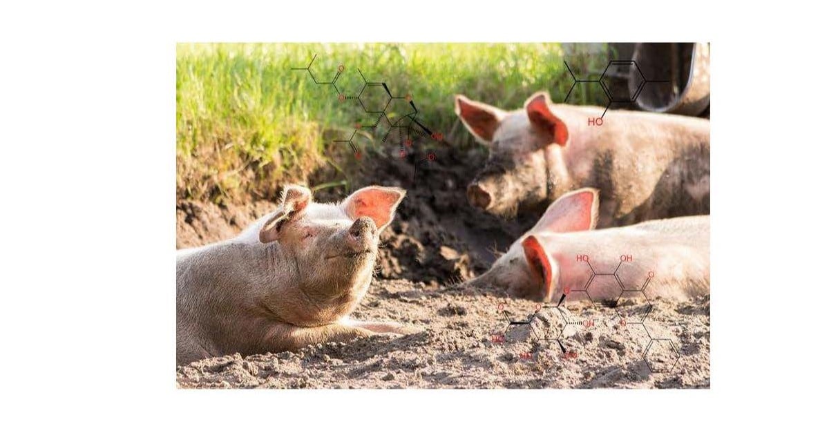
Nowadays, veterinary pharmacology and toxicology should be regarded as disciplines contributing to the “One Health” paradigm, reducing risk at the animal–human ecosystem. The use of antibiotics plays a crucial role in maintaining the safety, health, and rapid development of the animal husbandry industry. In the past 30 years, the fields of veterinary pharmacology and toxicology have experienced rapid development and achieved a series of important results. For example, in 2015, Chinese scientists from the fields of veterinary pharmacology and toxicology first discovered the colistin resistance gene MCR-1. Currently, the development of new veterinary drugs and the rational use of existing antibacterial drugs are hot topics in this field, and some progress has been made in recent years. Therefore, we call on scientists in this field to submit their latest research, including but not limited to bacterial resistance, combination therapy, drug toxicology and molecular mechanisms, and new drug development. We hope to attract more scholars to veterinary pharmacology and toxicology and promote the exchange and development of these disciplines through this Topic.

Prof. Dr. Chongshan Dai
Prof. Dr. Jichang Li
Topic Editors

Keywords

  • veterinary science
  • drug therapy
  • natural products
  • pharmacology
  • toxicology
  • antioxidants
  • combination therapy
  • cell death
  • bacterial infection

Participating Journals

Journal Name Impact Factor CiteScore Launched Year First Decision (median) APC
Antioxidants
antioxidants
6.0 10.6 2012 16.9 Days CHF 2900 Submit
Journal of Xenobiotics
jox
6.8 5.3 2011 28 Days CHF 1600 Submit
Metabolites
metabolites
3.5 5.7 2011 16.1 Days CHF 2700 Submit
Molecules
molecules
4.2 7.4 1996 15.1 Days CHF 2700 Submit
Toxics
toxics
3.9 4.5 2013 18.3 Days CHF 2600 Submit
Veterinary Sciences
vetsci
2.0 2.9 2014 21.2 Days CHF 2100 Submit
International Journal of Molecular Sciences
ijms
4.9 8.1 2000 16.8 Days CHF 2900 Submit
Biomolecules
biomolecules
4.8 9.4 2011 18.4 Days CHF 2700 Submit

Preprints.org is a multidisciplinary platform offering a preprint service designed to facilitate the early sharing of your research. It supports and empowers your research journey from the very beginning.

MDPI Topics is collaborating with Preprints.org and has established a direct connection between MDPI journals and the platform. Authors are encouraged to take advantage of this opportunity by posting their preprints at Preprints.org prior to publication:

  1. Share your research immediately: disseminate your ideas prior to publication and establish priority for your work.
  2. Safeguard your intellectual contribution: Protect your ideas with a time-stamped preprint that serves as proof of your research timeline.
  3. Boost visibility and impact: Increase the reach and influence of your research by making it accessible to a global audience.
  4. Gain early feedback: Receive valuable input and insights from peers before submitting to a journal.
  5. Ensure broad indexing: Web of Science (Preprint Citation Index), Google Scholar, Crossref, SHARE, PrePubMed, Scilit and Europe PMC.

Published Papers (11 papers)

Order results
Result details
Journals
Select all
Export citation of selected articles as:
22 pages, 13238 KiB  
Article
Euphorbia humifusa Willd. ex Schltdl. Mitigates Liver Injury via KEAP1-NFE2L2-Mediated Ferroptosis Regulation: Network Pharmacology and Experimental Validation
by Hongxu Du, Kunzhao Yang, Jingyi Yang, Junjie Wan, Yu Pan, Weijie Song, Shuang Xu, Cheng Chen and Jiahui Li
Vet. Sci. 2025, 12(4), 350; https://doi.org/10.3390/vetsci12040350 - 9 Apr 2025
Viewed by 163
Abstract
Liver injury poses major health risks in livestock, necessitating effective therapeutic interventions. This study elucidates the hepatoprotective mechanisms of Euphorbia humifusa Willd. ex Schltdl. (EHW) by integrating network pharmacology, molecular docking, and experimental validation. Using a CCl4-induced liver injury model mimicking [...] Read more.
Liver injury poses major health risks in livestock, necessitating effective therapeutic interventions. This study elucidates the hepatoprotective mechanisms of Euphorbia humifusa Willd. ex Schltdl. (EHW) by integrating network pharmacology, molecular docking, and experimental validation. Using a CCl4-induced liver injury model mimicking veterinary clinical scenarios, EHW markedly alleviated hepatic damage, demonstrated by reduced liver index, serum ALT and AST levels, histopathological lesions, iron accumulation, inflammatory cytokines, and ferroptosis-associated gene expression. Network pharmacology identified EHW’s core bioactive components (quercetin, kaempferol, and β-sitosterol) and critical targets (IL-6, STAT3, HIF-1α, PTGS2, NFE2L2, and KEAP1) which were linked to ferroptosis and oxidative stress. Molecular docking revealed robust binding affinities between these compounds and ferroptosis-related proteins. In vivo validation confirmed that EHW inhibited KEAP1, activated NFE2L2-mediated antioxidant defenses (upregulating SOD1 and NQO1), restored iron homeostasis (lowering TFR1, elevating FTH1), and attenuated phospholipid peroxidation by suppressing ACSL4 and ALOX12. These results indicate that EHW mitigates ferroptosis-driven liver injury via KEAP1-NFE2L2 signaling to restore iron homeostasis and reduce oxidative stress, offering a mechanistic foundation for its clinical application in veterinary hepatoprotection. Full article
(This article belongs to the Topic Recent Advances in Veterinary Pharmacology and Toxicology)
Show Figures

Figure 1

20 pages, 4095 KiB  
Article
Baicalin, Amoxicillin, and Probenecid Provide Protection in Mice Against Glaesserella parasuis Challenge
by Jingyang Li, Ronghui Luo, Yunjian Fu, Siyu Liu, Qiaoli Dong, Yamin Sun, Xinyue Tian, Yi Zhu, Peiyi Wang, Ling Guo, Qirong Lu, Chun Ye, Shulin Fu and Yinsheng Qiu
Biomolecules 2025, 15(4), 507; https://doi.org/10.3390/biom15040507 - 31 Mar 2025
Viewed by 125
Abstract
Glaesserella parasuis (G. parasuis) causes Glässer’s disease and systemic inflammatory responses in the host. The currently available therapies have limited efficacy and fail to achieve a balance between anti-inflammatory and antibacterial effects. In this study, we investigated the effects of baicalin, [...] Read more.
Glaesserella parasuis (G. parasuis) causes Glässer’s disease and systemic inflammatory responses in the host. The currently available therapies have limited efficacy and fail to achieve a balance between anti-inflammatory and antibacterial effects. In this study, we investigated the effects of baicalin, amoxicillin, and probenecid on blood biochemical parameters, routine blood indicators, survival rate, bacterial burden, and pathological tissue damage in G. parasuis-challenged mice. Treatment with baicalin, amoxicillin, and probenecid significantly modified the blood biochemical parameters and routine blood test indicators, increased the survival rate, attenuated the bacterial burden, and alleviated pathological tissue damage in G. parasuis-challenged mice. Treatment with baicalin, amoxicillin, and probenecid also increased the number of CD3+, CD3+CD4+, and CD3+CD8+ T cells as measured by flow cytometry, and restored the intensity of the CD3, CD4, and CD8 protein expression in the blood vessels of G. parasuis-challenged mice by immunohistochemistry. These compounds reduced interleukin 1β (IL-1β), IL-18, tumor necrosis factor alpha (TNF-α), and high mobility group box 1 protein (HMGB1) expression in the spleen of G. parasuis-challenged mice. Furthermore, baicalin, amoxicillin, and probenecid inhibited activation of the family pyrin domain containing 3 (NLRP3) inflammasome and apoptosis in the spleen of G. parasuis-challenged mice. This study showed the important roles of baicalin, amoxicillin, and probenecid in the modulation of the inflammatory response of Glässer’s disease. The findings might provide new strategies for combination therapy using antibiotics and anti-inflammatory drugs to control G. parasuis infection. Full article
(This article belongs to the Topic Recent Advances in Veterinary Pharmacology and Toxicology)
Show Figures

Figure 1

31 pages, 2066 KiB  
Review
Recent Advances in Pretreatment Methods and Detection Techniques for Veterinary Drug Residues in Animal-Derived Foods
by Qing Dai, Shusheng Tang and Chongshan Dai
Metabolites 2025, 15(4), 233; https://doi.org/10.3390/metabo15040233 - 28 Mar 2025
Viewed by 246
Abstract
Veterinary drugs are extensively employed in livestock, poultry, and aquaculture, playing a crucial role in preventing and treating animal diseases, facilitating growth, and enhancing feed conversion rates. Nevertheless, veterinary drug residues in animal-derived foods pose a direct or potential threat to human life [...] Read more.
Veterinary drugs are extensively employed in livestock, poultry, and aquaculture, playing a crucial role in preventing and treating animal diseases, facilitating growth, and enhancing feed conversion rates. Nevertheless, veterinary drug residues in animal-derived foods pose a direct or potential threat to human life and health. Precise detection of these residues in animal-derived foods to ensure their safety has become an important mission. In this review, we sum up the current progress of applied pretreatment methods and detection techniques for veterinary drug residues in animal-derived foods. At present, sample pretreatment methods mainly consist of the following: liquid–liquid extraction; solid-phase extraction; immunoaffinity chromatography; Quick, Easy, Cheap, Effective, Rugged, and Safe (QuEChERS) method; and molecular imprinting technology. Detection techniques mainly involve chromatographic techniques, immunoassay techniques, fluorescence polarization immunoassay, and surface-enhanced Raman scattering. We also discussed the advantages and limitations of these technologies. Moreover, we point out the development direction and tendency of detection techniques in the future, providing references for the detection of veterinary drug residues in animal-derived foods. Full article
(This article belongs to the Topic Recent Advances in Veterinary Pharmacology and Toxicology)
Show Figures

Figure 1

26 pages, 1598 KiB  
Review
The Intestinal Macrophage–Intestinal Stem Cell Axis in Inflammatory Bowel Diseases: From Pathogenesis to Therapy
by Tao Quan, Ran Li and Ting Gao
Int. J. Mol. Sci. 2025, 26(7), 2855; https://doi.org/10.3390/ijms26072855 - 21 Mar 2025
Viewed by 233
Abstract
The gut plays a crucial role in digestion and immunity, so its balance is essential to overall health. This balance relies on dynamic interactions between intestinal epithelial cells, immune cells, and crypt stem cells. Inflammatory bowel disease (IBD), which consists of ulcerative colitis [...] Read more.
The gut plays a crucial role in digestion and immunity, so its balance is essential to overall health. This balance relies on dynamic interactions between intestinal epithelial cells, immune cells, and crypt stem cells. Inflammatory bowel disease (IBD), which consists of ulcerative colitis and Crohn’s disease, is a chronic relapsing inflammatory disease of the gastrointestinal tract closely related to immune dysfunction. Stem cells, known for their ability to self-renew and differentiate, play an important role in repairing damaged intestinal epithelium and maintaining homeostasis in vivo. Macrophages are key gatekeepers of intestinal immune homeostasis and have a significant impact on IBD. Current research has focused on the link between epithelial cells and stem cells, but interactions with macrophages, which have been recognized as attractive targets for the development of new therapeutic approaches to disease, have been less explored. Recently, the developing field of immunometabolism has reinforced that metabolic reprogramming is a key determinant of macrophage function and subsequent disease progression. The aim of this review is to explore the role of the macrophage–stem cell axis in the maintenance of intestinal homeostasis and to summarize potential approaches to treating IBD by manipulating the cellular metabolism of macrophages, as well as the main opportunities and challenges faced. In summary, our overview provides a framework for understanding the critical role of macrophage immunometabolism in maintaining gut health and potential therapeutic targets. Full article
(This article belongs to the Topic Recent Advances in Veterinary Pharmacology and Toxicology)
Show Figures

Figure 1

20 pages, 1244 KiB  
Review
Role of Mitochondrial Dynamics in Skin Homeostasis: An Update
by Tao Quan, Ran Li and Ting Gao
Int. J. Mol. Sci. 2025, 26(5), 1803; https://doi.org/10.3390/ijms26051803 - 20 Feb 2025
Viewed by 736
Abstract
Skin aging is the most prominent phenotype of host aging and is the consequence of a combination of genes and environment. Improving skin aging is essential for maintaining the healthy physiological function of the skin and the mental health of the human body. [...] Read more.
Skin aging is the most prominent phenotype of host aging and is the consequence of a combination of genes and environment. Improving skin aging is essential for maintaining the healthy physiological function of the skin and the mental health of the human body. Mitochondria are vital organelles that play important roles in cellular mechanisms, including energy production and free radical balance. However, mitochondrial metabolism, mitochondrial dynamics, biogenesis, and degradation processes vary greatly in various cells in the skin. It is well known that mitochondrial dysfunction can promote the aging and its associated diseases of the skin, resulting in the damage of skin physiology and the occurrence of skin pathology. In this review, we summarize the important role of mitochondria in various skin cells, review the cellular responses to vital steps in mitochondrial quality regulation, mitochondrial dynamics, mitochondrial biogenesis, and mitochondrial phagocytosis, and describe their importance and specific pathways in skin aging. Full article
(This article belongs to the Topic Recent Advances in Veterinary Pharmacology and Toxicology)
Show Figures

Figure 1

23 pages, 50146 KiB  
Article
Isorhamnetin Ameliorates Non-Esterified Fatty Acid-Induced Apoptosis, Lipid Accumulation, and Oxidative Stress in Bovine Endometrial Epithelial Cells via Inhibiting the MAPK Signaling Pathway
by Haimiao Lv, Lijuan Liu, Wenna Zou, Ying Yang, Yuan Li, Shengji Yang, Aixin Liang and Liguo Yang
Antioxidants 2025, 14(2), 156; https://doi.org/10.3390/antiox14020156 - 28 Jan 2025
Viewed by 775
Abstract
High concentrations of non-esterified fatty acids (NEFA) in the blood contribute to various metabolic disorders and are linked to endometritis in dairy cows. Isorhamnetin (ISO), a flavonoid found in many plants, is known for its antioxidant, anti-inflammatory, and anti-obesity properties. This study systematically [...] Read more.
High concentrations of non-esterified fatty acids (NEFA) in the blood contribute to various metabolic disorders and are linked to endometritis in dairy cows. Isorhamnetin (ISO), a flavonoid found in many plants, is known for its antioxidant, anti-inflammatory, and anti-obesity properties. This study systematically assessed NEFA-induced damage in bovine endometrial epithelial cells (bEECs) and investigated whether ISO alleviates NEFA-induced cell damage and its underlying molecular mechanisms. Our observations revealed that excessive NEFA inhibited proliferation and induced apoptosis in bEECs, accompanied by an increase in the expression of BAX and cleaved caspase-3. We further observed that NEFA could induce lipid accumulation, reactive oxygen species (ROS) generation, and the release of pro-inflammatory factors IL-1β, IL-6, and TNF-α in bEECs. RNA sequencing and Western blot analysis revealed that NEFA induced damage in bEECs by activating MAPK signaling pathway. Notably, ISO treatment ameliorated these effects induced by NEFA, as evidenced by decreased protein levels of BAX, cleaved caspase-3, and PPAR-γ, along with reductions in triglyceride content, ROS generation, and levels of IL-1β, IL-6, and TNF-α. Mechanistically, our experimental results demonstrated that ISO inhibited NEFA-induced activation of MAPK signaling. Overall, ISO shows promise for therapeutic development to address NEFA-related endometritis in dairy cows. Full article
(This article belongs to the Topic Recent Advances in Veterinary Pharmacology and Toxicology)
Show Figures

Figure 1

11 pages, 3373 KiB  
Communication
Tea Polyphenols Relieve the Fluoride-Induced Oxidative Stress in the Intestinal Porcine Epithelial Cell Model
by Chunyan Xie, Shuyi Niu and Wen Tian
Toxics 2025, 13(2), 83; https://doi.org/10.3390/toxics13020083 - 24 Jan 2025
Viewed by 686
Abstract
Prolonged excessive intake of fluoride (F) can result in fluorosis, leading to a range of tissue oxidative damages. Therefore, mitigating the oxidative stress induced by fluorosis has become a significant research concern. Consequently, how to relieve oxidative stress caused by fluorosis is an [...] Read more.
Prolonged excessive intake of fluoride (F) can result in fluorosis, leading to a range of tissue oxidative damages. Therefore, mitigating the oxidative stress induced by fluorosis has become a significant research concern. Consequently, how to relieve oxidative stress caused by fluorosis is an urgent matter. In the present study, intestinal porcine epithelial (IPEC-J2) cells were chosen to explore the underlying mechanism of tea polyphenols (TPs) on F-induced oxidative stress. The results show that the cytotoxicity of IPEC-J2 cells induced by F presented a dose-dependent manner according to cell viability. Additionally, F treatment inhibited the activity of T-SOD, CAT, and GSH-Px as well as their transcription levels, increased the reactive oxygen (ROS) formation and cell damage rates, and then promoted cell apoptosis through the results of TUNEL and mitochondrial membrane potential detection when compared with the IPEC-J2 cells from the control group. As the main antioxidant ingredient in tea, TPs alleviated F-induced cell oxidation and apoptosis via blocking F-induced ROS generation and LDH’s release, as well as promoting the transcription of tight junction (TJ) proteins and the activities of antioxidant enzymes in IPEC-J2 cells. These results provide a new treatment strategy for F-induced intestinal oxidative impairment. Full article
(This article belongs to the Topic Recent Advances in Veterinary Pharmacology and Toxicology)
Show Figures

Graphical abstract

19 pages, 4894 KiB  
Article
Extraction and Identification of Polysaccharide from Lentinus edodes and Its Effect on Immunosuppression and Intestinal Barrier Injury Induced by Cyclophosphamide
by Xiaodi Jin, Zhiyong Wu, Hao Chen, Weiqi Liu, Fuhua Gu and Jichang Li
Int. J. Mol. Sci. 2024, 25(22), 12432; https://doi.org/10.3390/ijms252212432 - 19 Nov 2024
Cited by 1 | Viewed by 1125
Abstract
Lentinus edodes serves as a significant source of both medicine and food, with its key component, lentinan (LNT), recognized as an effective immunomodulator. However, the mechanisms by which it regulates immune and intestinal functions under conditions of immunosuppression remain unclear. This study aims [...] Read more.
Lentinus edodes serves as a significant source of both medicine and food, with its key component, lentinan (LNT), recognized as an effective immunomodulator. However, the mechanisms by which it regulates immune and intestinal functions under conditions of immunosuppression remain unclear. This study aims to investigate the components of lentinan and examine its potential effects on countering cyclophosphamide (CP)-induced immunosuppression, intestinal barrier damage, and dysregulation of gut microbiota. In this study, the effects of LNT were evaluated by serological indicators, histopathological changes in ileum, tight-junction-related protein expression, cytokine expression levels, and gut microbiota 16S rRNA gene sequencing. We found that LNT was effective in mitigating the abnormalities in body weight, immune organ index, and serum levels of IL-6, IL-2, IFN-γ, and IgG in mice induced by CP (p < 0.05). Furthermore, LNT demonstrated the ability to alleviate intestinal barrier damage induced by CP by increasing the mRNA levels of TNF-α, IL-1β, IFN-γ, Occludin, and ZO-1 (p < 0.05). Additionally, 16S rRNA gene sequencing revealed that LNT also normalized the disrupted abundance of Firmicutes, Proteobacteria, and Bacteroidets caused by CP. This restoration brought the gut microbiota back to normal levels and increased the abundance of certain tumor-inhibiting bacteria, such as Alistipes. Overall, lentinan demonstrated the ability to reverse the immunosuppressive effects induced by cyclophosphamide and modulate gut microbiota to restore a healthy microbial balance. Full article
(This article belongs to the Topic Recent Advances in Veterinary Pharmacology and Toxicology)
(This article belongs to the Section Bioactives and Nutraceuticals)
Show Figures

Graphical abstract

18 pages, 4460 KiB  
Article
Ellagic Acid Reduces Cadmium Exposure-Induced Apoptosis in HT22 Cells via Inhibiting Oxidative Stress and Mitochondrial Dysfunction and Activating Nrf2/HO-1 Pathway
by Yue Liu, Chunhong Chen, Zhihui Hao, Jianzhong Shen, Shusheng Tang and Chongshan Dai
Antioxidants 2024, 13(11), 1296; https://doi.org/10.3390/antiox13111296 - 25 Oct 2024
Cited by 1 | Viewed by 1341
Abstract
Exposure to cadmium sulfate (CdSO4) can lead to neurotoxicity. Nevertheless, the precise molecular mechanisms underlying this phenomenon remain unclear, and effective treatment strategies are scarce. This study explored the protective effects of ellagic acid (EA), a natural polyphenolic compound, against CdSO [...] Read more.
Exposure to cadmium sulfate (CdSO4) can lead to neurotoxicity. Nevertheless, the precise molecular mechanisms underlying this phenomenon remain unclear, and effective treatment strategies are scarce. This study explored the protective effects of ellagic acid (EA), a natural polyphenolic compound, against CdSO4 exposure-induced neurotoxicity in HT22 cells and the underlying molecular mechanisms. Our findings demonstrated that exposure of HT22 cells to CdSO4 resulted in apoptosis, which was effectively reversed by EA in a dose-dependent manner. EA supplementation also decreased reactive oxygen species (ROS) and mitochondrial ROS production, reduced malondialdehyde (MDA) levels, and restored the activities of superoxide dismutase (SOD) and catalase (CAT). Additionally, EA supplementation at 5–20 μM significantly counteracted Cd-induced the loss of mitochondrial membrane potential and the decrease of ATP and reduced the ratio of Bax/Bcl-2 and cleaved-caspase-3 protein expression. Furthermore, EA supplementation resulted in the upregulation of Nrf2 and HO-1 protein and mRNAs while simultaneously downregulating the phosphorylation of JNK and p38 proteins. The pharmacological inhibition of c-Jun N-terminal kinase (JNK) partially attenuated the activation of the Nrf2/HO-1 pathway induced by CdSO4 and exacerbated its cytotoxic effects. In conclusion, our findings suggest that ethyl acetate (EA) supplementation offers protective effects against CdSO4-induced apoptosis in HT22 cells by inhibiting oxidative stress and activating the Nrf2 signaling pathway. Furthermore, the activation of the JNK pathway appears to play a protective role in CdSO4-induced apoptosis in HT22 cells. Full article
(This article belongs to the Topic Recent Advances in Veterinary Pharmacology and Toxicology)
Show Figures

Figure 1

24 pages, 5596 KiB  
Article
Rosmarinic Acid Attenuates Salmonella enteritidis-Induced Inflammation via Regulating TLR9/NF-κB Signaling Pathway and Intestinal Microbiota
by Dandan Yi, Menghui Wang, Xia Liu, Lanqian Qin, Yu Liu, Linyi Zhao, Ying Peng, Zhengmin Liang and Jiakang He
Antioxidants 2024, 13(10), 1265; https://doi.org/10.3390/antiox13101265 - 18 Oct 2024
Cited by 2 | Viewed by 1411
Abstract
Salmonella enteritidis (SE) infection disrupts the homeostasis of the intestinal microbiota, causing an intestinal inflammatory response and posing a great threat to human and animal health. The unreasonable use of antibiotics has led to an increase in the prevalence of drug-resistant [...] Read more.
Salmonella enteritidis (SE) infection disrupts the homeostasis of the intestinal microbiota, causing an intestinal inflammatory response and posing a great threat to human and animal health. The unreasonable use of antibiotics has led to an increase in the prevalence of drug-resistant SE, increasing the difficulty of controlling SE. Therefore, new drug strategies and research are urgently needed to control SE. Rosmarinic acid (RA) is a natural phenolic acid with various pharmacological activities, including antioxidant, anti-inflammatory and antibacterial properties. However, the protective effects and mechanism of RA on intestinal inflammation and the gut microbial disorders caused by SE have not been fully elucidated. In this study, RAW264.7 cells, MCECs and BALB/c mice were challenged with SE to assess the protective effects and mechanisms of RA. The results showed that RA enhanced the phagocytic ability of RAW264.7 cells, reduced the invasion and adhesion ability of SE in MCECs, and inhibited SE-induced inflammation in cells. Moreover, RA inhibited the activation of the NF-κB signaling pathway by upregulating TLR9 expression. Importantly, we found that RA provided protection against SE and increased the diversity and abundance of the intestinal microbiota in mice. Compared with infection control, RA significantly increased the abundance of Firmicutes and Acidibacteria and decreased the abundance of Proteobacteria, Epsilonbacteraeota and Bacteroidota. However, RA failed to alleviate SE-induced inflammation and lost its regulatory effects on the TLR9/NF-κB signaling pathway after destroying the gut microbiota with broad-spectrum antibiotics. These results indicated that RA attenuated SE-induced inflammation by regulating the TLR9/NF-κB signaling pathway and maintaining the homeostasis of the gut microbiota. Our study provides a new strategy for preventing SE-induced intestinal inflammation. Full article
(This article belongs to the Topic Recent Advances in Veterinary Pharmacology and Toxicology)
Show Figures

Graphical abstract

21 pages, 2419 KiB  
Article
Ozone Therapy in the Integrated Treatment of Female Dogs with Mammary Cancer: Oxidative Profile and Quality of Life
by Laís Pereira Silva, Ricardo Wagner Portela, Marília Carneiro Machado, Gisele André Baptista Canuto, João Moreira Costa-Neto, Vitor de Moraes Pina de Carvalho, Hanna Carvalho de Sá, Karine Araújo Damasceno, Vinicius Ricardo Cuña de Souza, Clarisse Simões Coelho and Alessandra Estrela-Lima
Antioxidants 2024, 13(6), 673; https://doi.org/10.3390/antiox13060673 - 30 May 2024
Cited by 1 | Viewed by 2149
Abstract
Considering the high frequency of malignant breast tumors, there is a growing search for new therapeutic strategies that control neoplastic growth and dissemination, combined with fewer adverse reactions. Therefore, this study evaluated the effects of ozone therapy in female dogs with mammary cancer [...] Read more.
Considering the high frequency of malignant breast tumors, there is a growing search for new therapeutic strategies that control neoplastic growth and dissemination, combined with fewer adverse reactions. Therefore, this study evaluated the effects of ozone therapy in female dogs with mammary cancer undergoing chemotherapy treatment. Twenty-five canines diagnosed with malignant mammary neoplasia were divided into two groups: one treated with carboplatin alone (n = 11) and the other with carboplatin associated with ozone therapy (n = 14). Clinical and laboratory evaluations, mastectomy, analysis of the oxidative profile based on total antioxidant capacity (TAC) and serum concentrations of malondialdehyde (MDA), survival rate, and quality of life were performed. Animals in the ozone therapy group had higher concentrations of red blood cells and platelets, significantly improving the survival rate and quality of life. Furthermore, adverse reactions were less intense and frequent in this group, which was associated with an increase in TAC and a reduction in MDA. These results indicate that the combination of carboplatin and ozone therapy represents a promising complementary treatment for female dogs with mammary cancer, as it was associated with fewer adverse reactions and a better oxidative profile. Full article
(This article belongs to the Topic Recent Advances in Veterinary Pharmacology and Toxicology)
Show Figures

Figure 1

Back to TopTop