Next Issue
Volume 13, March-2
Previous Issue
Volume 13, February-2


Cancers, Volume 13, Issue 5 (March-1 2021) – 256 articles

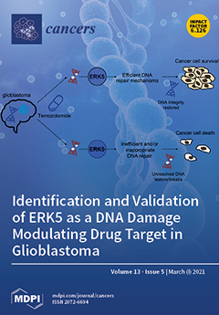
Cover Story (view full-size image): Brain tumours kill children and adults under 40 than any other cancer, with approximately half of primary brain tumours diagnosed as high-grade glioblastomas that account for ~190,000 deaths/year globally. The mean survival for these patients of ~15 months has improved little over the last 40 years, with less than 10% of patients surviving over 5 years, highlighting an urgent need to identify new drug targets for this disease. In this issue, Carmell et al. identify ERK5/MAPK7 as a previously unappreciated factor in the cellular response to standard-of-care chemotherapy drug temozolomide (TMZ). They further show that ERK5 promotes the efficient repair of TMZ-induced DNA damage and cell survival and that high levels of ERK5 are associated with high-grade tumours and poor survival rates. This work therefore identifies ERK5 as a potential novel drug target in this currently incurable disease. View this [...] Read more.
  • Issues are regarded as officially published after their release is announced to the table of contents alert mailing list.
  • You may sign up for e-mail alerts to receive table of contents of newly released issues.
  • PDF is the official format for papers published in both, html and pdf forms. To view the papers in pdf format, click on the "PDF Full-text" link, and use the free Adobe Reader to open them.
Order results
Result details
Section
Select all
Export citation of selected articles as:
10 pages, 243 KiB  
Article
Aspirin and Statin Use and the Risk of Gallbladder Cancer
by Kritika Prasai, Sri Harsha Tella, Siddhartha Yadav, Anuhya Kommalapati, Kristin Mara, Mohamed Mady, Mohamed A. Hassan, Nicha Wongjarupong, Natalia Rodriguez-Payan, Mitesh Borad, Tushar Patel, Lewis R. Roberts and Amit Mahipal
Cancers 2021, 13(5), 1186; https://doi.org/10.3390/cancers13051186 - 9 Mar 2021
Cited by 5 | Viewed by 2588
Abstract
Aspirin and statin drugs have been associated with reduced risk of several gastrointestinal cancers, but their association with gallbladder cancer (GBC) has not been well established. We evaluated the association of aspirin and statins with the risk of GBC. Patients with GBC managed [...] Read more.
Aspirin and statin drugs have been associated with reduced risk of several gastrointestinal cancers, but their association with gallbladder cancer (GBC) has not been well established. We evaluated the association of aspirin and statins with the risk of GBC. Patients with GBC managed at Mayo Clinic between 2000 and 2019 were matched 1:2 with a general patient pool by age and sex. Univariable and multivariable logistic regression models were used to assess associations between GBC and aspirin or statin use. The analysis included 795 cases and 1590 controls, with a median age of 67 years. Aspirin or statin use alone or in combination was higher in controls (p < 0.001). Univariate analysis showed that the use of aspirin [odds ratio (OR): 0.11; 95%CI: 0.08–0.15] or statins (OR: 0.29; 95%CI: 0.20–0.40) and their combined use (OR: 0.18; 95%CI: 0.13–0.24) was associated with lower risk of GBC. Multivariable analysis revealed that aspirin (OR: 0.12; 95%CI: 0.09–0.16) and combined statins and aspirin (OR: 0.46; 95%CI: 0.31–0.67) were associated with lower risk of GBC. Aspirin alone or in combination with statins is associated with a strongly reduced risk of GBC. Further prospective studies are needed to confirm these results and to elucidate their mechanisms. Full article
(This article belongs to the Special Issue Research Progress of Biliary Tract Cancers)
16 pages, 11709 KiB  
Article
Silk Fibroin Nanoparticle Functionalization with Arg-Gly-Asp Cyclopentapeptide Promotes Active Targeting for Tumor Site-Specific Delivery
by Elia Bari, Massimo Serra, Mayra Paolillo, Eric Bernardi, Sara Tengattini, Filippo Piccinini, Cristina Lanni, Marzio Sorlini, Giovanni Bisbano, Enrica Calleri, Maria Luisa Torre and Sara Perteghella
Cancers 2021, 13(5), 1185; https://doi.org/10.3390/cancers13051185 - 9 Mar 2021
Cited by 21 | Viewed by 3801
Abstract
Arg-Gly-Asp (RGD)-based cyclopentapeptides (cRGDs) have a high affinity towards integrin αvβ3 and αvβ5, which are overexpressed by many tumor cells. Here, curcumin-loaded silk fibroin nanoparticles (SFNs) have been functionalized on the surface with cRGD to provide active targeting towards tumor cells; a “click [...] Read more.
Arg-Gly-Asp (RGD)-based cyclopentapeptides (cRGDs) have a high affinity towards integrin αvβ3 and αvβ5, which are overexpressed by many tumor cells. Here, curcumin-loaded silk fibroin nanoparticles (SFNs) have been functionalized on the surface with cRGD to provide active targeting towards tumor cells; a “click reaction” between the RGD-based cyclopentapeptide carrying an azide group and triple-bond-functionalized nanoparticles has been exploited. Both naked and functionalized SFNs were less than 200 nm in diameter and showed a round-shaped morphology but, after functionalization, SFNs increased in size and protein molecular weight. The functionalization of SFNs’ surfaces with cRGD provided active internalization by cells overexpressing integrin receptors. At the lowest concentration tested (0.01 mg/mL), functionalized SFNs showed more effective uptake with respect to the naked by tumor cells that overexpress integrin receptors (but not for non-overexpressing ones). In contrast, at higher concentrations, the non-specific cell membrane protein–particle interactions are promoted and coupled to specific and target mediated uptake. Visual observations by fluorescence microscopy suggested that SFNs bind to integrin receptors on the cell surface and are then internalized by endocytosis. Overall, SFN functionalization provided in vitro active targeting for site-specific delivery of anticancer drugs, boosting activity and sparing healthy organs. Full article
Show Figures

Figure 1

19 pages, 10754 KiB  
Article
Integrin α10-Antibodies Reduce Glioblastoma Tumor Growth and Cell Migration
by Katarzyna Chmielarska Masoumi, Xiaoli Huang, Wondossen Sime, Anna Mirkov, Matilda Munksgaard Thorén, Ramin Massoumi and Evy Lundgren-Åkerlund
Cancers 2021, 13(5), 1184; https://doi.org/10.3390/cancers13051184 - 9 Mar 2021
Cited by 4 | Viewed by 3521
Abstract
Glioblastoma (GB) is the most common and the most aggressive form of brain tumor in adults, which currently lacks efficient treatment strategies. In this study, we investigated the therapeutic effect of function-blocking antibodies targeting integrin α10β1 on patient-derived-GB cell lines in vitro and [...] Read more.
Glioblastoma (GB) is the most common and the most aggressive form of brain tumor in adults, which currently lacks efficient treatment strategies. In this study, we investigated the therapeutic effect of function-blocking antibodies targeting integrin α10β1 on patient-derived-GB cell lines in vitro and in vivo. The in vitro studies demonstrated significant inhibiting effects of the integrin α10 antibodies on the adhesion, migration, proliferation, and sphere formation of GB cells. In a xenograft mouse model, the effect of the antibodies on tumor growth was investigated in luciferase-labeled and subcutaneously implanted GB cells. As demonstrated by in vivo imaging analysis and caliper measurements, the integrin α10-antibodies significantly suppressed GB tumor growth compared to control antibodies. Immunohistochemical analysis of the GB tumors showed lower expression of the proliferation marker Ki67 and an increased expression of cleaved caspase-3 after treatment with integrin α10 antibodies, further supporting a therapeutic effect. Our results suggest that function-blocking antibody targeting integrin α10β1 is a promising therapeutic strategy for the treatment of glioblastoma. Full article
(This article belongs to the Special Issue Targeted Therapies for the Treatment of Glioblastoma)
Show Figures

Figure 1

13 pages, 2779 KiB  
Article
Combined De-Repression of Chemoresistance Associated Mitogen-Activated Protein Kinase 14 and Activating Transcription Factor 2 by Loss of microRNA-622 in Hepatocellular Carcinoma
by Valerie Fritz, Lara Malek, Anne Gaza, Laura Wormser, Majken Appel, Andreas E. Kremer, Wolfgang E. Thasler, Jürgen Siebler, Markus F. Neurath, Claus Hellerbrand, Anja K. Bosserhoff and Peter Dietrich
Cancers 2021, 13(5), 1183; https://doi.org/10.3390/cancers13051183 - 9 Mar 2021
Cited by 5 | Viewed by 2357
Abstract
Chemoresistance is a major hallmark driving the progression and poor prognosis of hepatocellular carcinoma (HCC). Limited chemoresponse of HCC was demonstrated to be mediated by mitogen-activated protein kinase 14 (MAPK14) and activating transcription factor 2 (ATF2). Recently, we have demonstrated loss of control [...] Read more.
Chemoresistance is a major hallmark driving the progression and poor prognosis of hepatocellular carcinoma (HCC). Limited chemoresponse of HCC was demonstrated to be mediated by mitogen-activated protein kinase 14 (MAPK14) and activating transcription factor 2 (ATF2). Recently, we have demonstrated loss of control of RAS-RAF-ERK-signaling as a consequence of miR-622 downregulation in HCC. However, the majority of target genes of this potent tumorsuppressive microRNA had remained elusive. The MAPK14-ATF2-axis represents a collateral pathway ensuring persisting ERK-activation in the presence of sorafenib-mediated RAF-inhibition. In contrast to the function of the MAPK14-ATF2-axis, both the expression and regulation of MAPK14 and ATF2 in human HCC remained to be clarified. We found combined overexpression of MAPK14 and ATF2 in human HCC cells, tissues and in sorafenib resistant cell lines. High expression of MAPK14 and ATF2 was associated with reduced overall survival in HCC patients. Deciphering the molecular mechanism promoting combined upregulation of MAPK14 and ATF2 in HCC, we revealed that miR-622 directly targets both genes, resulting in combined de-repression of the MAPK14-ATF2-axis. Together, miR-622 represents a superior regulator of both RAS-RAF-ERK as well as MAPK14-ATF2-signaling pathways in liver cancer. Full article
(This article belongs to the Special Issue Primary and Secondary Liver Tumors)
Show Figures

Figure 1

21 pages, 3379 KiB  
Review
Radiobiological Studies of Microvascular Damage through In Vitro Models: A Methodological Perspective
by Luca Possenti, Laura Mecchi, Andrea Rossoni, Veronica Sangalli, Simone Bersini, Alessandro Cicchetti, Maria Laura Costantino, Christian Candrian, Chiara Arrigoni, Tiziana Rancati and Matteo Moretti
Cancers 2021, 13(5), 1182; https://doi.org/10.3390/cancers13051182 - 9 Mar 2021
Cited by 7 | Viewed by 4621
Abstract
Ionizing radiation (IR) is used in radiotherapy as a treatment to destroy cancer. Such treatment also affects other tissues, resulting in the so-called normal tissue complications. Endothelial cells (ECs) composing the microvasculature have essential roles in the microenvironment’s homeostasis (ME). Thus, detrimental effects [...] Read more.
Ionizing radiation (IR) is used in radiotherapy as a treatment to destroy cancer. Such treatment also affects other tissues, resulting in the so-called normal tissue complications. Endothelial cells (ECs) composing the microvasculature have essential roles in the microenvironment’s homeostasis (ME). Thus, detrimental effects induced by irradiation on ECs can influence both the tumor and healthy tissue. In-vitro models can be advantageous to study these phenomena. In this systematic review, we analyzed in-vitro models of ECs subjected to IR. We highlighted the critical issues involved in the production, irradiation, and analysis of such radiobiological in-vitro models to study microvascular endothelial cells damage. For each step, we analyzed common methodologies and critical points required to obtain a reliable model. We identified the generation of a 3D environment for model production and the inclusion of heterogeneous cell populations for a reliable ME recapitulation. Additionally, we highlighted how essential information on the irradiation scheme, crucial to correlate better observed in vitro effects to the clinical scenario, are often neglected in the analyzed studies, limiting the translation of achieved results. Full article
(This article belongs to the Special Issue Cancer-on-a-Chip: Applications and Challenges)
Show Figures

Graphical abstract

14 pages, 4934 KiB  
Article
Hepatitis B Virus X Protein (HBx) Suppresses Transcription Factor EB (TFEB) Resulting in Stabilization of Integrin Beta 1 (ITGB1) in Hepatocellular Carcinoma Cells
by Chunyan Zhang, Huan Yang, Liwei Pan, Guangfu Zhao, Ruofei Zhang, Tianci Zhang, Zhixiong Xiao, Ying Tong, Yi Zhang, Richard Hu, Stephen J. Pandol and Yuan-Ping Han
Cancers 2021, 13(5), 1181; https://doi.org/10.3390/cancers13051181 - 9 Mar 2021
Cited by 12 | Viewed by 3559
Abstract
Hepatitis B virus (HBV) infection is a major etiological risk for the incidence of hepatocellular carcinoma (HCC), and HBV X protein (HBx) is essential for oncogenic transformation. It is not known that if HBx can sabotage the lysosomal system for transformation and tumorigenesis, [...] Read more.
Hepatitis B virus (HBV) infection is a major etiological risk for the incidence of hepatocellular carcinoma (HCC), and HBV X protein (HBx) is essential for oncogenic transformation. It is not known that if HBx can sabotage the lysosomal system for transformation and tumorigenesis, or its mechanism if it does have an effect. Examining clinical data, we observed that the downregulation of lysosomal components and transcription factor EB (TFEB) was associated with a poor prognosis of HCC patients. In HCC cells, we found that expression of HBx suppressed TFEB, impaired biogenesis of autophagic-lysosome, and promoted cellular dissemination. HBx mediated downregulation of TFEB led to impairment of autophagic/lysosomal biogenesis and flux, and consequently, accumulation of integrin beta 1 (ITGB1) for motility of HCC cells. Conversely, TFEB, in a steady-state condition, through induction of lysosomal biogenesis restrained ITGB1 levels and limited mobility of HCC cells. Specifically, overexpression of TFEB upregulated and activated the cysteine proteases including cathepsin L (CTSL) to degrade ITGB1. Conversely, expression of cystatin A (CSTA) or cystatin B (CSTB), the cellular inhibitors of lysosomal cysteine proteinases, spared ITGB1 from degradation and promoted dissemination of HCC cells. Taken together, this study suggests a potential mechanism for HBV-mediated malignancy, showing that HBx mediated downregulation of TFEB leads to accumulation of ITGB1 for HCC cell migration. Full article
(This article belongs to the Section Molecular Cancer Biology)
Show Figures

Figure 1

16 pages, 2033 KiB  
Article
Concurrent Cetuximab and Nivolumab as a Second-Line or beyond Treatment of Patients with Recurrent and/or Metastatic Head and Neck Squamous Cell Carcinoma: Results of Phase I/II Study
by Christine H. Chung, Marcelo Bonomi, Conor E. Steuer, Jiannong Li, Priyanka Bhateja, Matthew Johnson, Jude Masannat, Feifei Song, Juan C. Hernandez-Prera, Bruce M. Wenig, Helen Molina, Joaquim M. Farinhas, Caitlin P. McMullen, J. Trad Wadsworth, Krupal B. Patel, Julie A. Kish, Jameel Muzaffar, Kedar Kirtane, James W. Rocco, Michael J. Schell and Nabil F. Sabaadd Show full author list remove Hide full author list
Cancers 2021, 13(5), 1180; https://doi.org/10.3390/cancers13051180 - 9 Mar 2021
Cited by 36 | Viewed by 5025
Abstract
We hypothesized the combination of cetuximab and nivolumab would improve survival in recurrent and/or metastatic (R/M) HNSCC by providing synergy in cancer control and evaluated toxicities and efficacy of the combination. Effects of sequential administration of cetuximab and anti-Programmed Cell Death-1 checkpoint inhibitors [...] Read more.
We hypothesized the combination of cetuximab and nivolumab would improve survival in recurrent and/or metastatic (R/M) HNSCC by providing synergy in cancer control and evaluated toxicities and efficacy of the combination. Effects of sequential administration of cetuximab and anti-Programmed Cell Death-1 checkpoint inhibitors (CPI) were also explored. Patients who failed at least one line of palliative treatment for incurable HNSCC were treated with cetuximab 500 mg/m2 IV on Day (D)-14 as a lead-in followed by cetuximab 500 mg/m2 IV and nivolumab 240 mg/m2 IV on D1 and D15 every 28-D cycle. Electronic health record-derived real-world data (RWD) were used to explore sequential treatment effects of CPI and cetuximab. A total of 45 evaluable patients were analyzed, and 31/45 (69%) patients had prior exposure to either CPI or cetuximab. The only grade 4 treatment-related adverse event was cetuximab infusion reaction in one patient. The 1-year progression-free survival (PFS) and overall survival (OS) rates were 19% and 44%, respectively. Although patients with no prior CPI (23/45, 51%) showed a trend for more favorable PFS relative to patients with prior CPI (22/45, 49%), the improvement in the 1-year OS did not reach the statistical threshold. For evaluation of sequential CPI and cetuximab treatment effects, we selected RWD-cetuximab cohort with 173 patients and RWD-CPI cohort with 658 patients from 6862 R/M HNSCC. Our result suggested patients treated with RWD-cetuximab after RWD-CPI had worse OS compared to no prior RWD-CPI (HR 1.81, 95% CI 1.02–3.16). Our data suggest the combination of cetuximab and nivolumab is well tolerated. Optimal sequencing of cetuximab and CPI may have an impact in prognosis and requires further evaluation. Full article
(This article belongs to the Section Clinical Research of Cancer)
Show Figures

Figure 1

13 pages, 2100 KiB  
Article
Conversion Therapy of Intrahepatic Cholangiocarcinoma Is Associated with Improved Prognosis and Verified by a Case of Patient-Derived Organoid
by Zhiwei Wang, Yun Jin, Yinghao Guo, Zhenhua Tan, Xiaoxiao Zhang, Dan Ye, Yuanquan Yu, Shuyou Peng, Lei Zheng and Jiangtao Li
Cancers 2021, 13(5), 1179; https://doi.org/10.3390/cancers13051179 - 9 Mar 2021
Cited by 13 | Viewed by 3427
Abstract
This study was performed to determine the efficacy of conversion therapy in intrahepatic cholangiocarcinoma (IHCC) and explore the feasibility of cancer organoid to direct the conversion therapy of IHCC. Patient data were retrospectively reviewed in this study and cancer organoids were established using [...] Read more.
This study was performed to determine the efficacy of conversion therapy in intrahepatic cholangiocarcinoma (IHCC) and explore the feasibility of cancer organoid to direct the conversion therapy of IHCC. Patient data were retrospectively reviewed in this study and cancer organoids were established using tissues obtained from two patients. A total of 42 patients with IHCC received conversion therapy, 9 of whom were downstaged successfully, and another 157 patients were initially resectable. Kaplan–Meier curves showed that the successfully downstaged patients had a significantly improved overall survival compared to those in whom downstaging was unsuccessful (p = 0.017), and had a similar overall survival to that of initially resectable patients (p = 0.965). The IHCC organoid was successfully established from one of two obtained tissues. Routine hematoxylin and eosin staining and immunohistological staining found the organoid retained the histopathological characteristics of the original tissues. Whole exome sequencing results indicated the IHCC organoid retained appropriately 87% of the variants in the original tissue. Gemcitabine and paclitaxel exhibited the strongest inhibitory effects on the cancer organoid as determined using drug screening tests, consistent with the levels of efficacy observed in the patient from whom it was derived. This study indicates that conversion therapy could improve the survival of patients with IHCC despite its low success rate, and it may be directed by cancer organoids though this is merely a proof of feasibility. Full article
(This article belongs to the Special Issue Therapy in Gastrointestinal Oncology)
Show Figures

Graphical abstract

16 pages, 1371 KiB  
Article
Evolution of Surgical Treatment of Colorectal Liver Metastases in the Real World: Single Center Experience in 1212 Cases
by Francesca Ratti, Federica Cipriani, Guido Fiorentini, Valentina Burgio, Monica Ronzoni, Angelo Della Corte, Stefano Cascinu, Francesco De Cobelli and Luca Aldrighetti
Cancers 2021, 13(5), 1178; https://doi.org/10.3390/cancers13051178 - 9 Mar 2021
Cited by 7 | Viewed by 3195
Abstract
Background: In recent years, the treatment of colorectal liver metastases (CRLM) has undergone significant evolution thanks to technical improvements as well as oncological advances, which have been the subject of targeted studies aimed at understanding the details of this heterogeneous disease. The purpose [...] Read more.
Background: In recent years, the treatment of colorectal liver metastases (CRLM) has undergone significant evolution thanks to technical improvements as well as oncological advances, which have been the subject of targeted studies aimed at understanding the details of this heterogeneous disease. The purpose of this study is to put together pieces of this complex scenario by providing an overview of the evolution that has occurred in the context of a single center within a multidisciplinary management approach. Methods: Between 2005 and 2020, 1212 resections for CRLM were performed at the Hepatobiliary Surgery Division of San Raffaele Hospital, Milan. The series was divided into three historical periods, which were compared in terms of disease characteristics and short- and long-term outcomes: Period 1, 2005–2009 (293 cases); Period 2, 2010–2014 (353 cases); Period 3, 2015–2020 (566 cases). The trends for surgical technical complexity, oncological burden of the disease, use of the laparoscopic approach and use of techniques for hepatic hypertrophy were analyzed year by year. Uni- and multivariate analyses were performed to identify factors associated with inclusion to a laparoscopic approach and with long-term prognosis. Results: The number of resections performed over the years progressively increased, with an increase in the number of cases with a high Clinical Risk Score and a high profile of technical complexity. The proportion of cases performed laparoscopically increased, but less rapidly compared to other malignant tumors. The risk of postoperative morbidity and mortality was similar in the three analyzed periods. Long-term survival, stratified by Clinical Risk Score, improved in Period 3, while overall survival remained unchanged. Conclusion: The cultural background, the maturation of technical expertise and the consolidation of the multidisciplinary team have resulted in safe expansion of the possibility to offer a curative opportunity to patients, while continuously implementing into clinical practice evidence provided by the literature. Full article
(This article belongs to the Special Issue Theranostic Advances in Hepatobiliary Tumors)
Show Figures

Figure 1

12 pages, 547 KiB  
Article
Changes in Metabolic Syndrome Status and Breast Cancer Risk: A Nationwide Cohort Study
by In Young Choi, Sohyun Chun, Dong Wook Shin, Kyungdo Han, Keun Hye Jeon, Jonghan Yu, Byung Joo Chae, Mina Suh and Yong-Moon Park
Cancers 2021, 13(5), 1177; https://doi.org/10.3390/cancers13051177 - 9 Mar 2021
Cited by 14 | Viewed by 3098
Abstract
Objective: To our knowledge, no studies have yet looked at how the risk of developing breast cancer (BC) varies with changes in metabolic syndrome (MetS) status. This study aimed to investigate the association between changes in MetS and subsequent BC occurrence. Research Design [...] Read more.
Objective: To our knowledge, no studies have yet looked at how the risk of developing breast cancer (BC) varies with changes in metabolic syndrome (MetS) status. This study aimed to investigate the association between changes in MetS and subsequent BC occurrence. Research Design and Methods: We enrolled 930,055 postmenopausal women aged 40–74 years who participated in a biennial National Health Screening Program in 2009–2010 and 2011–2012. Participants were categorized into four groups according to change in MetS status during the two-year interval screening: sustained non-MetS, transition to MetS, transition to non-MetS, and sustained MetS. We calculated multivariable-adjusted hazard ratios (aHRs) and 95% confidence intervals (CIs) for BC incidence using the Cox proportional hazards models. Results: At baseline, MetS was associated with a significantly increased risk of BC (aHR 1.11, 95% CI 1.06–1.17) and so were all of its components. The risk of BC increased as the number of the components increased (aHR 1.46, 95% CI 1.26–1.61 for women with all five components). Compared to the sustained non-MetS group, the aHR (95% CI) for BC was 1.11 (1.04–1.19) in the transition to MetS group, 1.05 (0.96–1.14) in the transition to non-MetS group, and 1.18 (1.12–1.25) in the sustained MetS group. Conclusions: Significantly increased BC risk was observed in the sustained MetS and transition to MetS groups. These findings are clinically meaningful in that efforts to recover from MetS may lead to reduced risk of BC. Full article
Show Figures

Figure 1

9 pages, 1046 KiB  
Communication
Value of the Lung Immune Prognostic Index in Patients with Non-Small Cell Lung Cancer Initiating First-Line Atezolizumab Combination Therapy: Subgroup Analysis of the IMPOWER150 Trial
by Ashley M. Hopkins, Ganessan Kichenadasse, Ahmad Y. Abuhelwa, Ross A. McKinnon, Andrew Rowland and Michael J. Sorich
Cancers 2021, 13(5), 1176; https://doi.org/10.3390/cancers13051176 - 9 Mar 2021
Cited by 20 | Viewed by 4216
Abstract
The lung immune prognostic index (LIPI) is proposed to differentiate prognosis and treatment benefit from immune checkpoint inhibitors (ICIs) in advanced non-small cell lung cancer (NSCLC). There is minimal information on the predictive importance with first-line, combination ICI approaches. In post-hoc analysis of [...] Read more.
The lung immune prognostic index (LIPI) is proposed to differentiate prognosis and treatment benefit from immune checkpoint inhibitors (ICIs) in advanced non-small cell lung cancer (NSCLC). There is minimal information on the predictive importance with first-line, combination ICI approaches. In post-hoc analysis of IMpower150, Cox-proportional hazard analysis assessed the association between LIPI groups and overall survival (OS)/progression free survival (PFS). IMpower150 involved chemotherapy-naïve, metastatic non-squamous NSCLC participants randomized atezolizumab-carboplatin-paclitaxel (ACP), bevacizumab-carboplatin-paclitaxel (BCP), or atezolizumab-BCP (ABCP). Good (0 factors), intermediate (1 factor), and poor LIPI (2 factors) were defined via derived neutrophil-to-lymphocyte ratio >3, and lactate dehydrogenase >upper limit of normal. Of 1148 participants, 548 had good, 479 intermediate, and 121 poor LIPI. In 385 participants randomised ABCP, a significant association between LIPI and OS (HR (95%CI): intermediate LIPI = 2.16 (1.47–3.18), poor LIPI = 5.28 (3.20–8.69), p < 0.001) and PFS (HR (95%CI): intermediate LIPI = 1.47 (1.11–1.95), poor LIPI = 3.02 (2.03–4.50), p < 0.001) was identified. Median OS was 24, 16, and 7 months for good, intermediate, and poor LIPI, respectively. ACP associations were similar. Relative OS treatment effect (HR 95%CI) of ABCP vs. BCP was 0.78 (0.53–1.15), 0.67 (0.49–0.91), and 0.87 (0.51–1.47) for the good, intermediate, and poor LIPI groups, respectively (P(interaction) = 0.66), with no benefit in median OS observed in the poor LIPI group. LIPI identified subgroups with significantly different survival following ABCP and ACP initiation for chemotherapy-naïve, metastatic non-squamous NSCLC. There was insufficient evidence that LIPI identifies patients unlikely to benefit from ABCP treatment. Full article
Show Figures

Figure 1

20 pages, 4362 KiB  
Article
CD11c-CD8 Spatial Cross Presentation: A Novel Approach to Link Immune Surveillance and Patient Survival in Soft Tissue Sarcoma
by Yanhong Su, Panagiotis Tsagkozis, Andri Papakonstantinou, Nicholas P. Tobin, Okan Gultekin, Anna Malmerfelt, Katrine Ingelshed, Shi Yong Neo, Johanna Lundquist, Wiem Chaabane, Maya H. Nisancioglu, Lina W. Leiss, Arne Östman, Jonas Bergh, Saikiran Sedimbi, Kaisa Lehti, Andreas Lundqvist, Christina L. Stragliotto, Felix Haglund and Monika Ehnman
Cancers 2021, 13(5), 1175; https://doi.org/10.3390/cancers13051175 - 9 Mar 2021
Cited by 1 | Viewed by 4562
Abstract
Checkpoint inhibitors are slowly being introduced in the care of specific sarcoma subtypes such as undifferentiated pleomorphic sarcoma, alveolar soft part sarcoma, and angiosarcoma even though formal indication is lacking. Proper biomarkers to unravel potential immune reactivity in the tumor microenvironment are therefore [...] Read more.
Checkpoint inhibitors are slowly being introduced in the care of specific sarcoma subtypes such as undifferentiated pleomorphic sarcoma, alveolar soft part sarcoma, and angiosarcoma even though formal indication is lacking. Proper biomarkers to unravel potential immune reactivity in the tumor microenvironment are therefore expected to be highly warranted. In this study, intratumoral spatial cross presentation was investigated as a novel concept where immune cell composition in the tumor microenvironment was suggested to act as a proxy for immune surveillance. Double immunohistochemistry revealed a prognostic role of direct spatial interactions between CD11c+ antigen-presenting cells (APCs) and CD8+ cells in contrast to each marker alone in a soft tissue sarcoma (STS) cohort of 177 patients from the Karolinska University Hospital (MFS p = 0.048, OS p = 0.025). The survival benefit was verified in multivariable analysis (MFS p = 0.012, OS p = 0.004). Transcriptomics performed in the TCGA sarcoma cohort confirmed the prognostic value of combining CD11c with CD8 (259 patients, p = 0.005), irrespective of FOXP3 levels and in a CD274 (PD-LI)-rich tumor microenvironment. Altogether, this study presents a histopathological approach to link immune surveillance and patient survival in STS. Notably, spatial cross presentation as a prognostic marker is distinct from therapy response-predictive biomarkers such as immune checkpoint molecules of the PD-L1/PD1 pathway. Full article
(This article belongs to the Special Issue Sarcomas: New Biomarkers and Therapeutic Strategies)
Show Figures

Graphical abstract

13 pages, 2596 KiB  
Article
Carbonic Anhydrase IX Promotes Human Cervical Cancer Cell Motility by Regulating PFKFB4 Expression
by Min-Chieh Hsin, Yi-Hsien Hsieh, Yi-Hsuan Hsiao, Pei-Ni Chen, Po-Hui Wang and Shun-Fa Yang
Cancers 2021, 13(5), 1174; https://doi.org/10.3390/cancers13051174 - 9 Mar 2021
Cited by 28 | Viewed by 3700
Abstract
Carbonic anhydrase IX (CAIX) is a hypoxia-induced protein that is highly expressed in numerous human cancers. However, the molecular mechanisms involved in CAIX and human cervical cancer metastasis remain poorly understood. In this study, CAIX overexpression in SiHa cells increased cell migration and [...] Read more.
Carbonic anhydrase IX (CAIX) is a hypoxia-induced protein that is highly expressed in numerous human cancers. However, the molecular mechanisms involved in CAIX and human cervical cancer metastasis remain poorly understood. In this study, CAIX overexpression in SiHa cells increased cell migration and epithelial-to-mesenchymal transition (EMT). Silencing CAIX in the Caski cell line decreased the motility of cells and EMT. Furthermore, the RNA-sequencing analysis identified a target gene, bifunctional 6-phosphofructo-2-kinase/fructose-2,6-bisphosphatase (PFKFB4), which is influenced by CAIX overexpression and knockdown. A positive correlation was found between CAIX expression and PFKFB4 levels in the cervical cancer of the TCGA database. Mechanistically, CAIX overexpression activated the phosphorylation of extracellular signal-regulated kinases (ERKs) to induce EMT and promote cell migration. In clinical results, human cervical cancer patients with CAIXhigh/PFKFB4high expression in the late stage had higher rates of lymph node metastasis and the shortest survival time. Our study found that CAIX overexpression increases PFKFB4 expression and EMT, promoting cervical cancer cell migration. CAIX could contribute to cervical cancer cell metastasis and its inhibition could be a cervical cancer treatment strategy. Full article
(This article belongs to the Special Issue Molecular Pathogenesis of Cervical Cancer)
Show Figures

Figure 1

22 pages, 1105 KiB  
Review
The Role of circRNAs in Human Papillomavirus (HPV)-Associated Cancers
by Patrizia Bonelli, Antonella Borrelli, Franca Maria Tuccillo, Franco Maria Buonaguro and Maria Lina Tornesello
Cancers 2021, 13(5), 1173; https://doi.org/10.3390/cancers13051173 - 9 Mar 2021
Cited by 14 | Viewed by 3531
Abstract
Circular RNAs (circRNAs) are a new class of “non-coding RNAs” that originate from non-sequential back-splicing of exons and/or introns of precursor messenger RNAs (pre-mRNAs). These molecules are generally produced at low levels in a cell-type-specific manner in mammalian tissues, but due to their [...] Read more.
Circular RNAs (circRNAs) are a new class of “non-coding RNAs” that originate from non-sequential back-splicing of exons and/or introns of precursor messenger RNAs (pre-mRNAs). These molecules are generally produced at low levels in a cell-type-specific manner in mammalian tissues, but due to their circular conformation they are unaffected by the cell mRNA decay machinery. circRNAs can sponge multiple microRNAs or RNA-binding proteins and play a crucial role in the regulation of gene expression and protein translation. Many circRNAs have been shown to be aberrantly expressed in several cancer types, and to sustain specific oncogenic processes. Particularly, in virus-associated malignancies such as human papillomavirus (HPV)-associated anogenital carcinoma and oropharyngeal and oral cancers, circRNAs have been shown to be involved in tumorigenesis and cancer progression, as well as in drug resistance, and some are useful diagnostic and prognostic markers. HPV-derived circRNAs, encompassing the HPV E7 oncogene, have been shown to be expressed and to serve as transcript for synthesis of the E7 oncoprotein, thus reinforcing the virus oncogenic activity in HPV-associated cancers. In this review, we summarize research advances in the biogenesis of cell and viral circRNAs, their features and functions in the pathophysiology of HPV-associated tumors, and their importance as diagnostic, prognostic, and therapeutic targets in anogenital and oropharyngeal and oral cancers. Full article
(This article belongs to the Special Issue Circular RNAs in Cancer)
Show Figures

Figure 1

17 pages, 1420 KiB  
Review
Cancer-Associated Fibroblast-Induced Resistance to Chemotherapy and Radiotherapy in Gastrointestinal Cancers
by In-Hye Ham, Dagyeong Lee and Hoon Hur
Cancers 2021, 13(5), 1172; https://doi.org/10.3390/cancers13051172 - 9 Mar 2021
Cited by 37 | Viewed by 5798
Abstract
In the past few decades, the role of cancer-associated fibroblasts (CAFs) in resistance to therapies for gastrointestinal (GI) cancers has emerged. Clinical studies focusing on GI cancers have revealed that the high expression of CAF-related molecules within tumors is significantly correlated with unfavorable [...] Read more.
In the past few decades, the role of cancer-associated fibroblasts (CAFs) in resistance to therapies for gastrointestinal (GI) cancers has emerged. Clinical studies focusing on GI cancers have revealed that the high expression of CAF-related molecules within tumors is significantly correlated with unfavorable therapeutic outcomes; however, the exact mechanisms whereby CAFs enhance resistance to chemotherapy and radiotherapy in GI cancers remain unclear. The cells of origin of CAFs in GI cancers include normal resident fibroblasts, mesenchymal stem cells, endothelial cells, pericytes, and even epithelial cells. CAFs accumulated within GI cancers produce cytokines, chemokines, and growth factors involved in resistance to therapies. CAF-derived exosomes can be engaged in stroma-related resistance to treatments, and several non-coding RNAs, such as miR-92a, miR-106b, CCAL, and H19, are present in CAF-derived exosomes and transferred to GI cancer cells. The CAF-induced desmoplastic reaction interferes with drug delivery to GI cancer cells, evoking resistance to chemotherapy. However, due to the heterogeneity of CAFs in GI cancers, identifying the exact mechanism underlying CAF-induced resistance may be difficult. Recent advancements in single-cell “omics” technologies could offer clues for revealing the specific subtypes and biomarkers related to resistance. Full article
(This article belongs to the Special Issue Cancer-Associated Fibroblast)
Show Figures

Figure 1

31 pages, 1928 KiB  
Review
Expanding the Spectrum of Pancreatic Cancers Responsive to Vesicular Stomatitis Virus-Based Oncolytic Virotherapy: Challenges and Solutions
by Molly C. Holbrook, Dakota W. Goad and Valery Z. Grdzelishvili
Cancers 2021, 13(5), 1171; https://doi.org/10.3390/cancers13051171 - 9 Mar 2021
Cited by 12 | Viewed by 5418
Abstract
Pancreatic ductal adenocarcinoma (PDAC) is a devastating malignancy with poor prognosis and a dismal survival rate, expected to become the second leading cause of cancer-related deaths in the United States. Oncolytic virus (OV) is an anticancer approach that utilizes replication-competent viruses to preferentially [...] Read more.
Pancreatic ductal adenocarcinoma (PDAC) is a devastating malignancy with poor prognosis and a dismal survival rate, expected to become the second leading cause of cancer-related deaths in the United States. Oncolytic virus (OV) is an anticancer approach that utilizes replication-competent viruses to preferentially infect and kill tumor cells. Vesicular stomatitis virus (VSV), one such OV, is already in several phase I clinical trials against different malignancies. VSV-based recombinant viruses are effective OVs against a majority of tested PDAC cell lines. However, some PDAC cell lines are resistant to VSV. Upregulated type I IFN signaling and constitutive expression of a subset of interferon-simulated genes (ISGs) play a major role in such resistance, while other mechanisms, such as inefficient viral attachment and resistance to VSV-mediated apoptosis, also play a role in some PDACs. Several alternative approaches have been shown to break the resistance of PDACs to VSV without compromising VSV oncoselectivity, including (i) combinations of VSV with JAK1/2 inhibitors (such as ruxolitinib); (ii) triple combinations of VSV with ruxolitinib and polycations improving both VSV replication and attachment; (iii) combinations of VSV with chemotherapeutic drugs (such as paclitaxel) arresting cells in the G2/M phase; (iv) arming VSV with p53 transgenes; (v) directed evolution approach producing more effective OVs. The latter study demonstrated impressive long-term genomic stability of complex VSV recombinants encoding large transgenes, supporting further clinical development of VSV as safe therapeutics for PDAC. Full article
(This article belongs to the Special Issue Oncolytic Virus Immunotherapy)
Show Figures

Figure 1

21 pages, 2411 KiB  
Article
ANO1 Expression Orchestrates p27Kip1/MCL1-Mediated Signaling in Head and Neck Squamous Cell Carcinoma
by Artemis Filippou, Henna Pehkonen, Piia-Riitta Karhemo, Juho Väänänen, Anni I. Nieminen, Juha Klefström, Reidar Grénman, Antti A. Mäkitie, Heikki Joensuu and Outi Monni
Cancers 2021, 13(5), 1170; https://doi.org/10.3390/cancers13051170 - 9 Mar 2021
Cited by 11 | Viewed by 3399
Abstract
Head and neck squamous cell carcinoma (HNSCC) is a heterogeneous group of tumors that derive from the mucosal epithelium of the upper aerodigestive tract and present high mortality rate. Lack of efficient targeted-therapies and biomarkers towards patients’ stratification are caveats in the disease [...] Read more.
Head and neck squamous cell carcinoma (HNSCC) is a heterogeneous group of tumors that derive from the mucosal epithelium of the upper aerodigestive tract and present high mortality rate. Lack of efficient targeted-therapies and biomarkers towards patients’ stratification are caveats in the disease treatment. Anoctamin 1 (ANO1) gene is amplified in 30% of HNSCC cases. Evidence suggests involvement of ANO1 in proliferation, migration, and evasion of apoptosis; however, the exact mechanisms remain elusive. Aim of this study was to unravel the ANO1-dependent transcriptional programs and expand the existing knowledge of ANO1 contribution to oncogenesis and drug response in HNSCC. We cultured two HNSCC cell lines established from primary tumors harboring amplification and high expression of ANO1 in three-dimensional collagen. Differential expression analysis of ANO1-depleted HNSCC cells demonstrated downregulation of MCL1 and simultaneous upregulation of p27Kip1 expression. Suppressing ANO1 expression led to redistribution of p27Kip1 from the cytoplasm to the nucleus and associated with a cell cycle arrested phenotype. ANO1 silencing or pharmacological inhibition resulted in reduction of cell viability and ANO1 protein levels, as well as suppression of pro-survival BCL2 family proteins. Collectively, these data provide insights of ANO1 involvement in HNSCC carcinogenesis and support the rationale that ANO1 is an actionable drug target. Full article
(This article belongs to the Special Issue Advances in Head and Neck Squamous Cell Carcinoma (HNSCC))
Show Figures

Figure 1

17 pages, 3968 KiB  
Article
Preclinical Safety Evaluation of Intranasally Delivered Human Mesenchymal Stem Cells in Juvenile Mice
by Yolanda Aguilera, Nuria Mellado-Damas, Laura Olmedo-Moreno, Víctor López, Concepción Panadero-Morón, Marina Benito, Hugo Guerrero-Cázares, Catalina Márquez-Vega, Alejandro Martín-Montalvo and Vivian Capilla-González
Cancers 2021, 13(5), 1169; https://doi.org/10.3390/cancers13051169 - 9 Mar 2021
Cited by 13 | Viewed by 4762
Abstract
Mesenchymal stem cell (MSC)-based therapy is a promising therapeutic approach in the management of several pathologies, including central nervous system diseases. Previously, we demonstrated the therapeutic potential of human adipose-derived MSCs for neurological sequelae of oncological radiotherapy using the intranasal route as a [...] Read more.
Mesenchymal stem cell (MSC)-based therapy is a promising therapeutic approach in the management of several pathologies, including central nervous system diseases. Previously, we demonstrated the therapeutic potential of human adipose-derived MSCs for neurological sequelae of oncological radiotherapy using the intranasal route as a non-invasive delivery method. However, a comprehensive investigation of the safety of intranasal MSC treatment should be performed before clinical applications. Here, we cultured human MSCs in compliance with quality control standards and administrated repeated doses of cells into the nostrils of juvenile immunodeficient mice, mimicking the design of a subsequent clinical trial. Short- and long-term effects of cell administration were evaluated by in vivo and ex vivo studies. No serious adverse events were reported on mouse welfare, behavioral performances, and blood plasma analysis. Magnetic resonance study and histological analysis did not reveal tumor formation or other abnormalities in the examined organs of mice receiving MSCs. Biodistribution study reveals a progressive disappearance of transplanted cells that was further supported by an absent expression of human GAPDH gene in the major organs of transplanted mice. Our data indicate that the intranasal application of MSCs is a safe, simple and non-invasive strategy and encourage its use in future clinical trials. Full article
(This article belongs to the Special Issue Stem Cell in Cancer Therapy)
Show Figures

Graphical abstract

16 pages, 1289 KiB  
Review
The Role of microRNAs in the Cisplatin- and Radio-Resistance of Cervical Cancer
by Rina Masadah, Syahrul Rauf, Muhammad Yogi Pratama, Claudio Tiribelli and Devis Pascut
Cancers 2021, 13(5), 1168; https://doi.org/10.3390/cancers13051168 - 9 Mar 2021
Cited by 30 | Viewed by 3878
Abstract
Cervical cancer is the fourth leading cause of cancer-related death among women worldwide. The chemotherapeutical agent cisplatin, a small platinum-based compound, is considered as the standard therapy for locally advanced cervical cancer or recurrent cancers, sometimes in combination with radiotherapy or other drugs. [...] Read more.
Cervical cancer is the fourth leading cause of cancer-related death among women worldwide. The chemotherapeutical agent cisplatin, a small platinum-based compound, is considered as the standard therapy for locally advanced cervical cancer or recurrent cancers, sometimes in combination with radiotherapy or other drugs. However, drug resistance and radio-resistance phenomena could reduce the life expectancy of cervical cancer patients. Resistance mechanisms are complex and often involve multiple cellular pathways in which microRNAs (miRNAs) play a fundamental role. miRNAs are a class of endogenous non-coding small RNAs responsible for post-transcriptional gene regulation. Convincing evidence demonstrates that several deregulated miRNAs are important regulators in the onset of drug and radioresistance in cervical cancer, thus underlying their potential applications in a clinical setting. In this review, we summarized the mechanisms by which miRNAs affect both cisplatin and radioresistance in cervical cancer. We also described the regulatory loops between miRNAs and lncRNAs promoting drug resistance. Besides, we reported evidence for the role of miRNAs in sensitizing cancer cells to cisplatin-based chemotherapy, and provided some suggestions for the development of new combined therapies for cervical cancer. Full article
(This article belongs to the Section Cancer Biomarkers)
Show Figures

Figure 1

17 pages, 2991 KiB  
Article
Relevance of 2′-O-Methylation and Pseudouridylation for the Malignant Melanoma
by Simon Jasinski-Bergner, Juliane Blümke, Claudia Wickenhauser and Barbara Seliger
Cancers 2021, 13(5), 1167; https://doi.org/10.3390/cancers13051167 - 9 Mar 2021
Cited by 8 | Viewed by 2619
Abstract
The two RNA modifications 2′-O-methylation and pseudouridylation occur on several RNA species including ribosomal RNAs leading to an increased translation as well as cell proliferation associated with distinct functions. Using malignant melanoma (MM) as a model system the proteins mediating these RNA modifications [...] Read more.
The two RNA modifications 2′-O-methylation and pseudouridylation occur on several RNA species including ribosomal RNAs leading to an increased translation as well as cell proliferation associated with distinct functions. Using malignant melanoma (MM) as a model system the proteins mediating these RNA modifications were for the first time analyzed by different bioinformatics tools and public available databases regarding their expression and histological localization. Next to this, the impact of these RNA-modifying factors on prognostic relevant processes and marker genes of malignant melanoma was investigated and correlated to immune surveillance and evasion strategies. The RNA modifying factors exerted statistically significant positive correlations to the expression of genes involved in cell proliferation and were statistically significant negative correlated to the expression of human leukocyte antigen class I genes as well as of components of the antigen processing machinery in malignant melanoma. Upregulation of the RNA modifying proteins was of prognostic relevance in this tumor disease with a negative impact on the overall survival of melanoma patients. Furthermore, the expression of known oncogenic miRs, which are induced in malignant melanoma, directly correlated to the expression of factors involved in these two RNA modifications. Full article
(This article belongs to the Special Issue MicroRNA and Cancer)
Show Figures

Figure 1

14 pages, 16693 KiB  
Article
Variability in Breast Cancer Biomarker Assessment and the Effect on Oncological Treatment Decisions: A Nationwide 5-Year Population-Based Study
by Balazs Acs, Irma Fredriksson, Caroline Rönnlund, Catharina Hagerling, Anna Ehinger, Anikó Kovács, Rasmus Røge, Jonas Bergh and Johan Hartman
Cancers 2021, 13(5), 1166; https://doi.org/10.3390/cancers13051166 - 9 Mar 2021
Cited by 33 | Viewed by 4499
Abstract
We compared estrogen receptor (ER), progesterone receptor (PR), human epidermal growth-factor receptor 2 (HER2), Ki67, and grade scores among the pathology departments in Sweden. We investigated how ER and HER2 positivity rates affect the distribution of endocrine and HER2-targeted treatments among oncology departments. [...] Read more.
We compared estrogen receptor (ER), progesterone receptor (PR), human epidermal growth-factor receptor 2 (HER2), Ki67, and grade scores among the pathology departments in Sweden. We investigated how ER and HER2 positivity rates affect the distribution of endocrine and HER2-targeted treatments among oncology departments. All breast cancer patients diagnosed between 2013 and 2018 in Sweden were identified in the National Quality Register for Breast Cancer. Cases with data on ER, PR, HER2, Ki67, grade, and treatment were selected (43,261 cases from 29 departments following the guidelines for biomarker testing). The ER positivity rates ranged from 84.2% to 97.6% with 6/29 labs out of the overall confidence intervals (CIs), while PR rates varied between 64.8% and 86.6% with 7/29 labs out of the CIs. HER2 positivity rates ranged from 9.4% to 16.3%, with 3/29 labs out of the overall CIs. Median Ki67 varied between 15% and 30%, where 19/29 labs showed significant intra-laboratory variability. The proportion of grade-II cases varied between 42.9% and 57.1%, and 13/29 labs were outside of the CI. Adjusting for patient characteristics, the proportion of endocrine and anti-HER2 treatments followed the rate of ER and HER2 positivity, illustrating the clinical effect of inter- and intra-laboratory variability. There was limited variability among departments in ER, PR, and HER2 testing. However, even a few outlier pathology labs affected endocrine and HER2-targeted treatment rates in a clinically relevant proportion, suggesting the need for improvement. High variability was found in grading and Ki67 assessment, illustrating the need for the adoption of new technologies in practice. Full article
Show Figures

Graphical abstract

12 pages, 7920 KiB  
Article
Epithelial to Mesenchymal Transition Regulates Surface PD-L1 via CMTM6 and CMTM7 Induction in Breast Cancer
by Malina Xiao, Meriem Hasmim, Audrey Lequeux, Kris Van Moer, Tuan Zea Tan, Christine Gilles, Brett G. Hollier, Jean Paul Thiery, Guy Berchem, Bassam Janji and Muhammad Zaeem Noman
Cancers 2021, 13(5), 1165; https://doi.org/10.3390/cancers13051165 - 9 Mar 2021
Cited by 26 | Viewed by 4040
Abstract
CMTM6 is a critical regulator of cell surface expression of PD-L1 in tumor cells, but little is known about the transcriptional regulation of CMTM6. Here we report that the expression of CMTM6 positively correlates with the epithelial to mesenchymal transition (EMT) score in [...] Read more.
CMTM6 is a critical regulator of cell surface expression of PD-L1 in tumor cells, but little is known about the transcriptional regulation of CMTM6. Here we report that the expression of CMTM6 positively correlates with the epithelial to mesenchymal transition (EMT) score in breast cancer cell lines and with the major EMT marker Vimentin in triple-negative breast cancers (TNBC). We showed that CMTM6 is concomitantly overexpressed with PD-L1 in breast mesenchymal compared with the epithelial cells. Driving a mesenchymal phenotype in SNAI1-inducible MCF-7 cells (MCF-7Mes cells) increased both PD-L1 and CMTM6. CMTM6 silencing in MCF-7Mes cells partially reduced cell surface expression of PD-L1, indicating that a proportion of the PD-L1 on the surface of MCF-7Mes cells depends on CMTM6. We also found a positive correlation between CMTM3 and CMTM7 expression with EMT score in breast cancer cells, and with Vimentin in TNBC patients. Dual knockdown of CMTM6 and CMTM7 significantly decreased PD-L1 surface expression in MCF-7Mes cells, indicating that both CMTM6 and CMTM7 regulate the expression of PD-L1. This study highlights the importance of CMTM6 and CMTM7 in EMT-induced PD-L1 and suggests that EMT, CMTM6 or CMTM7 modulators can be combined with anti-PD-L1 in patients with highly aggressive breast cancer. Full article
(This article belongs to the Collection Mechanism of Immunotherapy in Cancers)
Show Figures

Figure 1

17 pages, 5378 KiB  
Review
Stimuli-Responsive Hydrogels for Cancer Treatment: The Role of pH, Light, Ionic Strength and Magnetic Field
by Fernanda Andrade, Maria Mercé Roca-Melendres, Esteban F. Durán-Lara, Diana Rafael and Simó Schwartz, Jr.
Cancers 2021, 13(5), 1164; https://doi.org/10.3390/cancers13051164 - 9 Mar 2021
Cited by 112 | Viewed by 7942
Abstract
Cancer remains as the second leading cause of death, worldwide. Despite the enormous important advances observed in the last decades, advanced stages of the disease remain incurable. The severe side effects associated to systemic high doses of chemotherapy and the development of drug [...] Read more.
Cancer remains as the second leading cause of death, worldwide. Despite the enormous important advances observed in the last decades, advanced stages of the disease remain incurable. The severe side effects associated to systemic high doses of chemotherapy and the development of drug resistance impairs a safe and efficiency anticancer therapy. Therefore, new formulations are continuously under research and development to improve anticancer drugs therapeutic index through localized delivery at tumor sites. Among a wide range of possibilities, hydrogels have recently gained special attention due to their potential to allow in situ sustained and controlled anticancer drug release. In particular, stimuli-responsive hydrogels which are able to change their physical state from liquid to gel accordingly to external factors such as temperature, pH, light, ionic strength, and magnetic field, among others. Some of these formulations presented promising results for the localized control and treatment of cancer. The present work aims to discuss the main properties and application of stimuli-responsive hydrogels in cancer treatment and summarize the most important advances observed in the last decades focusing on the use of pH-, light-, ionic strength-, and magnetic-responsive hydrogels. Full article
(This article belongs to the Section Cancer Therapy)
Show Figures

Graphical abstract

16 pages, 6153 KiB  
Article
Genetic Variation in the Vascular Endothelial Growth Factor (VEGFA) Gene at rs13207351 Is Associated with Overall Survival of Patients with Head and Neck Cancer
by Foteinos-Ioannis Dimitrakopoulos, Georgia-Angeliki Koliou, Vassiliki Kotoula, Kyriaki Papadopoulou, Konstantinos Markou, Konstantinos Vlachtsis, Nikolaos Angouridakis, Ilias Karasmanis, Angelos Nikolaou, Amanda Psyrri, Anastasios Visvikis, Paris Kosmidis, George Fountzilas and Angelos Koutras
Cancers 2021, 13(5), 1163; https://doi.org/10.3390/cancers13051163 - 8 Mar 2021
Cited by 4 | Viewed by 2436
Abstract
Head and neck cancer (HNC) is a significantly heterogeneous disease and includes malignancies arising from different anatomical sites, such as nasopharyngeal cancer (NPC) and laryngeal cancer (LC). In the current study, polymorphisms located in angiogenesis- and apoptosis-related genes (VEGFA, FAS, [...] Read more.
Head and neck cancer (HNC) is a significantly heterogeneous disease and includes malignancies arising from different anatomical sites, such as nasopharyngeal cancer (NPC) and laryngeal cancer (LC). In the current study, polymorphisms located in angiogenesis- and apoptosis-related genes (VEGFA, FAS, EDNRA and NBS1) were evaluated regarding their clinical significance in HNC patients. In total, 333 HNC patients were enrolled in this study and 34 variants located on the aforementioned genes were genotyped via Sanger sequencing. LC patients, homozygous A for VEGFA rs13207351, had shorter overall survival (OS) as opposed to homozygous G (Hazard ratio (HR) = 2.06, Wald’s p = 0.017) upon adjustment for age, disease stage, and surgery. Following the dominant model, LC patients carrying the A allele had a marginally significantly higher risk for death (HR = 1.72, p = 0.059). NPC patients heterozygous (CT) for FAS rs2234768 had a marginal but significantly higher risk of death compared to those with homozygosity for the T allele (HR = 2.22, p = 0.056). In conclusion, rs13207351 (VEGFA) and rs2234768 (FAS) polymorphisms seem to have prognostic significance in HNC, with VEGFA rs13207351 showing the most promise in this subgroup of LC patients. Full article
(This article belongs to the Section Clinical Research of Cancer)
Show Figures

Figure 1

20 pages, 1016 KiB  
Review
Immune-Related Mutational Landscape and Gene Signatures: Prognostic Value and Therapeutic Impact for Head and Neck Cancer
by Bohai Feng and Jochen Hess
Cancers 2021, 13(5), 1162; https://doi.org/10.3390/cancers13051162 - 8 Mar 2021
Cited by 14 | Viewed by 3731
Abstract
Immunotherapy by immune checkpoint inhibition has become a main pillar in the armamentarium to treat head and neck cancer and is based on the premise that the host immune system can be reactivated to successfully eliminate cancer cells. However, the response rate remains [...] Read more.
Immunotherapy by immune checkpoint inhibition has become a main pillar in the armamentarium to treat head and neck cancer and is based on the premise that the host immune system can be reactivated to successfully eliminate cancer cells. However, the response rate remains low and only a small subset of head and neck cancer patients achieves a durable clinical benefit. The availability of multi-omics data and emerging computational technologies facilitate not only a deeper understanding of the cellular composition in the tumor immune microenvironment but also enables the study of molecular principles in the complex regulation of immune surveillance versus tolerance. These knowledges will pave the way to apply immunotherapy more precisely and effectively. This review aims to provide a holistic view on how the immune landscape dictates the tumor fate and vice versa, and how integrative analysis of multi-omics data contribute to our current knowledge on the accuracy of predictive biomarkers and on a broad range of factors influencing the response to immunotherapy in head and neck cancer. Full article
(This article belongs to the Section Cancer Pathophysiology)
Show Figures

Graphical abstract

17 pages, 6659 KiB  
Article
Patterns of Sequelae in Women with a History of Localized Breast Cancer: Results from the French VICAN Survey
by Lidia Delrieu, Liacine Bouaoun, Douae El Fatouhi, Elise Dumas, Anne-Deborah Bouhnik, Hugo Noelle, Emmanuelle Jacquet, Anne-Sophie Hamy, Florence Coussy, Fabien Reyal, Pierre-Etienne Heudel, Marc-Karim Bendiane, Baptiste Fournier, Mauricette Michallet, Béatrice Fervers, Guy Fagherazzi and Olivia Pérol
Cancers 2021, 13(5), 1161; https://doi.org/10.3390/cancers13051161 - 8 Mar 2021
Cited by 5 | Viewed by 3229
Abstract
Breast cancer (BC) remains complex for women both physically and psychologically. The objectives of this study were to (1) assess the evolution of the main sequelae and treatment two and five years after diagnosis in women with early-stage breast cancer, (2) explore patterns [...] Read more.
Breast cancer (BC) remains complex for women both physically and psychologically. The objectives of this study were to (1) assess the evolution of the main sequelae and treatment two and five years after diagnosis in women with early-stage breast cancer, (2) explore patterns of sequelae associated with given sociodemographic, clinical, and lifestyle factors. The current analysis was based on 654 localized BC patients enrolled in the French nationwide longitudinal survey “vie après cancer” VICAN (January–June 2010). Information about study participants was collected at enrollment, two and five years after diagnosis. Changes over time of the main sequelae were analyzed and latent class analysis was performed to identify patterns of sequelae related to BC five years after diagnosis. The mean age (±SD) of study participants at inclusion was 49.7 (±10.5) years old. Six main classes of sequelae were identified two years and five years post-diagnosis (functional, pain, esthetic, fatigue, psychological, and gynecological). A significant decrease was observed for fatigue (p = 0.03) and an increase in cognitive sequelae was reported (p = 0.03). Two latent classes were identified—functional and esthetic patterns. Substantial sequelae remain up to five years after BC diagnosis. Changes in patient care pathways are needed to identify BC patients at a high risk. Full article
Show Figures

Figure 1

14 pages, 1403 KiB  
Review
The Emerging Role of Extracellular Vesicles in Endocrine Resistant Breast Cancer
by Giusi La Camera, Luca Gelsomino, Amanda Caruso, Salvatore Panza, Ines Barone, Daniela Bonofiglio, Sebastiano Andò, Cinzia Giordano and Stefania Catalano
Cancers 2021, 13(5), 1160; https://doi.org/10.3390/cancers13051160 - 8 Mar 2021
Cited by 11 | Viewed by 3118
Abstract
Breast cancer is the most common solid malignancy diagnosed in females worldwide, and approximately 70% of these tumors express estrogen receptor α (ERα), the main biomarker of endocrine therapy. Unfortunately, despite the use of long-term anti-hormone adjuvant treatment, which has significantly reduced patient [...] Read more.
Breast cancer is the most common solid malignancy diagnosed in females worldwide, and approximately 70% of these tumors express estrogen receptor α (ERα), the main biomarker of endocrine therapy. Unfortunately, despite the use of long-term anti-hormone adjuvant treatment, which has significantly reduced patient mortality, resistance to the endocrine treatments often develops, leading to disease recurrence and limiting clinical benefits. Emerging evidence indicates that extracellular vesicles (EVs), nanosized particles that are released by all cell types and responsible for local and systemic intercellular communications, might represent a newly identified mechanism underlying endocrine resistance. Unraveling the role of EVs, released by transformed cells during the tumor evolution under endocrine therapy, is still an open question in the cancer research area and the molecular mechanisms involved should be better defined to discover alternative therapeutic approaches to overcome resistance. In this review, we will provide an overview of recent findings on the involvement of EVs in sustaining hormonal resistance in breast cancer and discuss opportunities for their potential use as biomarkers to monitor the therapeutic response and disease progression. Full article
(This article belongs to the Special Issue Estrogen Receptor-Positive (ER+) Breast Cancers)
Show Figures

Figure 1

20 pages, 15449 KiB  
Article
Tumor Suppressor miR-584-5p Inhibits Migration and Invasion in Smoking Related Non-Small Cell Lung Cancer Cells by Targeting YKT6
by Saet Byeol Lee, Young Soo Park, Jae Sook Sung, Jong Won Lee, Boyeon Kim and Yeul Hong Kim
Cancers 2021, 13(5), 1159; https://doi.org/10.3390/cancers13051159 - 8 Mar 2021
Cited by 20 | Viewed by 3073
Abstract
Cigarette smoke (CS) affects the expression of microRNAs (miRNAs), which are important regulators of gene expression by inducing DNA methylation. However, the effects of smoking on miRNA expression have not been fully elucidated in smoking-related lung carcinogenesis. Therefore, in this study, to investigate [...] Read more.
Cigarette smoke (CS) affects the expression of microRNAs (miRNAs), which are important regulators of gene expression by inducing DNA methylation. However, the effects of smoking on miRNA expression have not been fully elucidated in smoking-related lung carcinogenesis. Therefore, in this study, to investigate the change of miRNA expression pattern and to identify tumor suppressor miRNAs by smoking in lung carcinogenesis, we used lung carcinogenesis model cell lines that, derived from a murine xenograft model with human bronchial epithelial cells (BEAS-2B), exposed CS or not. The microarray analysis revealed that miR-584-5p expression was downregulated with cancer progression in lung carcinogenesis model cell lines. We confirmed by pyrosequencing that the methylation level of the miR-584-5p promoter increased with cancer progression. In vitro and in vivo experiments showed that miR-584-5p suppressed migration and invasion in non-small cell lung cancer (NSCLC) cells by targeting YKT6. Furthermore, we showed that high level of YKT6 was associated with a poor survival rate in NSCLC patients with a history of smoking. These results suggest that miR-584-5p acts as a tumor suppressor and is a potential molecular biomarker for smoking-related NSCLC. Full article
(This article belongs to the Special Issue MicroRNA and Cancer)
Show Figures

Graphical abstract

21 pages, 8368 KiB  
Article
Downregulation of Snail by DUSP1 Impairs Cell Migration and Invasion through the Inactivation of JNK and ERK and Is Useful as a Predictive Factor in the Prognosis of Prostate Cancer
by Desirée Martínez-Martínez, María-Val Toledo Lobo, Pablo Baquero, Santiago Ropero, Javier C. Angulo, Antonio Chiloeches and Marina Lasa
Cancers 2021, 13(5), 1158; https://doi.org/10.3390/cancers13051158 - 8 Mar 2021
Cited by 20 | Viewed by 3184
Abstract
Dual specificity phosphatase 1 (DUSP1) is crucial in prostate cancer (PC), since its expression is downregulated in advanced carcinomas. Here, we investigated DUSP1 effects on the expression of mesenchymal marker Snail, cell migration and invasion, analyzing the underlying mechanisms mediated by mitogen-activated protein [...] Read more.
Dual specificity phosphatase 1 (DUSP1) is crucial in prostate cancer (PC), since its expression is downregulated in advanced carcinomas. Here, we investigated DUSP1 effects on the expression of mesenchymal marker Snail, cell migration and invasion, analyzing the underlying mechanisms mediated by mitogen-activated protein kinases (MAPKs) inhibition. To this purpose, we used different PC cells overexpressing or lacking DUSP1 or incubated with MAPKs inhibitors. Moreover, we addressed the correlation of DUSP1 expression with Snail and activated MAPKs levels in samples from patients diagnosed with benign hyperplasia or prostate carcinoma, studying its implication in tumor prognosis and survival. We found that DUSP1 downregulates Snail expression and impairs migration and invasion in PC cells. Similar results were obtained following the inhibition of c-Jun N-terminal kinase (JNK) and extracellular-signal-regulated kinase (ERK). In clinical samples, we evidenced an inverse correlation between DUSP1 expression and Snail levels, which are further associated with JNK and ERK activation. Consequently, the pattern DUSP1high/activated JNKlow/activated ERKlow/Snaillow is associated with an overall extended survival of PC patients. In summary, the ratio between DUSP1 and Snail expression, with additional JNK and ERK activity measurement, may serve as a potential biomarker to predict the clinical outcome of PC patients. Furthermore, DUSP1 induction or inhibition of JNK and ERK pathways could be useful to treat PC. Full article
(This article belongs to the Special Issue Molecular Pathways in Cancers)
Show Figures

Figure 1

25 pages, 2338 KiB  
Review
Expedition into Exosome Biology: A Perspective of Progress from Discovery to Therapeutic Development
by Arif Tasleem Jan, Safikur Rahman, Raied Badierah, Eun Ju Lee, Ehab H. Mattar, Elrashdy M. Redwan and Inho Choi
Cancers 2021, 13(5), 1157; https://doi.org/10.3390/cancers13051157 - 8 Mar 2021
Cited by 34 | Viewed by 7001
Abstract
Exosomes are membrane-enclosed distinct cellular entities of endocytic origin that shuttle proteins and RNA molecules intercellularly for communication purposes. Their surface is embossed by a huge variety of proteins, some of which are used as diagnostic markers. Exosomes are being explored for potential [...] Read more.
Exosomes are membrane-enclosed distinct cellular entities of endocytic origin that shuttle proteins and RNA molecules intercellularly for communication purposes. Their surface is embossed by a huge variety of proteins, some of which are used as diagnostic markers. Exosomes are being explored for potential drug delivery, although their therapeutic utilities are impeded by gaps in knowledge regarding their formation and function under physiological condition and by lack of methods capable of shedding light on intraluminal vesicle release at the target site. Nonetheless, exosomes offer a promising means of developing systems that enable the specific delivery of therapeutics in diseases like cancer. This review summarizes information on donor cell types, cargoes, cargo loading, routes of administration, and the engineering of exosomal surfaces for specific peptides that increase target specificity and as such, therapeutic delivery. Full article
(This article belongs to the Special Issue Exosomes in Cancers Therapy)
Show Figures

Figure 1

Previous Issue
Next Issue
Back to TopTop