Next Issue
Volume 13, February-2
Previous Issue
Volume 13, January-2


Cells, Volume 13, Issue 3 (February-1 2024) – 88 articles

Cover Story (view full-size image): 
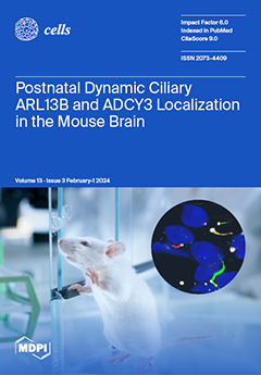
The dysfunction of primary cilia in the brain has been implicated in disease processes like obesity and learning and memory deficits. However, little is known about cilia within the central nervous system and their precise roles in behavior. This study focuses on fundamental questions about how cilia may differ by neuroanatomical regions, between the sexes, at different ages and under different physiological conditions. Understanding the dynamics and differences in cilia throughout the brain has implications for revealing their roles in signaling and cell–cell communication. On the cover, different cilia types in the adult mouse suprachiasmatic nucleus are observed via immunofluorescence with a green ADCY3 cilium, a red ARL13B cilium and two yellow cilia, which colocalize ADCY3 and ARL13B, and the basal body marker FOP labels the base of cilia. View this paper
  • Issues are regarded as officially published after their release is announced to the table of contents alert mailing list.
  • You may sign up for e-mail alerts to receive table of contents of newly released issues.
  • PDF is the official format for papers published in both, html and pdf forms. To view the papers in pdf format, click on the "PDF Full-text" link, and use the free Adobe Reader to open them.
Order results
Result details
Section
Select all
Export citation of selected articles as:
22 pages, 2754 KiB  
Review
Brain-Type Glycogen Phosphorylase (PYGB) in the Pathologies of Diseases: A Systematic Review
by Caiting Yang, Haojun Wang, Miaomiao Shao, Fengyu Chu, Yuyu He, Xiaoli Chen, Jiahui Fan, Jingwen Chen, Qianqian Cai and Changxin Wu
Cells 2024, 13(3), 289; https://doi.org/10.3390/cells13030289 - 5 Feb 2024
Viewed by 1003
Abstract
Glycogen metabolism is a form of crucial metabolic reprogramming in cells. PYGB, the brain-type glycogen phosphorylase (GP), serves as the rate-limiting enzyme of glycogen catabolism. Evidence is mounting for the association of PYGB with diverse human diseases. This review covers the advancements in [...] Read more.
Glycogen metabolism is a form of crucial metabolic reprogramming in cells. PYGB, the brain-type glycogen phosphorylase (GP), serves as the rate-limiting enzyme of glycogen catabolism. Evidence is mounting for the association of PYGB with diverse human diseases. This review covers the advancements in PYGB research across a range of diseases, including cancer, cardiovascular diseases, metabolic diseases, nervous system diseases, and other diseases, providing a succinct overview of how PYGB functions as a critical factor in both physiological and pathological processes. We present the latest progress in PYGB in the diagnosis and treatment of various diseases and discuss the current limitations and future prospects of this novel and promising target. Full article
Show Figures

Figure 1

13 pages, 4568 KiB  
Review
Considering Caenorhabditis elegans Aging on a Temporal and Tissue Scale: The Case of Insulin/IGF-1 Signaling
by Paola Fabrizio, Allan Alcolei and Florence Solari
Cells 2024, 13(3), 288; https://doi.org/10.3390/cells13030288 - 5 Feb 2024
Cited by 1 | Viewed by 1211
Abstract
The aging process is inherently complex, involving multiple mechanisms that interact at different biological scales. The nematode Caenorhabditis elegans is a simple model organism that has played a pivotal role in aging research following the discovery of mutations extending lifespan. Longevity pathways identified [...] Read more.
The aging process is inherently complex, involving multiple mechanisms that interact at different biological scales. The nematode Caenorhabditis elegans is a simple model organism that has played a pivotal role in aging research following the discovery of mutations extending lifespan. Longevity pathways identified in C. elegans were subsequently found to be conserved and regulate lifespan in multiple species. These pathways intersect with fundamental hallmarks of aging that include nutrient sensing, epigenetic alterations, proteostasis loss, and mitochondrial dysfunction. Here we summarize recent data obtained in C. elegans highlighting the importance of studying aging at both the tissue and temporal scale. We then focus on the neuromuscular system to illustrate the kinetics of changes that take place with age. We describe recently developed tools that enabled the dissection of the contribution of the insulin/IGF-1 receptor ortholog DAF-2 to the regulation of worm mobility in specific tissues and at different ages. We also discuss guidelines and potential pitfalls in the use of these new tools. We further highlight the opportunities that they present, especially when combined with recent transcriptomic data, to address and resolve the inherent complexity of aging. Understanding how different aging processes interact within and between tissues at different life stages could ultimately suggest potential intervention points for age-related diseases. Full article
(This article belongs to the Special Issue Caenorhabditis elegans: A Model Organism, Endless Possibilities)
Show Figures

Figure 1

15 pages, 3737 KiB  
Article
Green-Synthesized Silver and Selenium Nanoparticles Using Berberine: A Comparative Assessment of In Vitro Anticancer Potential on Human Hepatocellular Carcinoma Cell Line (HepG2)
by Azza M. Khaled, Mohamed S. Othman, Sofian T. Obeidat, Ghada M. Aleid, Shimaa M. Aboelnaga, Alaa Fehaid, Heba M. R. Hathout, Ashraf A. Bakkar, Ahmed E. Abdel Moneim, Islam M. El-Garawani and Dalia S. Morsi
Cells 2024, 13(3), 287; https://doi.org/10.3390/cells13030287 - 5 Feb 2024
Viewed by 1177
Abstract
A well-known natural ingredient found in several medicinal plants, berberine (Ber), has been shown to have anticancer properties against a range of malignancies. The limited solubility and bioavailability of berberine can be addressed using Ber-loaded nanoparticles. In this study, we compared the in [...] Read more.
A well-known natural ingredient found in several medicinal plants, berberine (Ber), has been shown to have anticancer properties against a range of malignancies. The limited solubility and bioavailability of berberine can be addressed using Ber-loaded nanoparticles. In this study, we compared the in vitro cytotoxic effects of both Ber-loaded silver nanoparticles (Ber-AgNPs) and Ber-loaded selenium nanoparticles (Ber-SeNPs) in the human liver cancer cell line (HepG2) and mouse normal liver cells (BNL). The IC50 values in HepG2 for berberine, Ber-AgNPs, Ber-SeNPs, and cisplatin were 26.69, 1.16, 0.04, and 0.33 µg/mL, respectively. Our results show that Ber and its Ag and Se nanoparticles exerted a good antitumor effect against HepG2 cells by inducing apoptosis via upregulating p53, Bax, cytosolic cytochrome C levels, and caspase-3 activity, and the down-regulation of Bcl-2 levels. Similarly, incubation with Ber and both Ber-NPs (Ag and Se) led to a significant dose-dependent elevation in inflammatory markers’ (TNF-α, NF-κB, and COX-2) levels compared to the control group. In addition, it led to the arrest of the G1 cell cycle by depleting the expression of cyclin D1 and CDK-2 mRNA. Furthermore, Ber and both Ber-NPs (Ag and Se) caused a significant dose-dependent increase in LDH activity in HepG2 cells. Furthermore, our findings offer evidence that Ber and its nanoparticles intensified oxidative stress in HepG2 cells. Furthermore, the migration rate of cells subjected to berberine and its nanoforms was notably decreased compared to that of control cells. It can be inferred that Ber nanoparticles exhibited superior anticancer efficacy against HepG2 compared to unprocessed Ber, perhaps due to their improved solubility and bioavailability. Furthermore, Ber-SeNPs exhibited greater efficacy than Ber-AgNPs, possibly as a result of the inherent anticancer characteristics of selenium. Full article
Show Figures

Figure 1

16 pages, 1670 KiB  
Review
Glymphatic System Pathology and Neuroinflammation as Two Risk Factors of Neurodegeneration
by Stanisław Szlufik, Kamila Kopeć, Stanisław Szleszkowski and Dariusz Koziorowski
Cells 2024, 13(3), 286; https://doi.org/10.3390/cells13030286 - 5 Feb 2024
Cited by 1 | Viewed by 1853
Abstract
The key to the effective treatment of neurodegenerative disorders is a thorough understanding of their pathomechanism. Neurodegeneration and neuroinflammation are mutually propelling brain processes. An impairment of glymphatic system function in neurodegeneration contributes to the progression of pathological processes. The question arises as [...] Read more.
The key to the effective treatment of neurodegenerative disorders is a thorough understanding of their pathomechanism. Neurodegeneration and neuroinflammation are mutually propelling brain processes. An impairment of glymphatic system function in neurodegeneration contributes to the progression of pathological processes. The question arises as to how neuroinflammation and the glymphatic system are related. This review highlights the direct and indirect influence of these two seemingly independent processes. Protein aggregates, a characteristic feature of neurodegeneration, are correlated with glymphatic clearance and neuroinflammation. Glial cells cannot be overlooked when considering the neuroinflammatory processes. Astrocytes are essential for the effective functioning of the glymphatic system and play a crucial role in the inflammatory responses in the central nervous system. It is imperative to acknowledge the significance of AQP4, a protein that exhibits a high degree of polarization in astrocytes and is crucial for the functioning of the glymphatic system. AQP4 influences inflammatory processes that have not yet been clearly delineated. Another interesting issue is the gut–brain axis and microbiome, which potentially impact the discussed processes. A discussion of the correlation between the functioning of the glymphatic system and neuroinflammation may contribute to exploring the pathomechanism of neurodegeneration. Full article
Show Figures

Figure 1

24 pages, 5252 KiB  
Article
Shear Stress and Sub-Femtomolar Levels of Ligand Synergize to Activate ALK1 Signaling in Endothelial Cells
by Ya-Wen Cheng, Anthony R. Anzell, Stefanie A. Morosky, Tristin A. Schwartze, Cynthia S. Hinck, Andrew P. Hinck, Beth L. Roman and Lance A. Davidson
Cells 2024, 13(3), 285; https://doi.org/10.3390/cells13030285 - 5 Feb 2024
Viewed by 1007
Abstract
Endothelial cells (ECs) respond to concurrent stimulation by biochemical factors and wall shear stress (SS) exerted by blood flow. Disruptions in flow-induced responses can result in remodeling issues and cardiovascular diseases, but the detailed mechanisms linking flow-mechanical cues and biochemical signaling remain unclear. [...] Read more.
Endothelial cells (ECs) respond to concurrent stimulation by biochemical factors and wall shear stress (SS) exerted by blood flow. Disruptions in flow-induced responses can result in remodeling issues and cardiovascular diseases, but the detailed mechanisms linking flow-mechanical cues and biochemical signaling remain unclear. Activin receptor-like kinase 1 (ALK1) integrates SS and ALK1-ligand cues in ECs; ALK1 mutations cause hereditary hemorrhagic telangiectasia (HHT), marked by arteriovenous malformation (AVM) development. However, the mechanistic underpinnings of ALK1 signaling modulation by fluid flow and the link to AVMs remain uncertain. We recorded EC responses under varying SS magnitudes and ALK1 ligand concentrations by assaying pSMAD1/5/9 nuclear localization using a custom multi-SS microfluidic device and a custom image analysis pipeline. We extended the previously reported synergy between SS and BMP9 to include BMP10 and BMP9/10. Moreover, we demonstrated that this synergy is effective even at extremely low SS magnitudes (0.4 dyn/cm2) and ALK1 ligand range (femtogram/mL). The synergistic response to ALK1 ligands and SS requires the kinase activity of ALK1. Moreover, ALK1’s basal activity and response to minimal ligand levels depend on endocytosis, distinct from cell–cell junctions, cytoskeleton-mediated mechanosensing, or cholesterol-enriched microdomains. However, an in-depth analysis of ALK1 receptor trafficking’s molecular mechanisms requires further investigation. Full article
Show Figures

Figure 1

14 pages, 1828 KiB  
Article
Imeglimin Exhibits Novel Anti-Inflammatory Effects on High-Glucose-Stimulated Mouse Microglia through ULK1-Mediated Suppression of the TXNIP–NLRP3 Axis
by Hisashi Kato, Kaori Iwashita, Masayo Iwasa, Sayaka Kato, Hajime Yamakage, Takayoshi Suganami, Masashi Tanaka and Noriko Satoh-Asahara
Cells 2024, 13(3), 284; https://doi.org/10.3390/cells13030284 - 5 Feb 2024
Viewed by 1499
Abstract
Type 2 diabetes mellitus (T2DM) is an epidemiological risk factor for dementia and has been implicated in multifactorial pathologies, including neuroinflammation. In the present study, we aimed to elucidate the potential anti-inflammatory effects of imeglimin, a novel antidiabetic agent, on high-glucose (HG)-stimulated microglia. [...] Read more.
Type 2 diabetes mellitus (T2DM) is an epidemiological risk factor for dementia and has been implicated in multifactorial pathologies, including neuroinflammation. In the present study, we aimed to elucidate the potential anti-inflammatory effects of imeglimin, a novel antidiabetic agent, on high-glucose (HG)-stimulated microglia. Mouse microglial BV2 cells were stimulated with HG in the presence or absence of imeglimin. We examined the effects of imeglimin on the levels of proinflammatory cytokines, intracellular reactive oxygen species (ROS), mitochondrial integrity, and components related to the inflammasome or autophagy pathways in these cells. Our results showed that imeglimin suppressed the HG-induced production of interleukin-1beta (IL-1β) by reducing the intracellular ROS levels, ameliorating mitochondrial dysfunction, and inhibiting the activation of the thioredoxin-interacting protein (TXNIP)–NOD-like receptor family pyrin domain containing 3 (NLRP3) axis. Moreover, the inhibitory effects of imeglimin on the TXNIP–NLRP3 axis depended on the imeglimin-induced activation of ULK1, which also exhibited novel anti-inflammatory effects without autophagy induction. These findings suggest that imeglimin exerted novel suppressive effects on HG-stimulated microglia through the ULK1–TXNIP–NLRP3 axis, and may, thereby, contribute to the development of innovative strategies to prevent T2DM-associated cognitive impairment. Full article
Show Figures

Figure 1

15 pages, 3728 KiB  
Article
Human β-Defensin 3 Inhibition of P. gingivalis LPS-Induced IL-1β Production by BV-2 Microglia through Suppression of Cathepsins B and L
by Erika Inoue, Shiyo Minatozaki, Sachi Shimizu, Sayaka Miyamoto, Misato Jo, Junjun Ni, Hidetoshi Tozaki-Saitoh, Kosuke Oda, Saori Nonaka and Hiroshi Nakanishi
Cells 2024, 13(3), 283; https://doi.org/10.3390/cells13030283 - 4 Feb 2024
Viewed by 1105
Abstract
Cathepsin B (CatB) is thought to be essential for the induction of Porphyromonas gingivalis lipopolysaccharide (Pg LPS)-induced Alzheimer’s disease-like pathologies in mice, including interleukin-1β (IL-1β) production and cognitive decline. However, little is known about the role of CatB in Pg virulence factor-induced [...] Read more.
Cathepsin B (CatB) is thought to be essential for the induction of Porphyromonas gingivalis lipopolysaccharide (Pg LPS)-induced Alzheimer’s disease-like pathologies in mice, including interleukin-1β (IL-1β) production and cognitive decline. However, little is known about the role of CatB in Pg virulence factor-induced IL-1β production by microglia. We first subjected IL-1β-luciferase reporter BV-2 microglia to inhibitors of Toll-like receptors (TLRs), IκB kinase, and the NLRP3 inflammasome following stimulation with Pg LPS and outer membrane vesicles (OMVs). To clarify the involvement of CatB, we used several known CatB inhibitors, including CA-074Me, ZRLR, and human β-defensin 3 (hBD3). IL-1β production in BV-2 microglia induced by Pg LPS and OMVs was significantly inhibited by the TLR2 inhibitor C29 and the IκB kinase inhibitor wedelolactonne, but not by the NLRPs inhibitor MCC950. Both hBD3 and CA-074Me significantly inhibited Pg LPS-induced IL-1β production in BV-2 microglia. Although CA-074Me also suppressed OMV-induced IL-1β production, hBD3 did not inhibit it. Furthermore, both hBD3 and CA-074Me significantly blocked Pg LPS-induced nuclear NF-κB p65 translocation and IκBα degradation. In contrast, hBD3 and CA-074Me did not block OMV-induced nuclear NF-κB p65 translocation or IκBα degradation. Furthermore, neither ZRLR, a specific CatB inhibitor, nor shRNA-mediated knockdown of CatB expression had any effect on Pg virulence factor-induced IL-1β production. Interestingly, phagocytosis of OMVs by BV-2 microglia induced IL-1β production. Finally, the structural models generated by AlphaFold indicated that hBD3 can bind to the substrate-binding pocket of CatB, and possibly CatL as well. These results suggest that Pg LPS induces CatB/CatL-dependent synthesis and processing of pro-IL-1β without activation of the NLRP3 inflammasome. In contrast, OMVs promote the synthesis and processing of pro-IL-1β through CatB/CatL-independent phagocytic mechanisms. Thus, hBD3 can improve the IL-1β-associated vicious inflammatory cycle induced by microglia through inhibition of CatB/CatL. Full article
Show Figures

Graphical abstract

14 pages, 3732 KiB  
Article
In Vitro Lipid Overload Affects Cellular Proliferation, Apoptosis, and Senescence in a Time-Dependent Manner in HepG2 Hepatocytes and LX-2 Hepatic Stellate Cells
by Adriana Campos-Espinosa, Carolina Guzmán, Karla Zaira Medina-Ávila and Gabriela Gutierrez-Reyes
Cells 2024, 13(3), 282; https://doi.org/10.3390/cells13030282 - 4 Feb 2024
Viewed by 890
Abstract
Different cellular mechanisms influence steatotic liver disease (SLD) progression. The influence of different levels of steatogenic inputs has not been studied in hepatocytes and hepatic stellate cells (HSCs). Methods: HepG2 hepatocytes and LX-2 HSCs were cultured in mild (MS) and severe (SS) steatogenic [...] Read more.
Different cellular mechanisms influence steatotic liver disease (SLD) progression. The influence of different levels of steatogenic inputs has not been studied in hepatocytes and hepatic stellate cells (HSCs). Methods: HepG2 hepatocytes and LX-2 HSCs were cultured in mild (MS) and severe (SS) steatogenic conditions. TGF-β stimulation was also tested for HSCs in control (T) and steatogenic conditions (MS-T and SS-T). Steatosis was stained with Oil Red, and the proliferation was assayed via WST-8 reduction, apoptosis via flow cytometry, and senescence via SA-β-galactosidase activity. Results: Regarding hepatocytes, steatosis progressively increased; proliferation was lower in MS and SS; and the viability of both conditions significantly decreased at 72 h. Apoptosis increased in MS at 72 h, while it decreased in SS. Senescence increased in MS and diminished in SS. Regarding HSCs, the SS and SS-T groups showed no proliferation, and the viability was reduced in MS at 72 h and in SS and SS-T. The LX-2 cells showed increased apoptosis in SS and SS-T at 24 h, and in MS and MS-T at 72 h. Senescence decreased in MS, SS, and SS-T. Conclusions: Lipid overload induces differential effects depending on the cell type, the steatogenic input level, and the exposure time. Hepatocytes are resilient to mild steatosis but susceptible to high lipotoxicity. HSCs are sensitive to lipid overload, undergoing apoptosis and lowering senescence and proliferation. Collectively, these data may help explain the development of steatosis and fibrosis in SLD. Full article
(This article belongs to the Special Issue Nonalcoholic Fatty Liver Disease: From Mechanisms to Therapeutics)
Show Figures

Graphical abstract

25 pages, 1137 KiB  
Review
Recent Achievements in the Heterogeneity of Mammalian and Human Retinal Pigment Epithelium: In Search of a Stem Cell
by Lyubov A. Rzhanova, Yuliya V. Markitantova and Maria A. Aleksandrova
Cells 2024, 13(3), 281; https://doi.org/10.3390/cells13030281 - 4 Feb 2024
Viewed by 940
Abstract
Retinal pigment epithelium (RPE) cells are important fundamentally for the development and function of the retina. In this regard, the study of the morphological and molecular properties of RPE cells, as well as their regenerative capabilities, is of particular importance for biomedicine. However, [...] Read more.
Retinal pigment epithelium (RPE) cells are important fundamentally for the development and function of the retina. In this regard, the study of the morphological and molecular properties of RPE cells, as well as their regenerative capabilities, is of particular importance for biomedicine. However, these studies are complicated by the fact that, despite the external morphological similarity of RPE cells, the RPE is a population of heterogeneous cells, the molecular genetic properties of which have begun to be revealed by sequencing methods only in recent years. This review carries out an analysis of the data from morphological and molecular genetic studies of the heterogeneity of RPE cells in mammals and humans, which reveals the individual differences in the subpopulations of RPE cells and the possible specificity of their functions. Particular attention is paid to discussing the properties of “stemness,” proliferation, and plasticity in the RPE, which may be useful for uncovering the mechanisms of retinal diseases associated with pathologies of the RPE and finding new ways of treating them. Full article
Show Figures

Graphical abstract

16 pages, 3155 KiB  
Brief Report
GPR55 Inactivation Diminishes Splenic Responses and Improves Neurological Outcomes in the Mouse Ischemia/Reperfusion Stroke Model
by Sachin Gajghate, Hongbo Li and Slava Rom
Cells 2024, 13(3), 280; https://doi.org/10.3390/cells13030280 - 3 Feb 2024
Viewed by 920
Abstract
Although strokes are frequent and severe, treatment options are scarce. Plasminogen activators, the only FDA-approved agents for clot treatment (tissue plasminogen activators (tPAs)), are used in a limited patient group. Moreover, there are few approaches for handling the brain’s inflammatory reactions to a [...] Read more.
Although strokes are frequent and severe, treatment options are scarce. Plasminogen activators, the only FDA-approved agents for clot treatment (tissue plasminogen activators (tPAs)), are used in a limited patient group. Moreover, there are few approaches for handling the brain’s inflammatory reactions to a stroke. The orphan G protein-coupled receptor 55 (GPR55)’s connection to inflammatory processes has been recently reported; however, its role in stroke remains to be discovered. Post-stroke neuroinflammation involves the central nervous system (CNS)’s resident microglia activation and the infiltration of leukocytes from circulation into the brain. Additionally, splenic responses have been shown to be detrimental to stroke recovery. While lymphocytes enter the brain in small numbers, they regularly emerge as a very influential leukocyte subset that causes secondary inflammatory cerebral damage. However, an understanding of how this limited lymphocyte presence profoundly impacts stroke outcomes remains largely unclear. In this study, a mouse model for transient middle cerebral artery occlusion (tMCAO) was used to mimic ischemia followed by a reperfusion (IS/R) stroke. GPR55 inactivation, with a potent GPR55-specific antagonist, ML-193, starting 6 h after tMCAO or the absence of the GPR55 in mice (GPR55 knock out (GPR55ko)) resulted in a reduced infarction volume, improved neurological outcomes, and decreased splenic responses. The inhibition of GPR55 with ML-193 diminished CD4+T-cell spleen egress and attenuated CD4+T-cell brain infiltration. Additionally, ML-193 treatment resulted in an augmented number of regulatory T cells (Tregs) in the brain post-tMCAO. Our report offers documentation and the functional evaluation of GPR55 in the brain–spleen axis and lays the foundation for refining therapeutics for patients after ischemic attacks. Full article
(This article belongs to the Special Issue Stroke Immunology: Mechanisms and Therapeutic Prospects)
Show Figures

Figure 1

16 pages, 6002 KiB  
Article
An Efficient Method for Isolating and Purifying Nuclei from Mice Brain for Single-Molecule Imaging Using High-Speed Atomic Force Microscopy
by Yujia Qiu, Elma Sakinatus Sajidah, Sota Kondo, Shinnosuke Narimatsu, Muhammad Isman Sandira, Yoshiki Higashiguchi, Goro Nishide, Azuma Taoka, Masaharu Hazawa, Yuka Inaba, Hiroshi Inoue, Ayami Matsushima, Yuki Okada, Mitsutoshi Nakada, Toshio Ando, Keesiang Lim and Richard W. Wong
Cells 2024, 13(3), 279; https://doi.org/10.3390/cells13030279 - 2 Feb 2024
Viewed by 1394
Abstract
Nuclear pore complexes (NPCs) on the nuclear membrane surface have a crucial function in controlling the movement of small molecules and macromolecules between the cell nucleus and cytoplasm through their intricate core channel resembling a spiderweb with several layers. Currently, there are few [...] Read more.
Nuclear pore complexes (NPCs) on the nuclear membrane surface have a crucial function in controlling the movement of small molecules and macromolecules between the cell nucleus and cytoplasm through their intricate core channel resembling a spiderweb with several layers. Currently, there are few methods available to accurately measure the dynamics of nuclear pores on the nuclear membranes at the nanoscale. The limitation of traditional optical imaging is due to diffraction, which prevents achieving the required resolution for observing a diverse array of organelles and proteins within cells. Super-resolution techniques have effectively addressed this constraint by enabling the observation of subcellular components on the nanoscale. Nevertheless, it is crucial to acknowledge that these methods often need the use of fixed samples. This also raises the question of how closely a static image represents the real intracellular dynamic system. High-speed atomic force microscopy (HS-AFM) is a unique technique used in the field of dynamic structural biology, enabling the study of individual molecules in motion close to their native states. Establishing a reliable and repeatable technique for imaging mammalian tissue at the nanoscale using HS-AFM remains challenging due to inadequate sample preparation. This study presents the rapid strainer microfiltration (RSM) protocol for directly preparing high-quality nuclei from the mouse brain. Subsequently, we promptly utilize HS-AFM real-time imaging and cinematography approaches to record the spatiotemporal of nuclear pore nano-dynamics from the mouse brain. Full article
(This article belongs to the Special Issue Nuclear Pore Complex in Nanomedicine 2.0)
Show Figures

Figure 1

16 pages, 6586 KiB  
Article
Filamin C Deficiency Impairs Sarcomere Stability and Activates Focal Adhesion Kinase through PDGFRA Signaling in Induced Pluripotent Stem Cell-Derived Cardiomyocytes
by Shanshan Gao, Lingaonan He, Chi Keung Lam, Matthew R. G. Taylor, Luisa Mestroni, Raffaella Lombardi and Suet Nee Chen
Cells 2024, 13(3), 278; https://doi.org/10.3390/cells13030278 - 2 Feb 2024
Cited by 1 | Viewed by 1094
Abstract
Truncating mutations in filamin C (FLNC) are associated with dilated cardiomyopathy and arrhythmogenic cardiomyopathy. FLNC is an actin-binding protein and is known to interact with transmembrane and structural proteins; hence, the ablation of FLNC in cardiomyocytes is expected to dysregulate cell [...] Read more.
Truncating mutations in filamin C (FLNC) are associated with dilated cardiomyopathy and arrhythmogenic cardiomyopathy. FLNC is an actin-binding protein and is known to interact with transmembrane and structural proteins; hence, the ablation of FLNC in cardiomyocytes is expected to dysregulate cell adhesion, cytoskeletal organization, sarcomere structural integrity, and likely nuclear function. Our previous study showed that the transcriptional profiles of FLNC homozygous deletions in human pluripotent stem cell-derived cardiomyocytes (hiPSC-CMs) are highly comparable to the transcriptome profiles of hiPSC-CMs from patients with FLNC truncating mutations. Therefore, in this study, we used CRISPR-Cas-engineered hiPSC-derived FLNC knockout cardiac myocytes as a model of FLNC cardiomyopathy to determine pathogenic mechanisms and to examine structural changes caused by FLNC deficiency. RNA sequencing data indicated the significant upregulation of focal adhesion signaling and the dysregulation of thin filament genes in FLNC-knockout (FLNCKO) hiPSC-CMs compared to isogenic hiPSC-CMs. Furthermore, our findings suggest that the complete loss of FLNC in cardiomyocytes led to cytoskeletal defects and the activation of focal adhesion kinase. Pharmacological inhibition of PDGFRA signaling using crenolanib (an FDA-approved drug) reduced focal adhesion kinase activation and partially normalized the focal adhesion signaling pathway. The findings from this study suggest the opportunity in repurposing FDA-approved drug as a therapeutic strategy to treat FLNC cardiomyopathy. Full article
Show Figures

Figure 1

16 pages, 1780 KiB  
Review
The Role of FNDC5/Irisin in Cardiovascular Disease
by Maciej Grzeszczuk, Piotr Dzięgiel and Katarzyna Nowińska
Cells 2024, 13(3), 277; https://doi.org/10.3390/cells13030277 - 2 Feb 2024
Cited by 1 | Viewed by 1278
Abstract
Disorders of cardiomyocyte metabolism play a crucial role in many cardiovascular diseases, such as myocardial infarction, heart failure and ischemia–reperfusion injury. In myocardial infarction, cardiomyocyte metabolism is regulated by mitochondrial changes and biogenesis, which allows energy homeostasis. There are many proteins in cells [...] Read more.
Disorders of cardiomyocyte metabolism play a crucial role in many cardiovascular diseases, such as myocardial infarction, heart failure and ischemia–reperfusion injury. In myocardial infarction, cardiomyocyte metabolism is regulated by mitochondrial changes and biogenesis, which allows energy homeostasis. There are many proteins in cells that regulate and control metabolic processes. One of them is irisin (Ir), which is released from the transmembrane protein FNDC5. Initial studies indicated that Ir is a myokine secreted mainly by skeletal muscles. Further studies showed that Ir was also present in various tissues. However, its highest levels were observed in cardiomyocytes. Ir is responsible for many processes, including the conversion of white adipose tissue (WAT) to brown adipose tissue (BAT) by increasing the expression of thermogenin (UCP1). In addition, Ir affects mitochondrial biogenesis. Therefore, the levels of FNDC5/Ir in the blood and myocardium may be important in cardiovascular disease. This review discusses the current knowledge about the role of FNDC5/Ir in cardiovascular disease. Full article
(This article belongs to the Special Issue Cardiovascular Biomarkers: Current Status and Future Directions)
Show Figures

Figure 1

12 pages, 965 KiB  
Article
Clinical Relevance and Interplay between miRNAs in Influencing Glioblastoma Multiforme Prognosis
by Samantha Epistolio, Giulia Dazio, Ismail Zaed, Nora Sahnane, Debora Cipriani, Francesco Polinelli, Jessica Barizzi, Paolo Spina, Federico Mattia Stefanini, Michele Cerati, Sergio Balbi, Luca Mazzucchelli, Fausto Sessa, Gianfranco Angelo Pesce, Michael Reinert, Andrea Cardia, Francesco Marchi and Milo Frattini
Cells 2024, 13(3), 276; https://doi.org/10.3390/cells13030276 - 2 Feb 2024
Viewed by 850
Abstract
Glioblastoma multiforme (GBM) is usually treated with surgery followed by adjuvant partial radiotherapy combined with temozolomide (TMZ) chemotherapy. Recent studies demonstrated a better survival and good response to TMZ in methylguanine-DNA methyltransferase (MGMT)-methylated GBM cases. However, approximately 20% of patients with [...] Read more.
Glioblastoma multiforme (GBM) is usually treated with surgery followed by adjuvant partial radiotherapy combined with temozolomide (TMZ) chemotherapy. Recent studies demonstrated a better survival and good response to TMZ in methylguanine-DNA methyltransferase (MGMT)-methylated GBM cases. However, approximately 20% of patients with MGMT-unmethylated GBM display an unexpectedly favorable outcome. Therefore, additional mechanisms related to the TMZ response need to be investigated. As such, we decided to investigate the clinical relevance of six miRNAs involved in brain tumorigenesis (miR-181c, miR-181d, miR-21, miR-195, miR-196b, miR-648) as additional markers of response and survival in patients receiving TMZ for GBM. We evaluated miRNA expression and the interplay between miRNAs in 112 IDH wt GBMs by applying commercial assays. Then, we correlated the miRNA expression with patients’ clinical outcomes. Upon bivariate analyses, we found a significant association between the expression levels of the miRNAs analyzed, but, more interestingly, the OS curves show that the combination of low miR-648 and miR-181c or miR-181d expressions is associated with a worse prognosis than cases with other low-expression miRNA pairs. To conclude, we found how specific miRNA pairs can influence survival in GBM cases treated with TMZ. Full article
Show Figures

Figure 1

21 pages, 4191 KiB  
Article
Deep-Learning-Based Analysis Reveals a Social Behavior Deficit in Mice Exposed Prenatally to Nicotine
by Mengyun Zhou, Wen Qiu, Nobuhiko Ohashi, Lihao Sun, Marie-Louis Wronski, Emi Kouyama-Suzuki, Yoshinori Shirai, Toru Yanagawa, Takuma Mori and Katsuhiko Tabuchi
Cells 2024, 13(3), 275; https://doi.org/10.3390/cells13030275 - 1 Feb 2024
Viewed by 1716
Abstract
Cigarette smoking during pregnancy is known to be associated with the incidence of attention-deficit/hyperactive disorder (ADHD). Recent developments in deep learning algorithms enable us to assess the behavioral phenotypes of animal models without cognitive bias during manual analysis. In this study, we established [...] Read more.
Cigarette smoking during pregnancy is known to be associated with the incidence of attention-deficit/hyperactive disorder (ADHD). Recent developments in deep learning algorithms enable us to assess the behavioral phenotypes of animal models without cognitive bias during manual analysis. In this study, we established prenatal nicotine exposure (PNE) mice and evaluated their behavioral phenotypes using DeepLabCut and SimBA. We optimized the training parameters of DeepLabCut for pose estimation and succeeded in labeling a single-mouse or two-mouse model with high fidelity during free-moving behavior. We applied the trained network to analyze the behavior of the mice and found that PNE mice exhibited impulsivity and a lessened working memory, which are characteristics of ADHD. PNE mice also showed elevated anxiety and deficits in social interaction, reminiscent of autism spectrum disorder (ASD). We further examined PNE mice by evaluating adult neurogenesis in the hippocampus, which is a pathological hallmark of ASD, and demonstrated that newborn neurons were decreased, specifically in the ventral part of the hippocampus, which is reported to be related to emotional and social behaviors. These results support the hypothesis that PNE is a risk factor for comorbidity with ADHD and ASD in mice. Full article
(This article belongs to the Section Cells of the Nervous System)
Show Figures

Figure 1

22 pages, 1773 KiB  
Review
Cellular, Molecular and Clinical Aspects of Aortic Aneurysm—Vascular Physiology and Pathophysiology
by Dominika Domagała, Krzysztof Data, Hubert Szyller, Maryam Farzaneh, Paul Mozdziak, Sławomir Woźniak, Maciej Zabel, Piotr Dzięgiel and Bartosz Kempisty
Cells 2024, 13(3), 274; https://doi.org/10.3390/cells13030274 - 1 Feb 2024
Cited by 1 | Viewed by 1439
Abstract
A disturbance of the structure of the aortic wall results in the formation of aortic aneurysm, which is characterized by a significant bulge on the vessel surface that may have consequences, such as distention and finally rupture. Abdominal aortic aneurysm (AAA) is a [...] Read more.
A disturbance of the structure of the aortic wall results in the formation of aortic aneurysm, which is characterized by a significant bulge on the vessel surface that may have consequences, such as distention and finally rupture. Abdominal aortic aneurysm (AAA) is a major pathological condition because it affects approximately 8% of elderly men and 1.5% of elderly women. The pathogenesis of AAA involves multiple interlocking mechanisms, including inflammation, immune cell activation, protein degradation and cellular malalignments. The expression of inflammatory factors, such as cytokines and chemokines, induce the infiltration of inflammatory cells into the wall of the aorta, including macrophages, natural killer cells (NK cells) and T and B lymphocytes. Protein degradation occurs with a high expression not only of matrix metalloproteinases (MMPs) but also of neutrophil gelatinase-associated lipocalin (NGAL), interferon gamma (IFN-γ) and chymases. The loss of extracellular matrix (ECM) due to cell apoptosis and phenotype switching reduces tissue density and may contribute to AAA. It is important to consider the key mechanisms of initiating and promoting AAA to achieve better preventative and therapeutic outcomes. Full article
(This article belongs to the Special Issue Understanding Biomarkers in Cardiology Volume II)
Show Figures

Figure 1

15 pages, 2037 KiB  
Article
Identification and Characterization of HIRIP3 as a Histone H2A Chaperone
by Maria Ignatyeva, Abdul Kareem Mohideen Patel, Abdulkhaleg Ibrahim, Raed S. Albiheyri, Ali T. Zari, Ahmed Bahieldin, Christian Bronner, Jamal S. M. Sabir and Ali Hamiche
Cells 2024, 13(3), 273; https://doi.org/10.3390/cells13030273 - 1 Feb 2024
Viewed by 941
Abstract
HIRIP3 is a mammalian protein homologous to the yeast H2A.Z deposition chaperone Chz1. However, the structural basis underlying Chz’s binding preference for H2A.Z over H2A, as well as the mechanism through which Chz1 modulates histone deposition or replacement, remains enigmatic. In this study, [...] Read more.
HIRIP3 is a mammalian protein homologous to the yeast H2A.Z deposition chaperone Chz1. However, the structural basis underlying Chz’s binding preference for H2A.Z over H2A, as well as the mechanism through which Chz1 modulates histone deposition or replacement, remains enigmatic. In this study, we aimed to characterize the function of HIRIP3 and to identify its interacting partners in HeLa cells. Our findings reveal that HIRIP3 is specifically associated in vivo with H2A–H2B dimers and CK2 kinase. While bacterially expressed HIRIP3 exhibited a similar binding affinity towards H2A and H2A.Z, the associated CK2 kinase showed a notable preference for H2A phosphorylation at serine 1. The recombinant HIRIP3 physically interacted with the H2A αC helix through an extended CHZ domain and played a crucial role in depositing the canonical core histones onto naked DNA. Our results demonstrate that mammalian HIRIP3 acts as an H2A histone chaperone, assisting in its selective phosphorylation by Ck2 kinase at serine 1 and facilitating its deposition onto chromatin. Full article
(This article belongs to the Special Issue Epigenetics, Chromatin Structure and Transcription Regulation)
Show Figures

Figure 1

13 pages, 2436 KiB  
Article
Hyperglycemic Stress Induces Expression, Degradation, and Nuclear Association of Rho GDP Dissociation Inhibitor 2 (RhoGDIβ) in Pancreatic β-Cells
by Noah Gleason and Anjaneyulu Kowluru
Cells 2024, 13(3), 272; https://doi.org/10.3390/cells13030272 - 1 Feb 2024
Viewed by 766
Abstract
Small G proteins (e.g., Rac1) play critical regulatory roles in islet β-cell function in health (physiological insulin secretion) and in metabolic stress (cell dysfunction and demise). Multiple regulatory factors for these G proteins, such as GDP dissociation inhibitors (GDIs), have been implicated in [...] Read more.
Small G proteins (e.g., Rac1) play critical regulatory roles in islet β-cell function in health (physiological insulin secretion) and in metabolic stress (cell dysfunction and demise). Multiple regulatory factors for these G proteins, such as GDP dissociation inhibitors (GDIs), have been implicated in the functional regulation of these G proteins. The current set of investigations is aimed at understanding impact of chronic hyperglycemic stress on the expression and subcellular distribution of three known isoforms of RhoGDIs (RhoGDIα, RhoGDIβ, and RhoGDIγ) in insulin-secreting β-cells. The data accrued in these studies revealed that the expression of RhoGDIβ, but not RhoGDIα or RhoGDIγ, is increased in INS-1 832/13 cells, rat islets, and human islets. Hyperglycemic stress also promoted the cleavage of RhoGDIβ, leading to its translocation to the nuclear compartment. We also report that RhoGDIα, but not RhoGDIγ, is associated with the nuclear compartment. However, unlike RhoGDIβ, hyperglycemic conditions exerted no effects on RhoGDIα’s association with nuclear fraction. Based on these observations, and our earlier findings of the translocation of Rac1 to the nuclear compartment under the duress of metabolic stress, we conclude that the RhoGDIβ-Rac1 signaling module promotes signals from the cytosolic to the nucleus, culminating in accelerated β-cell dysfunction under metabolic stress. Full article
(This article belongs to the Special Issue Signal Transduction in the Islets of Langerhans)
Show Figures

Figure 1

17 pages, 1097 KiB  
Review
The Contribution of Innate Immunity in Large-Vessel Vasculitis: Detangling New Pathomechanisms beyond the Onset of Vascular Inflammation
by Lidia La Barbera, Chiara Rizzo, Federica Camarda, Giuseppe Miceli, Antonino Tuttolomondo and Giuliana Guggino
Cells 2024, 13(3), 271; https://doi.org/10.3390/cells13030271 - 1 Feb 2024
Cited by 1 | Viewed by 1108
Abstract
Large-vessel vasculitis (LVV) are autoimmune and autoinflammatory diseases focused on vascular inflammation. The central core of the intricate immunological and molecular network resides in the disruption of the “privileged immune state” of the arterial wall. The outbreak, initially primed by dendritic cells (DC), [...] Read more.
Large-vessel vasculitis (LVV) are autoimmune and autoinflammatory diseases focused on vascular inflammation. The central core of the intricate immunological and molecular network resides in the disruption of the “privileged immune state” of the arterial wall. The outbreak, initially primed by dendritic cells (DC), is then continuously powered in a feed-forward loop by the intimate cooperation between innate and adaptive immunity. If the role of adaptive immunity has been largely elucidated, knowledge of the critical function of innate immunity in LVV is still fragile. A growing body of evidence has strengthened the active role of innate immunity players and their key signaling pathways in orchestrating the complex pathomechanisms underlying LVV. Besides DC, macrophages are crucial culprits in LVV development and participate across all phases of vascular inflammation, culminating in vessel wall remodeling. In recent years, the variety of potential pathogenic actors has expanded to include neutrophils, mast cells, and soluble mediators, including the complement system. Interestingly, new insights have recently linked the inflammasome to vascular inflammation, paving the way for its potential pathogenic role in LVV. Overall, these observations encourage a new conceptual approach that includes a more in-depth study of innate immunity pathways in LVV to guide future targeted therapies. Full article
(This article belongs to the Special Issue Cellular and Molecular Mechanisms Underlying Large-Vessel Vasculitis)
Show Figures

Figure 1

15 pages, 3777 KiB  
Article
Evidence for Involvement of ADP-Ribosylation Factor 6 in Intracellular Trafficking and Release of Murine Leukemia Virus Gag
by Hyokyun Kang, Taekwon Kang, Lauryn Jackson, Amaiya Murphy and Takayuki Nitta
Cells 2024, 13(3), 270; https://doi.org/10.3390/cells13030270 - 31 Jan 2024
Viewed by 918
Abstract
Murine leukemia viruses (MuLVs) are simple retroviruses that cause several diseases in mice. Retroviruses encode three basic genes: gag, pol, and env. Gag is translated as a polyprotein and moves to assembly sites where viral particles are shaped by cleavage of [...] Read more.
Murine leukemia viruses (MuLVs) are simple retroviruses that cause several diseases in mice. Retroviruses encode three basic genes: gag, pol, and env. Gag is translated as a polyprotein and moves to assembly sites where viral particles are shaped by cleavage of poly-Gag. Viral release depends on the intracellular trafficking of viral proteins, which is determined by both viral and cellular factors. ADP-ribosylation factor 6 (Arf6) is a small GTPase that regulates vesicular trafficking and recycling of different types of cargo in cells. Arf6 also activates phospholipase D (PLD) and phosphatidylinositol-4-phosphate 5-kinase (PIP5K) and produces phosphatidylinositol-4,5-bisphosphate (PI(4,5)P2). We investigated how Arf6 affected MuLV release with a constitutively active form of Arf6, Arf6Q67L. Expression of Arf6Q67L impaired Gag release by accumulating Gag at PI(4,5)P2-enriched compartments in the cytoplasm. Treatment of the inhibitors for PLD and PIP5K impaired or recovered MuLV Gag release in the cells expressing GFP (control) and Arf6Q67L, implying that regulation of PI(4,5)P2 through PLD and PIP5K affected MuLV release. Interference with the phosphoinositide 3-kinases, mammalian target of rapamycin (mTOR) pathway, and vacuolar-type ATPase activities showed further impairment of Gag release from the cells expressing Arf6Q67L. In contrast, mTOR inhibition increased Gag release in the control cells. The proteasome inhibitors reduced viral release in the cells regardless of Arf6Q67L expression. These data outline the differences in MuLV release under the controlled and overactivated Arf6 conditions and provide new insight into pathways for MuLV release. Full article
Show Figures

Figure 1

15 pages, 17227 KiB  
Article
Effects of Temperature Adaptation on the Metabolism and Physiological Properties of Sturgeon Fish Larvae Cell Line
by Philipp Lutze, Julia Brenmoehl, Stephanie Tesenvitz, Daniela Ohde, Heike Wanka, Zianka Meyer and Bianka Grunow
Cells 2024, 13(3), 269; https://doi.org/10.3390/cells13030269 - 31 Jan 2024
Cited by 1 | Viewed by 779
Abstract
This study investigated how Atlantic sturgeon cells respond to elevated temperatures, shedding light on the potential impacts of climate change on fish. Atlantic sturgeon (Acipenser oxyrinchus), an IUCN (International Union for Conservation of Nature) Red List species and evolutionarily related to [...] Read more.
This study investigated how Atlantic sturgeon cells respond to elevated temperatures, shedding light on the potential impacts of climate change on fish. Atlantic sturgeon (Acipenser oxyrinchus), an IUCN (International Union for Conservation of Nature) Red List species and evolutionarily related to paleonisiform species, may have considerable physiological adaptability, suggesting that this species may be able to cope with changing climatic conditions and higher temperatures. To test this hypothesis, the AOXlar7y cell line was examined at 20 °C (control) and at elevated temperatures of 25 °C and 28 °C. Parameters including proliferation, vitality, morphology, and gene expressions related to proliferation, stemness, and stress were evaluated. Additionally, to achieve a comprehensive understanding of cellular changes, mitochondrial and metabolic activities were assessed using Seahorse XF96. AOXlar7y cells adapted to 28 °C exhibited enhanced mitochondrial adaptability, plasticity, heightened cell proliferation, and increased hsp70 expression. Increased baseline respiration indicated elevated ATP demand, which is potentially linked to higher cell proliferation and heat stress defense. Cells at 28 °C also displayed elevated reserve respiration capacity, suggesting adaptation to energy demands. At 25 °C, AOXlar7y cells showed no changes in basal respiration or mitochondrial capacity, suggesting unchanged ATP demand compared to cells cultivated at 20 °C. Proliferation and glycolytic response to energy requirements were diminished, implying a connection between glycolysis inhibition and proliferation suppression. These research results indicate sturgeon cells are capable of withstanding and adapting to an 8 °C temperature increase. This cellular analysis lays a foundation for future studies aimed at a deeper understanding of fish cell physiological adaptations, which will contribute to a better knowledge of environmental threats facing Atlantic sturgeon and fish populations amid climate change. Full article
Show Figures

Figure 1

17 pages, 2329 KiB  
Article
Evaluating the Mechanism of Cell Death in Melanoma Induced by the Cannabis Extract PHEC-66
by Ava Bachari, Nazim Nassar, Srinivasareddy Telukutla, Roby Zomer, Terrence J. Piva and Nitin Mantri
Cells 2024, 13(3), 268; https://doi.org/10.3390/cells13030268 - 31 Jan 2024
Viewed by 11075
Abstract
Research suggests the potential of using cannabinoid-derived compounds to function as anticancer agents against melanoma cells. Our recent study highlighted the remarkable in vitro anticancer effects of PHEC-66, an extract from Cannabis sativa, on the MM418-C1, MM329, and MM96L melanoma cell lines. [...] Read more.
Research suggests the potential of using cannabinoid-derived compounds to function as anticancer agents against melanoma cells. Our recent study highlighted the remarkable in vitro anticancer effects of PHEC-66, an extract from Cannabis sativa, on the MM418-C1, MM329, and MM96L melanoma cell lines. However, the complete molecular mechanism behind this action remains to be elucidated. This study aims to unravel how PHEC-66 brings about its antiproliferative impact on these cell lines, utilising diverse techniques such as real-time polymerase chain reaction (qPCR), assays to assess the inhibition of CB1 and CB2 receptors, measurement of reactive oxygen species (ROS), apoptosis assays, and fluorescence-activated cell sorting (FACS) for apoptosis and cell cycle analysis. The outcomes obtained from this study suggest that PHEC-66 triggers apoptosis in these melanoma cell lines by increasing the expression of pro-apoptotic markers (BAX mRNA) while concurrently reducing the expression of anti-apoptotic markers (Bcl-2 mRNA). Additionally, PHEC-66 induces DNA fragmentation, halting cell progression at the G1 cell cycle checkpoint and substantially elevating intracellular ROS levels. These findings imply that PHEC-66 might have potential as an adjuvant therapy in the treatment of malignant melanoma. However, it is essential to conduct further preclinical investigations to delve deeper into its potential and efficacy. Full article
Show Figures

Graphical abstract

17 pages, 1132 KiB  
Review
Giant Cell Arteritis: Advances in Understanding Pathogenesis and Implications for Clinical Practice
by Marino Paroli, Rosalba Caccavale and Daniele Accapezzato
Cells 2024, 13(3), 267; https://doi.org/10.3390/cells13030267 - 31 Jan 2024
Cited by 1 | Viewed by 1247
Abstract
Giant cell arteritis (GCA) is a noninfectious granulomatous vasculitis of unknown etiology affecting individuals older than 50 years. Two forms of GCA have been identified: a cranial form involving the medium-caliber temporal artery causing temporal arteritis (TA) and an extracranial form involving the [...] Read more.
Giant cell arteritis (GCA) is a noninfectious granulomatous vasculitis of unknown etiology affecting individuals older than 50 years. Two forms of GCA have been identified: a cranial form involving the medium-caliber temporal artery causing temporal arteritis (TA) and an extracranial form involving the large vessels, mainly the thoracic aorta and its branches. GCA generally affects individuals with a genetic predisposition, but several epigenetic (micro)environmental factors are often critical for the onset of this vasculitis. A key role in the pathogenesis of GCA is played by cells of both the innate and adaptive immune systems, which contribute to the formation of granulomas that may include giant cells, a hallmark of the disease, and arterial tertiary follicular organs. Cells of the vessel wall cells, including vascular smooth muscle cells (VSMCs) and endothelial cells, actively contribute to vascular remodeling responsible for vascular stenosis and ischemic complications. This review will discuss new insights into the molecular and cellular pathogenetic mechanisms of GCA, as well as the implications of these findings for the development of new diagnostic biomarkers and targeted drugs that could hopefully replace glucocorticoids (GCs), still the backbone of therapy for this vasculitis. Full article
(This article belongs to the Special Issue Cellular and Molecular Mechanisms Underlying Large-Vessel Vasculitis)
Show Figures

Figure 1

13 pages, 1560 KiB  
Article
Optimization of Tissue Digestion Methods for Characterization of Photoaged Skin by Single Cell RNA Sequencing Reveals Preferential Enrichment of T Cell Subsets
by Terri Clister, Rosalyn M. Fey, Zachary R. Garrison, Cristian D. Valenzuela, Anna Bar, Justin J. Leitenberger and Rajan P. Kulkarni
Cells 2024, 13(3), 266; https://doi.org/10.3390/cells13030266 - 31 Jan 2024
Viewed by 1109
Abstract
Healthy human skin tissue is often used as a control for comparison to diseased skin in patients with skin pathologies, including skin cancers or other inflammatory conditions such as atopic dermatitis or psoriasis. Although non-affected skin from these patients is a more appropriate [...] Read more.
Healthy human skin tissue is often used as a control for comparison to diseased skin in patients with skin pathologies, including skin cancers or other inflammatory conditions such as atopic dermatitis or psoriasis. Although non-affected skin from these patients is a more appropriate choice for comparison, there is a paucity of studies examining such tissue. This lack is exacerbated by the difficulty of processing skin tissue for experimental analysis. In addition, choosing a processing protocol for skin tissue which preserves cell viability and identity while sufficiently dissociating cells for single-cell analysis is not a trivial task. Here, we compare three digestion methods for human skin tissue, evaluating the cell yield and viability for each protocol. We find that the use of a sequential dissociation method with multiple enzymatic digestion steps produces the highest cell viability. Using single-cell sequencing, we show this method results in a relative increase in the proportion of non-antigen-presenting mast cells and CD8 T cells as well as a relative decrease in the proportion of antigen-presenting mast cells and KYNU+ CD4 T cells. Overall, our findings support the use of this sequential digestion method on freshly processed human skin samples for optimal cell yield and viability. Full article
Show Figures

Figure 1

37 pages, 2318 KiB  
Review
Unraveling the Signaling Dynamics of Small Extracellular Vesicles in Cardiac Diseases
by Sheila Caño-Carrillo, Juan Manuel Castillo-Casas, Diego Franco and Estefanía Lozano-Velasco
Cells 2024, 13(3), 265; https://doi.org/10.3390/cells13030265 - 31 Jan 2024
Viewed by 1005
Abstract
Effective intercellular communication is essential for cellular and tissue balance maintenance and response to challenges. Cellular communication methods involve direct cell contact or the release of biological molecules to cover short and long distances. However, a recent discovery in this communication network is [...] Read more.
Effective intercellular communication is essential for cellular and tissue balance maintenance and response to challenges. Cellular communication methods involve direct cell contact or the release of biological molecules to cover short and long distances. However, a recent discovery in this communication network is the involvement of extracellular vesicles that host biological contents such as proteins, nucleic acids, and lipids, influencing neighboring cells. These extracellular vesicles are found in body fluids; thus, they are considered as potential disease biomarkers. Cardiovascular diseases are significant contributors to global morbidity and mortality, encompassing conditions such as ischemic heart disease, cardiomyopathies, electrical heart diseases, and heart failure. Recent studies reveal the release of extracellular vesicles by cardiovascular cells, influencing normal cardiac function and structure. However, under pathological conditions, extracellular vesicles composition changes, contributing to the development of cardiovascular diseases. Investigating the loading of molecular cargo in these extracellular vesicles is essential for understanding their role in disease development. This review consolidates the latest insights into the role of extracellular vesicles in diagnosis and prognosis of cardiovascular diseases, exploring the potential applications of extracellular vesicles in personalized therapies, shedding light on the evolving landscape of cardiovascular medicine. Full article
Show Figures

Figure 1

22 pages, 3643 KiB  
Article
Post-Anesthesia Cognitive Dysfunction in Mice Is Associated with an Age-Related Increase in Neuronal Intracellular [Ca2+]—Neuroprotective Effect of Reducing Intracellular [Ca2+]: In Vivo and In Vitro Studies
by Arkady Uryash, Alfredo Mijares, Carlos E. Lopez, Jose A. Adams, Paul D. Allen and Jose R. Lopez
Cells 2024, 13(3), 264; https://doi.org/10.3390/cells13030264 - 31 Jan 2024
Viewed by 861
Abstract
Background: Postoperative cognitive dysfunction (POCD) is a common disorder after general anesthesia in elderly patients, the precise mechanisms of which remain unclear. Methods: We investigated the effect of isoflurane with or without dantrolene pretreatment on intracellular calcium concentration ([Ca2+]i), [...] Read more.
Background: Postoperative cognitive dysfunction (POCD) is a common disorder after general anesthesia in elderly patients, the precise mechanisms of which remain unclear. Methods: We investigated the effect of isoflurane with or without dantrolene pretreatment on intracellular calcium concentration ([Ca2+]i), reactive oxygen species (ROS) production, cellular lactate dehydrogenase (LDH) leak, calpain activity, and cognitive function using the Morris water maze test of young (3 months), middle-aged (12–13 months), and aged (24–25 months) C57BL6/J mice. Results: Aged cortical and hippocampal neurons showed chronically elevated [Ca2+]i compared to young neurons. Furthermore, aged hippocampal neurons exhibited higher ROS production, increased LDH leak, and elevated calpain activity. Exposure to isoflurane exacerbated these markers in aged neurons, contributing to increased cognitive deficits in aged mice. Dantrolene pretreatment reduced [Ca2+]i for all age groups and prevented or significantly mitigated the effects of isoflurane on [Ca2+]i, ROS production, LDH leak, and calpain activity in aged neurons. Dantrolene also normalized or improved age-associated cognitive deficits and mitigated the cognitive deficits caused by isoflurane. Conclusions: These findings suggest that isoflurane-induced cytotoxicity and cognitive decline in aging are linked to disruptions in neuronal intracellular processes, highlighting the reduction of [Ca2+]i as a potential therapeutic intervention. Full article
Show Figures

Graphical abstract

16 pages, 3103 KiB  
Article
Suggesting Dictyostelium as a Model for Disease-Related Protein Studies through Myosin II Polymerization Pathway
by Xiong Liu and Shi Shu
Cells 2024, 13(3), 263; https://doi.org/10.3390/cells13030263 - 31 Jan 2024
Viewed by 691
Abstract
Dictyostelium myosin II displays remarkable dynamism within the cell, continually undergoing polymerization and depolymerization processes. Under low-ion conditions, it assumes a folded structure like muscle myosins and forms thick filaments through polymerization. In our study, we presented intermediate structures observed during the early [...] Read more.
Dictyostelium myosin II displays remarkable dynamism within the cell, continually undergoing polymerization and depolymerization processes. Under low-ion conditions, it assumes a folded structure like muscle myosins and forms thick filaments through polymerization. In our study, we presented intermediate structures observed during the early stages of polymerization of purified myosin via negative staining electron microscopy, immediately crosslinked with glutaraldehyde at the onset of polymerization. We identified folded monomers, dimers, and tetramers in the process. Our findings suggest that Dictyostelium myosin II follows a polymerization pathway in vitro akin to muscle myosin, with folded monomers forming folded parallel and antiparallel dimers that subsequently associate to create folded tetramers. These folded tetramers eventually unfold and associate with other tetramers to produce long filaments. Furthermore, our research revealed that ATP influences filament size, reducing it regardless of the status of RLC phosphorylation while significantly increasing the critical polymerization concentrations from 0.2 to 9 nM. In addition, we demonstrate the morphology of fully matured Dictyostelium myosin II filaments. Full article
Show Figures

Figure 1

22 pages, 2163 KiB  
Article
β2-Adrenoceptor Activation Favor Acquisition of Tumorigenic Properties in Non-Tumorigenic MCF-10A Breast Epithelial Cells
by Dany Silva, Clara Quintas, Jorge Gonçalves and Paula Fresco
Cells 2024, 13(3), 262; https://doi.org/10.3390/cells13030262 - 30 Jan 2024
Viewed by 858
Abstract
Noradrenaline and adrenaline, and their cognate receptors, are currently accepted to participate in cancer progression. They may also participate in cancer initiation, although their role in this phase is much less explored. The aim of this work was to study the influence of [...] Read more.
Noradrenaline and adrenaline, and their cognate receptors, are currently accepted to participate in cancer progression. They may also participate in cancer initiation, although their role in this phase is much less explored. The aim of this work was to study the influence of adrenergic stimulation in several processes related to breast cancer carcinogenesis, using several adrenergic agonists in the MCF-10A non-tumorigenic breast cells. Activation of the β-adrenoceptors promoted an epithelial phenotype in MCF-10A cells, revealed by an increased expression of the epithelial marker E-cadherin and a decrease in the mesenchymal markers, N-cadherin and vimentin. MCF-10A cell motility and migration were also impaired after the β-adrenoceptors activation. Concomitant with this effect, β-adrenoceptors decrease cell protrusions (lamellipodia and filopodia) while increasing cell adhesion. Activation of the β-adrenoceptors also decreases MCF-10A cell proliferation. When the MCF-10A cells were cultured under low attachment conditions, activation the of β- (likely β2) or of α2-adrenoceptors had protective effects against cell death, suggesting a pro-survival role of these adrenoceptors. Overall, our results showed that, in breast cells, adrenoceptor activation (mainly through β-adrenoceptors) may be a risk factor in breast cancer by inducing some cancer hallmarks, providing a mechanistic explanation for the increase in breast cancer incidences that may be associated with conditions that cause massive adrenergic stimulation, such as stress. Full article
(This article belongs to the Special Issue Advances in Cellular and Molecular Research in Breast Cancer)
Show Figures

Figure 1

16 pages, 1721 KiB  
Article
Systematic Comparison of Computational Tools for Sanger Sequencing-Based Genome Editing Analysis
by Kanae Aoki, Mai Yamasaki, Riku Umezono, Takanori Hamamoto and Yusuke Kamachi
Cells 2024, 13(3), 261; https://doi.org/10.3390/cells13030261 - 30 Jan 2024
Viewed by 1166
Abstract
Successful genome editing depends on the cleavage efficiency of programmable nucleases (PNs) such as the CRISPR–Cas system. Various methods have been developed to assess the efficiency of PNs, most of which estimate the occurrence of indels caused by PN-induced double-strand breaks. In these [...] Read more.
Successful genome editing depends on the cleavage efficiency of programmable nucleases (PNs) such as the CRISPR–Cas system. Various methods have been developed to assess the efficiency of PNs, most of which estimate the occurrence of indels caused by PN-induced double-strand breaks. In these methods, PN genomic target sites are amplified through PCR, and the resulting PCR products are subsequently analyzed using Sanger sequencing, high-throughput sequencing, or mismatch detection assays. Among these methods, Sanger sequencing of PCR products followed by indel analysis using online web tools has gained popularity due to its user-friendly nature. This approach estimates indel frequencies by computationally analyzing sequencing trace data. However, the accuracy of these computational tools remains uncertain. In this study, we compared the performance of four web tools, TIDE, ICE, DECODR, and SeqScreener, using artificial sequencing templates with predetermined indels. Our results demonstrated that these tools were able to estimate indel frequency with acceptable accuracy when the indels were simple and contained only a few base changes. However, the estimated values became more variable among the tools when the sequencing templates contained more complex indels or knock-in sequences. Moreover, although these tools effectively estimated the net indel sizes, their capability to deconvolute indel sequences exhibited variability with certain limitations. These findings underscore the importance of judiciously selecting and using an appropriate tool with caution, depending on the type of genome editing being performed. Full article
(This article belongs to the Section Cell Methods)
Show Figures

Figure 1

20 pages, 1336 KiB  
Review
Recapitulation of Structure–Function–Regulation of Blood–Brain Barrier under (Patho)Physiological Conditions
by Hin Fong, Botao Zhou, Haixiao Feng, Chuoying Luo, Boren Bai, John Zhang and Yuechun Wang
Cells 2024, 13(3), 260; https://doi.org/10.3390/cells13030260 - 30 Jan 2024
Viewed by 1645
Abstract
The blood–brain barrier (BBB) is a remarkable and intricate barrier that controls the exchange of molecules between the bloodstream and the brain. Its role in maintaining the stability of the central nervous system cannot be overstated. Over the years, advancements in neuroscience and [...] Read more.
The blood–brain barrier (BBB) is a remarkable and intricate barrier that controls the exchange of molecules between the bloodstream and the brain. Its role in maintaining the stability of the central nervous system cannot be overstated. Over the years, advancements in neuroscience and technology have enabled us to delve into the cellular and molecular components of the BBB, as well as its regulation. Yet, there is a scarcity of comprehensive reviews that follow a logical framework of structure–function–regulation, particularly focusing on the nuances of BBB regulation under both normal and pathological conditions. This review sets out to address this gap by taking a historical perspective on the discovery of the BBB and highlighting the major observations that led to its recognition as a distinct brain barrier. It explores the intricate cellular elements contributing to the formation of the BBB, including endothelial cells, pericytes, astrocytes, and neurons, emphasizing their collective role in upholding the integrity and functionality of the BBB. Furthermore, the review delves into the dynamic regulation of the BBB in physiological states, encompassing neural, humoral, and auto-regulatory mechanisms. By shedding light on these regulatory processes, a deeper understanding of the BBB’s response to various physiological cues emerges. This review also investigates the disruption of the BBB integrity under diverse pathological conditions, such as ischemia, infection, and toxin exposure. It elucidates the underlying mechanisms that contribute to BBB dysfunction and explores potential therapeutic strategies that aim to restore the BBB integrity and function. Overall, this recapitulation provides valuable insights into the structure, functions, and regulation of the BBB. By integrating historical perspectives, cellular elements, regulatory mechanisms, and pathological implications, this review contributes to a more comprehensive understanding of the BBB and paves the way for future research and therapeutic interventions. Full article
(This article belongs to the Special Issue Regulation of Blood-Brain Barrier in Health and Disease)
Show Figures

Figure 1

Previous Issue
Back to TopTop