Next Issue
Volume 18, August
Previous Issue
Volume 18, June
 
 
ijms-logo

Journal Browser

Journal Browser

Int. J. Mol. Sci., Volume 18, Issue 7 (July 2017) – 268 articles

Cover Story (view full-size image): 
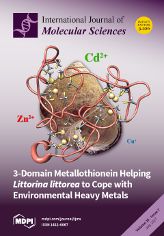
After determining the 3D structure of Littorina littorea metallothionein (Angew. Chem. Int. Ed., 2017, 56, 4617-4622) and its recognition in Science (2017, 356(6334), 150-151), in this work we analyze in depth the in vivo and in vitro metal binding capabilities of this 3-dominial metalloprotein and of two truncated mutants. We conclude that a Cd-specific metallothionein protein confers this snail a particular adaptive advantage in its changeable marine habitat. View this paper
  • Issues are regarded as officially published after their release is announced to the table of contents alert mailing list.
  • You may sign up for e-mail alerts to receive table of contents of newly released issues.
  • PDF is the official format for papers published in both, html and pdf forms. To view the papers in pdf format, click on the "PDF Full-text" link, and use the free Adobe Reader to open them.
Order results
Result details
Section
Select all
Export citation of selected articles as:
16 pages, 4134 KiB  
Article
FRET-Mediated Long-Range Wavelength Transformation by Photoconvertible Fluorescent Proteins as an Efficient Mechanism to Generate Orange-Red Light in Symbiotic Deep Water Corals
by Elena Bollati 1, Daniel Plimmer 1, Cecilia D’Angelo 1,2 and Jörg Wiedenmann 1,2,*
1 Coral Reef Laboratory, University of Southampton, Waterfront Campus, European Way, Southampton SO143ZH, UK
2 Institute for Life Sciences (IFLS), University of Southampton, Highfield Campus, Southampton SO171BJ, UK
Int. J. Mol. Sci. 2017, 18(7), 1174; https://doi.org/10.3390/ijms18071174 - 4 Jul 2017
Cited by 11 | Viewed by 8081
Abstract
Photoconvertible fluorescent proteins (pcRFPs) are a group of fluorophores that undergo an irreversible green-to-red shift in emission colour upon irradiation with near-ultraviolet (near-UV) light. Despite their wide application in biotechnology, the high-level expression of pcRFPs in mesophotic and depth-generalist coral species currently lacks [...] Read more.
Photoconvertible fluorescent proteins (pcRFPs) are a group of fluorophores that undergo an irreversible green-to-red shift in emission colour upon irradiation with near-ultraviolet (near-UV) light. Despite their wide application in biotechnology, the high-level expression of pcRFPs in mesophotic and depth-generalist coral species currently lacks a biological explanation. Additionally, reduced penetration of near-UV wavelengths in water poses the question whether light-driven photoconversion is relevant in the mesophotic zone, or whether a different mechanism is involved in the post-translational pigment modification in vivo. Here, we show in a long-term mesocosm experiment that photoconversion in vivo is entirely dependent on near-UV wavelengths. However, a near-UV intensity equivalent to the mesophotic underwater light field at 80 m depth is sufficient to drive the process in vitro, suggesting that photoconversion can occur near the lower distribution limits of these corals. Furthermore, live coral colonies showed evidence of efficient Förster Resonance Energy Transfer (FRET). Our simulated mesophotic light field maintained the pcRFP pool in a partially photoconverted state in vivo, maximising intra-tetrameric FRET and creating a long-range wavelength conversion system with higher quantum yield than other native RFPs. We hypothesise that efficient conversion of blue wavelengths, abundant at depth, into orange-red light could constitute an adaptation of corals to life in light-limited environments. Full article
(This article belongs to the Special Issue Fluorescent Proteins)
Show Figures

Graphical abstract

16 pages, 2104 KiB  
Article
Colonization and Maize Growth Promotion Induced by Phosphate Solubilizing Bacterial Isolates
by Yongbin Li, Xiaomeng Liu, Tianyi Hao and Sanfeng Chen *
State Key Laboratory of Agrobiotechnology and College of Biological Sciences, China Agricultural University, Beijing 100094, China
Int. J. Mol. Sci. 2017, 18(7), 1253; https://doi.org/10.3390/ijms18071253 - 29 Jun 2017
Cited by 84 | Viewed by 8876
Abstract
Phosphorus (P) limits the production of maize, one of the major food crops in China. Phosphate-solubilizing bacteria (PSB) have the capacity to solubilize phosphate complexes into plant absorbable and utilizable forms by the process of acidification, chelation, and exchange reactions. In this study, [...] Read more.
Phosphorus (P) limits the production of maize, one of the major food crops in China. Phosphate-solubilizing bacteria (PSB) have the capacity to solubilize phosphate complexes into plant absorbable and utilizable forms by the process of acidification, chelation, and exchange reactions. In this study, six bacteria, including one Paenibacillus sp. B1 strain, four Pseudomonas sp. strains (B10, B14, SX1, and SX2) and one Sphingobium sp. SX14 strain, were those isolated from the maize rhizosphere and identified based on their 16S rRNA sequences. All strains could solubilize inorganic P (Ca3(PO4)2, FePO4 and AlPO4), and only B1 and B10 organic P (lecithin). All strains, except of SX1, produced IAA, and SX14 and B1 showed the highest level. B1 incited the highest increase in root length and the second increase in shoot and total dry weight, shoot length, and total P and nitrogen (N), along with increased root length. In addition, by confocal laser scanning microscopy (CLSM), we found that green fluorescent protein (GFP)-labeled B1 mainly colonized root surfaces and in epidermal and cortical tissue. Importantly, B1 can survive through forming spores under adverse conditions and prolong quality guarantee period of bio-fertilizer. Therefore, it can act as a good substitute for bio-fertilizer to promote agricultural sustainability. Full article
(This article belongs to the Section Molecular Plant Sciences)
Show Figures

Graphical abstract

16 pages, 3115 KiB  
Article
Identification of Light-Independent Anthocyanin Biosynthesis Mutants Induced by Ethyl Methane Sulfonate in Turnip “Tsuda” (Brassica rapa)
by Jian-Fei Yang 1, Yun-Zhu Chen 1, Saneyuki Kawabata 2, Yu-Hua Li 1,3,* and Yu Wang 1,3,*
1 College of Life Science, Northeast Forestry University, Harbin 150040, China
2 Graduate School of Agricultural and Life Sciences, The University of Tokyo, Yayoi, Bunkyo Tokyo 113-8654, Japan
3 State Key Laboratory of Tree Genetics and Breeding, Northeast Forestry University, Harbin 150040, China
Int. J. Mol. Sci. 2017, 18(7), 1288; https://doi.org/10.3390/ijms18071288 - 22 Jun 2017
Cited by 20 | Viewed by 7692
Abstract
The epidermis of swollen storage roots in purple cultivars of turnip “Tsuda” (Brassica rapa) accumulates anthocyanin in a light-dependent manner, especially in response to UV-A light, of which the mechanism is unclear. In this study, we mutagenized 15,000 seeds by 0.5% [...] Read more.
The epidermis of swollen storage roots in purple cultivars of turnip “Tsuda” (Brassica rapa) accumulates anthocyanin in a light-dependent manner, especially in response to UV-A light, of which the mechanism is unclear. In this study, we mutagenized 15,000 seeds by 0.5% (v/v) ethyl methane sulfonate (EMS) and obtained 14 mutants with abnormal anthocyanin production in their epidermis of swollen storage roots. These mutants were classified into two groups: the red mutants with constitutive anthocyanin accumulation in their epidermis of storage roots even in underground parts in darkness and the white mutants without anthocyanin accumulation in the epidermis of storage roots in aboveground parts exposed to sunlight. Test cross analysis demonstrated that w9, w68, w204, r15, r21, r30 and r57 contained different mutations responsible for their phenotypic variations. Further genetic analysis of four target mutants (w9, w68, w204 and r15) indicated that each of them was controlled by a different recessive gene. Intriguingly, the expression profiles of anthocyanin biosynthesis genes, including structural and regulatory genes, coincided with their anthocyanin levels in the epidermis of storage roots in the four target mutants. We proposed that potential genes responsible for the mutations should be upstream factors of the anthocyanin biosynthesis pathway in turnips, which provided resources to further investigate the mechanisms of light-induced anthocyanin accumulation. Full article
(This article belongs to the Special Issue Anthocyanins)
Show Figures

Graphical abstract

12 pages, 2870 KiB  
Article
Moringa Leaves Prevent Hepatic Lipid Accumulation and Inflammation in Guinea Pigs by Reducing the Expression of Genes Involved in Lipid Metabolism
by Manal Mused Almatrafi 1, Marcela Vergara-Jimenez 2, Ana Gabriela Murillo 1, Gregory H. Norris 1, Christopher N. Blesso 1 and Maria Luz Fernandez 1,*
1 Department of Nutritional Sciences, University of Connecticut, Storrs, CT 06269, USA
2 Department of Nutrition, Universidad Autonoma de Sinaloa, Culiacán, Sinaloa 80019, Mexico
Int. J. Mol. Sci. 2017, 18(7), 1330; https://doi.org/10.3390/ijms18071330 - 22 Jun 2017
Cited by 48 | Viewed by 9015
Abstract
To investigate the mechanisms by which Moringa oleifera leaves (ML) modulate hepatic lipids, guinea pigs were allocated to either control (0% ML), 10% Low Moringa (LM) or 15% High Moringa (HM) diets with 0.25% dietary cholesterol to induce hepatic steatosis. After 6 weeks, [...] Read more.
To investigate the mechanisms by which Moringa oleifera leaves (ML) modulate hepatic lipids, guinea pigs were allocated to either control (0% ML), 10% Low Moringa (LM) or 15% High Moringa (HM) diets with 0.25% dietary cholesterol to induce hepatic steatosis. After 6 weeks, guinea pigs were sacrificed and liver and plasma were collected to determine plasma lipids, hepatic lipids, cytokines and the expression of genes involved in hepatic cholesterol (CH) and triglyceride (TG) metabolism. There were no differences in plasma lipids among groups. A dose-response effect of ML was observed in hepatic lipids (CH and TG) with the lowest concentrations in the HM group (p < 0.001), consistent with histological evaluation of lipid droplets. Hepatic gene expression of diglyceride acyltransferase-2 and peroxisome proliferator activated receptor-γ, as well as protein concentrations interleukin (IL)-1β and interferon-γ, were lowest in the HM group (p < 0.005). Hepatic gene expression of cluster of differentiation-68 and sterol regulatory element binding protein-1c were 60% lower in both the LM and HM groups compared to controls (p < 0.01). This study demonstrates that ML may prevent hepatic steatosis by affecting gene expression related to hepatic lipids synthesis resulting in lower concentrations of cholesterol and triglycerides and reduced inflammation in the liver. Full article
(This article belongs to the Special Issue Natural Anti-Inflammatory Agents)
Show Figures

Figure 1

17 pages, 1304 KiB  
Article
Pharmacogenomic Variants May Influence the Urinary Excretion of Novel Kidney Injury Biomarkers in Patients Receiving Cisplatin
by Cara Chang 1, Yichun Hu 2, Susan L. Hogan 2, Nickie Mercke 1, Madeleine Gomez 1, Cindy O’Bryant 1,3, Daniel W. Bowles 3, Blessy George 4, Xia Wen 4, Lauren M. Aleksunes 4 and Melanie S. Joy 1,3,5,*
1 Department of Pharmaceutical Sciences, Skaggs School of Pharmacy and Pharmaceutical Sciences, University of Colorado, Aurora, CO 80045, USA
2 Kidney Center, University of North Carolina School of Medicine, Division of Nephrology and Hypertension, Chapel Hill, NC 27599, USA
3 Cancer Center, University of Colorado, Aurora, CO 80045, USA
4 Department of Pharmacology and Toxicology, Ernest Mario School of Pharmacy, Rutgers University, Piscataway, NJ 08854, USA
5 Division of Renal Diseases and Hypertension, University of Colorado School of Medicine, Aurora, CO 80045, USA
Int. J. Mol. Sci. 2017, 18(7), 1333; https://doi.org/10.3390/ijms18071333 - 22 Jun 2017
Cited by 30 | Viewed by 7300
Abstract
Nephrotoxicity is a dose limiting side effect associated with the use of cisplatin in the treatment of solid tumors. The degree of nephrotoxicity is dictated by the selective accumulation of cisplatin in renal tubule cells due to: (1) uptake by organic cation transporter [...] Read more.
Nephrotoxicity is a dose limiting side effect associated with the use of cisplatin in the treatment of solid tumors. The degree of nephrotoxicity is dictated by the selective accumulation of cisplatin in renal tubule cells due to: (1) uptake by organic cation transporter 2 (OCT2) and copper transporter 1 (CTR1); (2) metabolism by glutathione S-transferases (GSTs) and γ-glutamyltransferase 1 (GGT1); and (3) efflux by multidrug resistance-associated protein 2 (MRP2) and multidrug and toxin extrusion protein 1 (MATE1). The purpose of this study was to determine the significance of single nucleotide polymorphisms that regulate the expression and function of transporters and metabolism genes implicated in development of acute kidney injury (AKI) in cisplatin treated patients. Changes in the kidney function were assessed using novel urinary protein biomarkers and traditional markers. Genotyping was conducted by the QuantStudio 12K Flex Real-Time PCR System using a custom open array chip with metabolism, transport, and transcription factor polymorphisms of interest to cisplatin disposition and toxicity. Traditional and novel biomarker assays for kidney toxicity were assessed for differences according to genotype by ANOVA. Allele and genotype frequencies were determined based on Caucasian population frequencies. The polymorphisms rs596881 (SLC22A2/OCT2), and rs12686377 and rs7851395 (SLC31A1/CTR1) were associated with renoprotection and maintenance of estimated glomerular filtration rate (eGFR). Polymorphisms in SLC22A2/OCT2, SLC31A1/CTRI, SLC47A1/MATE1, ABCC2/MRP2, and GSTP1 were significantly associated with increases in the urinary excretion of novel AKI biomarkers: KIM-1, TFF3, MCP1, NGAL, clusterin, cystatin C, and calbindin. Knowledge concerning which genotypes in drug transporters are associated with cisplatin-induced nephrotoxicity may help to identify at-risk patients and initiate strategies, such as using lower or fractionated cisplatin doses or avoiding cisplatin altogether, in order to prevent AKI. Full article
(This article belongs to the Special Issue Nephrotoxicity)
Show Figures

Figure 1

17 pages, 262 KiB  
Review
Lectins from Mycelia of Basidiomycetes
by Valentina E. Nikitina, Ekaterina A. Loshchinina * and Elena P. Vetchinkina
Laboratory of Microbiology, Institute of Biochemistry and Physiology of Plants and Microorganisms, Russian Academy of Sciences, 13 Prospekt Entuziastov, Saratov 410049, Russia
Int. J. Mol. Sci. 2017, 18(7), 1334; https://doi.org/10.3390/ijms18071334 - 22 Jun 2017
Cited by 14 | Viewed by 5745
Abstract
Lectins are proteins of a nonimmunoglobulin nature that are capable of specific recognition of and reversible binding to the carbohydrate moieties of complex carbohydrates, without altering the covalent structure of any of the recognized glycosyl ligands. They have a broad range of biological [...] Read more.
Lectins are proteins of a nonimmunoglobulin nature that are capable of specific recognition of and reversible binding to the carbohydrate moieties of complex carbohydrates, without altering the covalent structure of any of the recognized glycosyl ligands. They have a broad range of biological activities important for the functioning of the cell and the whole organism and, owing to the high specificity of reversible binding to carbohydrates, are valuable tools used widely in biology and medicine. Lectins can be produced by many living organisms, including basidiomycetes. Whereas lectins from the fruit bodies of basidiomycetes have been studied sufficiently well, mycelial lectins remain relatively unexplored. Here, we review and comparatively analyze what is currently known about lectins isolated from the vegetative mycelium of macrobasidiomycetes, including their localization, properties, and carbohydrate specificities. Particular attention is given to the physiological role of mycelial lectins in fungal growth and development. Full article
(This article belongs to the Special Issue Plant Lectins: From Model Species to Crop Plants)
11 pages, 860 KiB  
Article
Associations of Drug Lipophilicity and Extent of Metabolism with Drug-Induced Liver Injury
by Kristin McEuen 1,2, Jürgen Borlak 3, Weida Tong 1 and Minjun Chen 1,*
1 Division of Bioinformatics and Biostatistics, National Center for Toxicological Research, US Food and Drug Administration, Jefferson, AR 72079, USA
2 Department of Information Science, University of Arkansas at Little Rock, Little Rock, AR 72204, USA
3 Center of Pharmacology and Toxicology, Hannover Medical School, Hannover 30625, Germany
Int. J. Mol. Sci. 2017, 18(7), 1335; https://doi.org/10.3390/ijms18071335 - 22 Jun 2017
Cited by 50 | Viewed by 5353
Abstract
Drug-induced liver injury (DILI), although rare, is a frequent cause of adverse drug reactions resulting in warnings and withdrawals of numerous medications. Despite the research community’s best efforts, current testing strategies aimed at identifying hepatotoxic drugs prior to human trials are not sufficiently [...] Read more.
Drug-induced liver injury (DILI), although rare, is a frequent cause of adverse drug reactions resulting in warnings and withdrawals of numerous medications. Despite the research community’s best efforts, current testing strategies aimed at identifying hepatotoxic drugs prior to human trials are not sufficiently powered to predict the complex mechanisms leading to DILI. In our previous studies, we demonstrated lipophilicity and dose to be associated with increased DILI risk and, and in our latest work, we factored reactive metabolites into the algorithm to predict DILI. Given the inconsistency in determining the potential for drugs to cause DILI, the present study comprehensively assesses the relationship between DILI risk and lipophilicity and the extent of metabolism using a large published dataset of 1036 Food and Drug Administration (FDA)-approved drugs by considering five independent DILI annotations. We found that lipophilicity and the extent of metabolism alone were associated with increased risk for DILI. Moreover, when analyzed in combination with high daily dose (≥100 mg), lipophilicity was statistically significantly associated with the risk of DILI across all datasets (p < 0.05). Similarly, the combination of extensive hepatic metabolism (≥50%) and high daily dose (≥100 mg) was also strongly associated with an increased risk of DILI among all datasets analyzed (p < 0.05). Our results suggest that both lipophilicity and the extent of hepatic metabolism can be considered important risk factors for DILI in humans, and that this relationship to DILI risk is much stronger when considered in combination with dose. The proposed paradigm allows the convergence of different published annotations to a more uniform assessment. Full article
(This article belongs to the Special Issue Molecular Research on Drug Induced Liver Injury)
Show Figures

Figure 1

11 pages, 3223 KiB  
Article
Quinolin-6-Yloxyacetamides Are Microtubule Destabilizing Agents That Bind to the Colchicine Site of Tubulin
by Ashwani Sharma 1, Gonzalo Sáez-Calvo 2, Natacha Olieric 1, Francisco De Asís Balaguer 2, Isabel Barasoain 2, Clemens Lamberth 3, J. Fernando Díaz 2,* and Michel O. Steinmetz 1,*
1 Laboratory of Biomolecular Research, Division of Biology and Chemistry, Paul Scherrer Institut, CH-5232 Villigen, Switzerland
2 Centro de Investigaciones Biológicas, Consejo Superior de Investigaciones Científicas, Ramiro de Maeztu 9, 28040 Madrid, Spain
3 Chemical Research, Syngenta Crop Protection AG, Schaffhauserstrasse 101, CH-4332 Stein, Switzerland
Int. J. Mol. Sci. 2017, 18(7), 1336; https://doi.org/10.3390/ijms18071336 - 22 Jun 2017
Cited by 11 | Viewed by 5371
Abstract
Quinolin-6-yloxyacetamides (QAs) are a chemical class of tubulin polymerization inhibitors that were initially identified as fungicides. Here, we report that QAs are potent anti-proliferative agents against human cancer cells including ones that are drug-resistant. QAs act by disrupting the microtubule cytoskeleton and by [...] Read more.
Quinolin-6-yloxyacetamides (QAs) are a chemical class of tubulin polymerization inhibitors that were initially identified as fungicides. Here, we report that QAs are potent anti-proliferative agents against human cancer cells including ones that are drug-resistant. QAs act by disrupting the microtubule cytoskeleton and by causing severe mitotic defects. We further demonstrate that QAs inhibit tubulin polymerization in vitro. The high resolution crystal structure of the tubulin-QA complex revealed that QAs bind to the colchicine site on tubulin, which is targeted by microtubule-destabilizing agents such as colchicine and nocodazole. Together, our data establish QAs as colchicine-site ligands and explain the molecular mechanism of microtubule destabilization by this class of compounds. They further extend our structural knowledge on antitubulin agents and thus should aid in the development of new strategies for the rational design of ligands against multidrug-resistant cancer cells. Full article
(This article belongs to the Special Issue Microtubule-Targeting Agents)
Show Figures

Figure 1

18 pages, 3692 KiB  
Article
Nomegestrol Acetate Suppresses Human Endometrial Cancer RL95-2 Cells Proliferation In Vitro and In Vivo Possibly Related to Upregulating Expression of SUFU and Wnt7a
by A-ying Ma, Shu-wu Xie, Jie-yun Zhou and Yan Zhu *
1 Lab of Reproductive Pharmacology, Key Lab of Reproduction Regulation of NPFPC, SIPPR, IRD, Fudan University, Shanghai 200032, China
These authors contributed equally to this work.
Int. J. Mol. Sci. 2017, 18(7), 1337; https://doi.org/10.3390/ijms18071337 - 22 Jun 2017
Cited by 19 | Viewed by 6740
Abstract
Nomegestrol acetate (NOMAC) has been successfully used for the treatment of some gynecological disorders, and as a combined oral contraceptive with approval in many countries. In this study, we investigated the effects of NOMAC on human endometrial cancer cells in vitro and in [...] Read more.
Nomegestrol acetate (NOMAC) has been successfully used for the treatment of some gynecological disorders, and as a combined oral contraceptive with approval in many countries. In this study, we investigated the effects of NOMAC on human endometrial cancer cells in vitro and in vivo. The proliferation of human endometrial cancer cells (RL95-2 and KLE) were assessed using CCK-8 and EdU incorporation assays. Whole-genome cDNA microarray analysis was used to identify the effects of NOMAC on gene expression profiles in RL95-2 cells. RL95-2 xenograft nude mice were treated with NOMAC (50, 100, and 200 mg/kg) or medroxyprogesterone acetate (MPA; 100 and 200 mg/kg) for 28 consecutive days. The results showed that NOMAC significantly inhibited the growth of RL95-2 cells in a concentration-dependent manner, but not in KLE cells. Further investigation demonstrated that NOMAC produced a stronger inhibition of tumor growth (inhibition rates for 50, 100, and 200 mg/kg NOMAC were 24.74%, 47.04%, and 58.06%, respectively) than did MPA (inhibition rates for 100 and 200 mg/kg MPA were 41.06% and 27.01%, respectively) in the nude mice bearing the cell line of RL95-2. NOMAC altered the expression of several genes related to cancer cell proliferation, including SUFU and Wnt7a. The upregulation of SUFU and Wnt7a was confirmed using real-time quantitative polymerase chain reaction and Western blotting in RL95-2 cells and RL95-2 xenograft tumor tissues, but not in KLE cells. These data indicate that NOMAC can inhibit the proliferation of RL95-2 cell in vitro and suppress the growth of xenografts in the nude mice bearing the cell line of RL95-2 in vivo. This effect could be related to the upregulating expression of SUFU and Wnt7a. Full article
(This article belongs to the Special Issue Gynecologic Oncology: From Molecular Mechanisms to Targeted Therapies)
Show Figures

Graphical abstract

43 pages, 1523 KiB  
Review
Pancreatic Ductal Adenocarcinoma: Current and Evolving Therapies
by Aleksandra Adamska, Alice Domenichini and Marco Falasca *
Metabolic Signalling Group, School of Biomedical Sciences, Curtin Health Innovation Research Institute, Curtin University, Perth, WA 6102, Australia
Int. J. Mol. Sci. 2017, 18(7), 1338; https://doi.org/10.3390/ijms18071338 - 22 Jun 2017
Cited by 437 | Viewed by 19558
Abstract
Pancreatic ductal adenocarcinoma (PDAC), which constitutes 90% of pancreatic cancers, is the fourth leading cause of cancer-related deaths in the world. Due to the broad heterogeneity of genetic mutations and dense stromal environment, PDAC belongs to one of the most chemoresistant cancers. Most [...] Read more.
Pancreatic ductal adenocarcinoma (PDAC), which constitutes 90% of pancreatic cancers, is the fourth leading cause of cancer-related deaths in the world. Due to the broad heterogeneity of genetic mutations and dense stromal environment, PDAC belongs to one of the most chemoresistant cancers. Most of the available treatments are palliative, with the objective of relieving disease-related symptoms and prolonging survival. Currently, available therapeutic options are surgery, radiation, chemotherapy, immunotherapy, and use of targeted drugs. However, thus far, therapies targeting cancer-associated molecular pathways have not given satisfactory results; this is due in part to the rapid upregulation of compensatory alternative pathways as well as dense desmoplastic reaction. In this review, we summarize currently available therapies and clinical trials, directed towards a plethora of pathways and components dysregulated during PDAC carcinogenesis. Emerging trends towards targeted therapies as the most promising approach will also be discussed. Full article
(This article belongs to the Special Issue Pancreatic Disorders)
Show Figures

Figure 1

10 pages, 552 KiB  
Review
Oncogene-Induced Replication Stress Drives Genome Instability and Tumorigenesis
by Dan Sarni and Batsheva Kerem *
Department of Genetics, The Life Sciences Institute, The Hebrew University, 91904 Jerusalem, Israel
Int. J. Mol. Sci. 2017, 18(7), 1339; https://doi.org/10.3390/ijms18071339 - 22 Jun 2017
Cited by 20 | Viewed by 5894
Abstract
Genomic instability plays a key role in driving cancer development. It is already found in precancerous lesions and allows the acquisition of additional cancerous features. A major source of genomic instability in early stages of tumorigenesis is DNA replication stress. Normally, origin licensing [...] Read more.
Genomic instability plays a key role in driving cancer development. It is already found in precancerous lesions and allows the acquisition of additional cancerous features. A major source of genomic instability in early stages of tumorigenesis is DNA replication stress. Normally, origin licensing and activation, as well as replication fork progression, are tightly regulated to allow faithful duplication of the genome. Aberrant origin usage and/or perturbed replication fork progression leads to DNA damage and genomic instability. Oncogene activation is an endogenous source of replication stress, disrupting replication regulation and inducing DNA damage. Oncogene-induced replication stress and its role in cancer development have been studied comprehensively, however its molecular basis is still unclear. Here, we review the current understanding of replication regulation, its potential disruption and how oncogenes perturb the replication and induce DNA damage leading to genomic instability in cancer. Full article
(This article belongs to the Special Issue Mechanisms Leading to Genomic Instability)
Show Figures

Graphical abstract

13 pages, 3194 KiB  
Article
Reverse Gyrase Functions in Genome Integrity Maintenance by Protecting DNA Breaks In Vivo
by Wenyuan Han *, Xu Feng and Qunxin She *
Archaeal Centre, Department of Biology, University of Copenhagen, Ole Maaløes Vej 5, Copenhagen Biocenter, DK-2200 Copenhagen N, Denmark
Int. J. Mol. Sci. 2017, 18(7), 1340; https://doi.org/10.3390/ijms18071340 - 22 Jun 2017
Cited by 25 | Viewed by 5183
Abstract
Reverse gyrase introduces positive supercoils to circular DNA and is implicated in genome stability maintenance in thermophiles. The extremely thermophilic crenarchaeon Sulfolobus encodes two reverse gyrase proteins, TopR1 (topoisomerase reverse gyrase 1) and TopR2, whose functions in thermophilic life remain to be demonstrated. [...] Read more.
Reverse gyrase introduces positive supercoils to circular DNA and is implicated in genome stability maintenance in thermophiles. The extremely thermophilic crenarchaeon Sulfolobus encodes two reverse gyrase proteins, TopR1 (topoisomerase reverse gyrase 1) and TopR2, whose functions in thermophilic life remain to be demonstrated. Here, we investigated the roles of TopR1 in genome stability maintenance in S. islandicus in response to the treatment of methyl methanesulfonate (MMS), a DNA alkylation agent. Lethal MMS treatment induced two successive events: massive chromosomal DNA backbone breakage and subsequent DNA degradation. The former occurred immediately after drug treatment, leading to chromosomal DNA degradation that concurred with TopR1 degradation, followed by chromatin protein degradation and DNA-less cell formation. To gain a further insight into TopR1 function, the expression of the enzyme was reduced in S. islandicus cells using a CRISPR-mediated mRNA interference approach (CRISPRi) in which topR1 mRNAs were targeted for degradation by endogenous III-B CRISPR-Cas systems. We found that the TopR1 level was reduced in the S. islandicus CRISPRi cells and that the cells underwent accelerated genomic DNA degradation during MMS treatment, accompanied by a higher rate of cell death. Taken together, these results indicate that TopR1 probably facilitates genome integrity maintenance by protecting DNA breaks from thermo-degradation in vivo. Full article
(This article belongs to the Special Issue DNA Injury and Repair Systems)
Show Figures

Graphical abstract

13 pages, 1100 KiB  
Review
RNA Binding Proteins and Genome Integrity
by Kensei Nishida 1,*, Yuki Kuwano 1, Tatsuya Nishikawa 1, Kiyoshi Masuda 2 and Kazuhito Rokutan 1
1 Department of Pathophysiology, Institute of Biomedical Sciences, Tokushima University Graduate School, Tokushima 770-8503, Japan
2 Department of Human Genetics, Institute of Biomedical Sciences, Tokushima University Graduate School, Tokushima 770-8503, Japan
Int. J. Mol. Sci. 2017, 18(7), 1341; https://doi.org/10.3390/ijms18071341 - 23 Jun 2017
Cited by 44 | Viewed by 8972
Abstract
Genome integrity can be threatened by various endogenous or exogenous events. To counteract these stressors, the DNA damage response network contributes to the prevention and/or repair of genomic DNA damage and serves an essential function in cellular survival. DNA binding proteins are involved [...] Read more.
Genome integrity can be threatened by various endogenous or exogenous events. To counteract these stressors, the DNA damage response network contributes to the prevention and/or repair of genomic DNA damage and serves an essential function in cellular survival. DNA binding proteins are involved in this network. Recently, several RNA-binding proteins (RBPs) that are recruited to DNA damage sites have been shown to be direct players in the prevention or repair of DNA damage. In addition, non-coding RNAs, themselves, are involved in the RNA-mediated DNA repair system. Furthermore, RNA modification such as m6A methylation might also contribute to the ultraviolet-responsive DNA damage response. Accumulating evidence suggests that RNA metabolism is more deeply involved in diverse cellular functions than previously expected, and is also intricately associated with the maintenance of genome integrity. In this review, we highlight the roles of RBPs in the maintenance of genome integrity. Full article
(This article belongs to the Special Issue Mechanisms Leading to Genomic Instability)
Show Figures

Graphical abstract

11 pages, 1785 KiB  
Article
GC-MS Fingerprinting Combined with Chemometric Methods Reveals Key Bioactive Components in Acori Tatarinowii Rhizoma
by Wenbin Liu 1, Bingyang Zhang 2, Zhongquan Xin 1, Dabing Ren 1 and Lunzhao Yi 1,*
1 Yunnan Food Safety Research Institute, Kunming University of Science and Technology, Kunming 650500, China
2 School of Science, Kunming University of Science and Technology, Kunming 650500, China
Int. J. Mol. Sci. 2017, 18(7), 1342; https://doi.org/10.3390/ijms18071342 - 3 Jul 2017
Cited by 26 | Viewed by 6209
Abstract
This present study aims to identify the key bioactive components in acorus tatarinowii rhizoma (ATR), a traditional Chinese medicine (TCM) with various bioactivities. Partial least squares regression (PLSR) was employed to describe the relationship between the radical scavenging activity and the volatile components. [...] Read more.
This present study aims to identify the key bioactive components in acorus tatarinowii rhizoma (ATR), a traditional Chinese medicine (TCM) with various bioactivities. Partial least squares regression (PLSR) was employed to describe the relationship between the radical scavenging activity and the volatile components. The PLSR model was improved by outlier elimination and variable selection and was evaluated by 10-fold cross-validation and external validation in this study. Based on the PLSR model, eleven chemical components were identified as the key bioactive components by variable importance in projection. The final PLS regression model with these components has good predictive ability. The Q2 was 0.8284, and the root mean square error for prediction was 2.9641. The results indicated that the eleven components could be a pattern to predict the radical scavenging activity of ATR. In addition, we did not find any specific relationship between the radical scavenging ability and the habitat of the ATRs. This study proposed an efficient strategy to predict bioactive components using the combination of quantitative chromatography fingerprints and PLS regression, and has potential perspective for screening bioactive components in complex analytical systems, such as TCM. Full article
Show Figures

Graphical abstract

16 pages, 2358 KiB  
Article
Capsaicin Induces Autophagy and Apoptosis in Human Nasopharyngeal Carcinoma Cells by Downregulating the PI3K/AKT/mTOR Pathway
by Yu-Tsai Lin 1,2,3, Hung-Chen Wang 4, Yi-Chiang Hsu 5, Chung-Lung Cho 6, Ming-Yu Yang 1,2,* and Chih-Yen Chien 1,3,*
1 Department of Otolaryngology, Kaohsiung Chang Gung Memorial Hospital and Chang Gung University College of Medicine, Kaohsiung 83301, Taiwan
2 Graduate Institute of Clinical Medical Sciences, College of Medicine, Chang Gung University, Taoyuan City 33302, Taiwan
3 Kaohsiung Chang Gung Head and Neck Oncology Group, Cancer Center, Kaohsiung Chang Gung Memorial Hospital, Kaohsiung 83301, Taiwan
4 Department of Neurosurgery, Kaohsiung Chang Gung Memorial Hospital, Kaohsiung 83301, Taiwan
5 Graduate Institute of Medical Science and Innovative Research Center of Medicine, College of Health Sciences, Chang Jung Christian University, Tainan 71101, Taiwan
6 Department of Biological Sciences, National Sun Yat-sen University, Kaohsiung 80424, Taiwan
Int. J. Mol. Sci. 2017, 18(7), 1343; https://doi.org/10.3390/ijms18071343 - 23 Jun 2017
Cited by 96 | Viewed by 8618
Abstract
Capsaicin is a potential chemotherapeutic agent for different human cancers. In Southeast China, nasopharyngeal carcinoma (NPC) has the highest incidence of all cancers, but final treatment outcomes are unsatisfactory. However, there is a lack of information regarding the anticancer activity of capsaicin in [...] Read more.
Capsaicin is a potential chemotherapeutic agent for different human cancers. In Southeast China, nasopharyngeal carcinoma (NPC) has the highest incidence of all cancers, but final treatment outcomes are unsatisfactory. However, there is a lack of information regarding the anticancer activity of capsaicin in NPC cells, and its effects on the signaling transduction pathways related to apoptosis and autophagy remain unclear. In the present study, the precise mechanisms by which capsaicin exerts anti-proliferative effects, cell cycle arrest, autophagy and apoptosis were investigated in NPC-TW01 cells. Exposure to capsaicin inhibited cancer cell growth and increased G1 phase cell cycle arrest. Western blotting and quantitative real-time reverse transcription polymerase chain reaction (qRT-PCR) were used to measure capsaicin-induced autophagy via involvement of the class III PI3K/Beclin-1/Bcl-2 signaling pathway. Capsaicin induced autophagy by increasing levels of the autophagy markers LC3-II and Atg5, enhancing p62 and Fap-1 degradation and increasing caspase-3 activity to induce apoptosis, suggesting a correlation of blocking the PI3K/Akt/mTOR pathway with the above-mentioned anticancer activities. Taken together, these data confirm that capsaicin inhibited the growth of human NPC cells and induced autophagy, supporting its potential as a therapeutic agent for cancer. Full article
Show Figures

Figure 1

12 pages, 240 KiB  
Review
Chronic Respiratory Infection in Patients with Chronic Obstructive Pulmonary Disease: What Is the Role of Antibiotics?
by Marc Miravitlles 1 and Antonio Anzueto 2,3,*
1 Pneumology Department, Hospital Universitari Vall d’Hebron, Ciber de Enfermedades Respiratorias (CIBERES), 08035 Barcelona, Spain
2 Department of Medicine, Division of Pulmonary Diseases/Critical Care Medicine, The University of Texas Health Science Center at San Antonio, San Antonio, TX 78229, USA
3 Pulmonary Section, The South Texas Veterans Health Care System, Audie L. Murphy Memorial Veterans Hospital Division, Pulmonary Diseases Section (111E), 7400 Merton Minter Boulevard, San Antonio, TX 78229, USA
Int. J. Mol. Sci. 2017, 18(7), 1344; https://doi.org/10.3390/ijms18071344 - 23 Jun 2017
Cited by 39 | Viewed by 7249
Abstract
Chronic infections are associated with exacerbation in patients with chronic obstructive pulmonary disease (COPD). The major objective of the management of these patients is the prevention and effective treatment of exacerbations. Patients that have increased sputum production, associated with purulence and worsening shortness [...] Read more.
Chronic infections are associated with exacerbation in patients with chronic obstructive pulmonary disease (COPD). The major objective of the management of these patients is the prevention and effective treatment of exacerbations. Patients that have increased sputum production, associated with purulence and worsening shortness of breath, are the ones that will benefit from antibiotic therapy. It is important to give the appropriate antibiotic therapy to prevent treatment failure, relapse, and the emergence of resistant pathogens. In some patients, systemic corticosteroids are also indicated to improve symptoms. In order to identify which patients are more likely to benefit from these therapies, clinical guidelines recommend stratifying patients based on their risk factor associated with poor outcome or recurrence. It has been identified that patients with more severe disease, recurrent infection and presence of purulent sputum are the ones that will be more likely to benefit from this therapy. Another approach related to disease prevention could be the use of prophylactic antibiotics during steady state condition. Some studies have evaluated the continuous or the intermittent use of antibiotics in order to prevent exacerbations. Due to increased bacterial resistance to antibiotics and the presence of side effects, several antibiotics have been developed to be nebulized for both treatment and prevention of acute exacerbations. There is a need to design long-term studies to evaluate these interventions in the natural history of the disease. The purpose of this publication is to review our understanding of the role of bacterial infection in patients with COPD exacerbation, the role of antibiotics, and future interventions. Full article
(This article belongs to the Special Issue Lung Diseases: Chronic Respiratory Infections)
15 pages, 534 KiB  
Review
Development of Bone Targeting Drugs
by Molly Stapleton 1,2,†, Kazuki Sawamoto 2,†, Carlos J. Alméciga-Díaz 3,†, William G. Mackenzie 2, Robert W. Mason 1,2, Tadao Orii 4 and Shunji Tomatsu 1,2,4,5,*
1 Department of Biological Sciences, University of Delaware, Newark, DE 19716, USA
2 Nemours/Alfred I. duPont Hospital for Children, Wilmington, DE 19803, USA
3 Institute for the Study of Inborn Errors of Metabolism, Pontificia Universidad Javeriana, Bogotá D.C. 110231, Colombia
4 Department of Pediatrics, Graduate School of Medicine, Gifu University, Gifu 501-1193, Japan
5 Department of Pediatrics, Thomas Jefferson University, Philadelphia, PA 19107, USA
These authors contributed equally to this work.
Int. J. Mol. Sci. 2017, 18(7), 1345; https://doi.org/10.3390/ijms18071345 - 23 Jun 2017
Cited by 91 | Viewed by 10594
Abstract
The skeletal system, comprising bones, ligaments, cartilage and their connective tissues, is critical for the structure and support of the body. Diseases that affect the skeletal system can be difficult to treat, mainly because of the avascular cartilage region. Targeting drugs to the [...] Read more.
The skeletal system, comprising bones, ligaments, cartilage and their connective tissues, is critical for the structure and support of the body. Diseases that affect the skeletal system can be difficult to treat, mainly because of the avascular cartilage region. Targeting drugs to the site of action can not only increase efficacy but also reduce toxicity. Bone-targeting drugs are designed with either of two general targeting moieties, aimed at the entire skeletal system or a specific cell type. Most bone-targeting drugs utilize an affinity to hydroxyapatite, a major component of the bone matrix that includes a high concentration of positively-charged Ca2+. The strategies for designing such targeting moieties can involve synthetic and/or biological components including negatively-charged amino acid peptides or bisphosphonates. Efficient delivery of bone-specific drugs provides significant impact in the treatment of skeletal related disorders including infectious diseases (osteoarthritis, osteomyelitis, etc.), osteoporosis, and metabolic skeletal dysplasia. Despite recent advances, however, both delivering the drug to its target without losing activity and avoiding adverse local effects remain a challenge. In this review, we investigate the current development of bone-targeting moieties, their efficacy and limitations, and discuss future directions for the development of these specific targeted treatments. Full article
(This article belongs to the Special Issue Advances in Bone and Cartilage Research)
Show Figures

Figure 1

15 pages, 5962 KiB  
Article
Cotton Ascorbate Oxidase Promotes Cell Growth in Cultured Tobacco Bright Yellow-2 Cells through Generation of Apoplast Oxidation
by Rong Li 1,†, Shan Xin 1,†, Chengcheng Tao 1, Xiang Jin 2 and Hongbin Li 1,*
1 College of Life Sciences, Key laboratory of Agrobiotechnology, Shihezi University, Shihezi 832003, China
2 Institute of Tropical Biosciences and Biotechnology, Chinese Academy of Tropical Agricultural Sciences, Haikou 571101, China
These authors contributed equally to this work.
Int. J. Mol. Sci. 2017, 18(7), 1346; https://doi.org/10.3390/ijms18071346 - 23 Jun 2017
Cited by 28 | Viewed by 5846
Abstract
Ascorbate oxidase (AO) plays an important role in cell growth through the modulation of reduction/oxidation (redox) control of the apoplast. Here, a cotton (Gossypium hirsutum) apoplastic ascorbate oxidase gene (GhAO1) was obtained from fast elongating fiber tissues. GhAO1 belongs [...] Read more.
Ascorbate oxidase (AO) plays an important role in cell growth through the modulation of reduction/oxidation (redox) control of the apoplast. Here, a cotton (Gossypium hirsutum) apoplastic ascorbate oxidase gene (GhAO1) was obtained from fast elongating fiber tissues. GhAO1 belongs to the multicopper oxidase (MCO) family and includes a signal peptide and several transmembrane regions. Analyses of quantitative real-time polymerase chain reaction (QRT-PCR) and enzyme activity showed that GhAO1 was expressed abundantly in 15-day post-anthesis (dpa) wild-type (WT) fibers in comparison with fuzzless-lintless (fl) mutant ovules. Subcellular distribution analysis in onion cells demonstrated that GhAO1 is localized in the cell wall. In transgenic tobacco bright yellow-2 (BY-2) cells with ectopic overexpression of GhAO1, the enhancement of cell growth with 1.52-fold increase in length versus controls was indicated, as well as the enrichment of both total ascorbate in whole-cells and dehydroascorbate acid (DHA) in apoplasts. In addition, promoted activities of AO and monodehydroascorbate reductase (MDAR) in apoplasts and dehydroascorbate reductase (DHAR) in whole-cells were displayed in transgenic tobacco BY-2 cells. Accumulation of H2O2, and influenced expressions of Ca2+ channel genes with the activation of NtMPK9 and NtCPK5 and the suppression of NtTPC1B were also demonstrated in transgenic tobacco BY-2 cells. Finally, significant induced expression of the tobacco NtAO gene in WT BY-2 cells under indole-3-acetic acid (IAA) treatment appeared; however, the sensitivity of the NtAO gene expression to IAA disappeared in transgenic BY-2 cells, revealing that the regulated expression of the AO gene is under the control of IAA. Taken together, these results provide evidence that GhAO1 plays an important role in fiber cell elongation and may promote cell growth by generating the oxidation of apoplasts, via the auxin-mediated signaling pathway. Full article
(This article belongs to the Special Issue Selected Papers from the 6th National Plant Protein Research Congress)
Show Figures

Graphical abstract

17 pages, 2303 KiB  
Article
New MT2 Melatonin Receptor-Selective Ligands: Agonists and Partial Agonists
by Jean A. Boutin 1,2,*, Anne Bonnaud 1, Chantal Brasseur 1, Olivier Bruno 1, Nolwenn Lepretre 3, Peter Oosting 3, Sophie Coumailleau 1, Philippe Delagrange 4, Olivier Nosjean 1,2 and Céline Legros 1
1 Pôle d’Expertise Biotechnologie, Chimie, Biologie, Institut de Recherches SERVIER, 78290 Croissy-sur-Seine, France
2 Pôle d’Expertise Recherches & BioPharmacie, Institut de Recherches Internationales SERVIER, 92150 Suresnes, France
3 DIVERCHIM S.A., 95700 Roissy-en-France, France
4 Pôle d’Innovations Thérapeutiques en Neurosciences, Institut de Recherches, SERVIER, 78290 Croissy-sur-Seine, France
Int. J. Mol. Sci. 2017, 18(7), 1347; https://doi.org/10.3390/ijms18071347 - 23 Jun 2017
Cited by 12 | Viewed by 6851
Abstract
The search for melatonin receptor agonists and antagonists specific towards one of the receptor subtypes will extend our understanding of the role of this system in relaying circadian information to the body. A series of compounds derived from a hit compound discovered in [...] Read more.
The search for melatonin receptor agonists and antagonists specific towards one of the receptor subtypes will extend our understanding of the role of this system in relaying circadian information to the body. A series of compounds derived from a hit compound discovered in a screening process led to powerful agonists specific for one of the isoform of the melatonin receptor namely, MT2. The compounds are based on a poorly explored skeleton in the molecular pharmacology of melatonin. By changing the steric hindrance of one substituent (i.e., from a hydrogen atom to a tributylstannyl group), we identified a possible partial agonist that could lead to antagonist analogues. The functionalities of these compounds were measured with a series of assays, including the binding of GTPγS, the inhibition of the cyclic AMP production, the β-arrestin recruitment, and the cell shape changes as determined by cellular dielectric spectroscopy (CellKey®). The variations between the compounds are discussed. Full article
(This article belongs to the Special Issue Melatonin and Its Analogues: Experimental and Clinical Aspects)
Show Figures

Graphical abstract

16 pages, 5053 KiB  
Article
RUNX1 Plays an Important Role in Mediating BMP9-Induced Osteogenic Differentiation of Mesenchymal Stem Cells Line C3H10T1/2, Murine Multi-Lineage Cells Lines C2C12 and MEFs
by Caixia Ji, Xiaohua Liu, Li Xu, Tingting Yu, Chaoqun Dong and Jinyong Luo *
Department of Laboratory Medicine, M.O.E. Key Laboratory of Laboratory Medicine Diagnostics, Chongqing Medical University, Chongqing 400016,China
Int. J. Mol. Sci. 2017, 18(7), 1348; https://doi.org/10.3390/ijms18071348 - 23 Jun 2017
Cited by 26 | Viewed by 5713
Abstract
As one of the least studied bone morphogenetic proteins (BMPs), BMP9 is highly capable of promoting osteogenic differentiation. However, the underlying mechanism involved remains largely unknown. Recent studies have demonstrated that RUNX1 (runt-related transcription factor 1) is essential in osteoblast/chondrocyte maturation. In this [...] Read more.
As one of the least studied bone morphogenetic proteins (BMPs), BMP9 is highly capable of promoting osteogenic differentiation. However, the underlying mechanism involved remains largely unknown. Recent studies have demonstrated that RUNX1 (runt-related transcription factor 1) is essential in osteoblast/chondrocyte maturation. In this study, we investigated the function of RUNX1 in BMP9-induced osteogenic of murine mesenchymal stem cell line (C3H10T1/2) and murine multi-lineage cell lines (C2C12 and MEFs). Our data showed that BMP9 promoted the endogenous expression of RUNX1 in C3H10T1/2, C2C12 and MEFs. Moreover, RUNX1 was probably a direct target of BMP9/Smad signaling. BMP9-induced osteogenic differentiation was enhanced by overexpression of RUNX1, whereas inhibited by knockdown RUNX1 in C3H10T1/2, C2C12 and MEFs. Further mechanism studies demonstrated that RUNX1 might affect BMP9-induced phosphorylation of Smad1/5/8, but not the phosphorylation of p38 and ERK1/2.Our results suggest that RUNX1 may be an essential modulator in BMP9- induced osteogenic differentiation of MSCs (Mesenchymal stem cells). Full article
(This article belongs to the Special Issue Stem Cell Research)
Show Figures

Graphical abstract

12 pages, 984 KiB  
Article
Tear Film Steroid Profiling in Dry Eye Disease by Liquid Chromatography Tandem Mass Spectrometry
by Damiana Pieragostino 1,2,*, Luca Agnifili 3, Ilaria Cicalini 2,4, Roberta Calienno 3, Mirco Zucchelli 1,2, Leonardo Mastropasqua 3, Paolo Sacchetta 1,2, Piero Del Boccio 2,4 and Claudia Rossi 1,2
1 Department of Medical Oral and Biotechnological Sciences, University “G. d’Annunzio” of Chieti-Pescara, 66100 Chieti, Italy
2 Analytical Biochemistry and Proteomics Laboratory, Research Centre on Aging and Translational Medicine (Ce.S.I-MeT), University “G. d’Annunzio” of Chieti-Pescara, 66100 Chieti, Italy
3 Opthalmic Clinic, Department of Medicine and Aging Science, “G. d’Annunzio” University of Chieti-Pescara, 66100 Chieti, Italy
4 Department of Pharmacy, University “G. d’Annunzio” of Chieti-Pescara, 66100 Chieti, Italy
Int. J. Mol. Sci. 2017, 18(7), 1349; https://doi.org/10.3390/ijms18071349 - 24 Jun 2017
Cited by 48 | Viewed by 6827
Abstract
Dry eye disease (DED) is a multifactorial disorder of the ocular surface unit resulting in eye discomfort, visual disturbance, and ocular surface damage; the risk of DED increases with age in both sexes, while its incidence is higher among females caused by an [...] Read more.
Dry eye disease (DED) is a multifactorial disorder of the ocular surface unit resulting in eye discomfort, visual disturbance, and ocular surface damage; the risk of DED increases with age in both sexes, while its incidence is higher among females caused by an overall hormonal imbalance. The role of androgens has recently investigated and these hormones were considered to have a protective function on the ocular surface. In order to correlate DED to tear steroid levels, a robust, specific, and selective method for the simultaneous quantification of cortisol (CORT), corticosterone (CCONE), 11-deoxycortisol (11-DECOL), 4-androstene-3,17-dione (ADIONE), testosterone (TESTO), 17α-hydroxyprogesterone (17-OHP), and progesterone (PROG) was developed and applied for the analysis of tear samples. The method involves a simple extraction procedure of steroids from tears collected on Schirmer strips, followed by a high-performance liquid chromatography-tandem mass spectrometry (HPLC-MS/MS) analysis. In total, tear samples from 14 DED female patients and 13 healthy female controls were analysed and, CORT, ADIONE, and 17-OHP response levels resulted significantly decreased in dry eye patients respect to controls. The receiver operating characteristic (ROC) curve obtained by the combination of these three steroids (AUC = 0.964) demonstrated the good diagnostic power of the differential tear steroids in identifying DED. In conclusion, the present method made it possible, for the first time, to study steroid profiling directly in tear fluid. Full article
(This article belongs to the Special Issue Dry Eye and Ocular Surface Disorders)
Show Figures

Graphical abstract

10 pages, 809 KiB  
Article
Integrity and Quantity of Total Cell-Free DNA in the Diagnosis of Thyroid Cancer: Correlation with Cytological Classification
by Francesca Salvianti 1, Corinna Giuliani 2, Luisa Petrone 2, Irene Mancini 1, Vania Vezzosi 3, Cinzia Pupilli 4 and Pamela Pinzani 1,*
1 Department of Experimental and Clinical Biomedical Sciences, Molecular and Clinical Biochemistry Unit, Careggi Hospital and University of Florence, 50134 Florence, Italy
2 Department of Clinical, Experimental and Biomedical Sciences, Endocrinology Unit, Careggi Hospital and University of Florence, 50134 Florence, Italy
3 Division of Pathological Anatomy, University of Florence, 50121 Florence, Italy
4 Endocrinology Unit, Santa Maria Nuova Hospital, Florence, Azienda USL Toscana Centro, 50122 Florence, Italy
Int. J. Mol. Sci. 2017, 18(7), 1350; https://doi.org/10.3390/ijms18071350 - 24 Jun 2017
Cited by 45 | Viewed by 5932
Abstract
Cell-free DNA (cfDNA) quantity and quality in plasma has been investigated as a non-invasive biomarker in cancer. Previous studies have demonstrated increased cfDNA amount and length in different types of cancer with respect to healthy controls. The present study aims to test the [...] Read more.
Cell-free DNA (cfDNA) quantity and quality in plasma has been investigated as a non-invasive biomarker in cancer. Previous studies have demonstrated increased cfDNA amount and length in different types of cancer with respect to healthy controls. The present study aims to test the hypothesis that the presence of longer DNA strands circulating in plasma can be considered a biomarker for tumor presence in thyroid cancer. We adopted a quantitative real-time PCR (qPCR) approach based on the quantification of two amplicons of different length (67 and 180 bp respectively) to evaluate the integrity index 180/67. Cell-free DNA quantity and integrity were higher in patients affected by nodular thyroid diseases than in healthy controls. Importantly, cfDNA integrity index was higher in patients with cytological diagnosis of thyroid carcinoma (Thy4/Thy5) than in subjects with benign nodules (Thy2). Therefore, cfDNA integrity index 180/67 is a suitable parameter for monitoring cfDNA fragmentation in thyroid cancer patients and a promising circulating biomarker in the diagnosis of thyroid nodules. Full article
(This article belongs to the Special Issue Current Knowledge in Thyroid Cancer—From Bench to Bedside)
Show Figures

Figure 1

15 pages, 5472 KiB  
Communication
Differential Metabolic Profiles during the Developmental Stages of Plant-Parasitic Nematode Meloidogyne incognita
by Parthiban Subramanian 1, Byung-Ju Oh 1, Vimalraj Mani 1,2, Jae Kook Lee 3, Chang-Muk Lee 1, Joon-Soo Sim 1, Ja Choon Koo 2 and Bum-Soo Hahn 1,*
1 Department of Agricultural Biotechnology, National Institute of Agricultural Sciences, Rural Development Administration, Jeonju 54874, Korea
2 Division of Science Education and Institute of Fusion Science, Chonbuk National University, Jeonju 761-756, Korea
3 Department of Agro-Food Safety and Crop Protection, National Institute of Agricultural Sciences, Rural Development Administration, Jeonju 54874, Korea
Int. J. Mol. Sci. 2017, 18(7), 1351; https://doi.org/10.3390/ijms18071351 - 24 Jun 2017
Cited by 5 | Viewed by 5659
Abstract
Meloidogyne incognita is a common root-knot nematode with a wide range of plant hosts. We aimed to study the metabolites produced at each stage of the nematode life cycle to understand its development. Metabolites of Meloidogyne incognita were extracted at egg, J2, J3, [...] Read more.
Meloidogyne incognita is a common root-knot nematode with a wide range of plant hosts. We aimed to study the metabolites produced at each stage of the nematode life cycle to understand its development. Metabolites of Meloidogyne incognita were extracted at egg, J2, J3, J4, and female stages and 110 metabolites with available standards were quantified using CE-TOF/MS. Analyses indicated abundance of stage-specific metabolites with the exception of J3 and J4 stages which shared similar metabolic profiles. The egg stage showed increased abundance in glycolysis and energy metabolism related metabolites while the J2 metabolites are associated with tissue formation, motility, and neurotransmission. The J3 and J4 stages indicated amino acid metabolism and urea cycle- related metabolites. The female stage was characterized with polyamine synthesis, antioxidant activity, and synthesis of reproduction related metabolites. Such metabolic profiling helps us understand the dynamic physiological changes related to each developmental stage of the root-knot nematode life cycle. Full article
(This article belongs to the Section Biochemistry)
Show Figures

Graphical abstract

20 pages, 1042 KiB  
Review
Hypotheses on the Potential of Rice Bran Intake to Prevent Gastrointestinal Cancer through the Modulation of Oxidative Stress
by Bernard M. H. Law, Mary M. Y. Waye, Winnie K. W. So and Sek Ying Chair *
The Nethersole School of Nursing, The Chinese University of Hong Kong, Shatin, The New Territories, Hong Kong
Int. J. Mol. Sci. 2017, 18(7), 1352; https://doi.org/10.3390/ijms18071352 - 24 Jun 2017
Cited by 28 | Viewed by 7838
Abstract
Previous studies have suggested the potential involvement of oxidative stress in gastrointestinal cancers. In light of this, research efforts have been focused on the potential of dietary antioxidant intake to prevent gastrointestinal cancer through the modulation of oxidative stress. Rice bran, a by-product [...] Read more.
Previous studies have suggested the potential involvement of oxidative stress in gastrointestinal cancers. In light of this, research efforts have been focused on the potential of dietary antioxidant intake to prevent gastrointestinal cancer through the modulation of oxidative stress. Rice bran, a by-product of rice milling, has been shown to contain an abundance of phytochemicals, which are dietary antioxidants. To date, a number of studies have shown the antioxidative effect of rice bran intake, and some demonstrated that such an effect may contribute to gastrointestinal cancer prevention, largely through the antioxidative properties of rice bran phytochemicals. In addition, these phytochemicals were shown to provide protection against cancer through mechanisms linked to oxidative stress, including β-catenin-mediated cell proliferation and inflammation. The present article provides an overview of current evidence for the antioxidative properties of rice bran and its phytochemicals, and for the potential of such properties in cancer prevention through the oxidative-stress-linked mechanisms mentioned above. The article also highlights the need for an evaluation of the effectiveness of rice bran dietary interventions among cancer survivors in ameliorating oxidative stress and reducing the level of gastrointestinal cancer biomarkers, thereby establishing the potential of such interventions among these individuals in the prevention of cancer recurrence. Full article
(This article belongs to the Special Issue Correlation between Nutrition, Oxidative Stress and Disease)
Show Figures

Graphical abstract

17 pages, 5099 KiB  
Article
Montelukast Induces Apoptosis-Inducing Factor-Mediated Cell Death of Lung Cancer Cells
by Ming-Ju Tsai 1,2,3,4, Wei-An Chang 1,3, Pei-Hsun Tsai 1,3,5, Cheng-Ying Wu 3, Ya-Wen Ho 3, Meng-Chi Yen 3,6, Yi-Shiuan Lin 3, Po-Lin Kuo 3,* and Ya-Ling Hsu 5,*
1 Division of Pulmonary and Critical Care Medicine, Department of Internal Medicine, Kaohsiung Medical University Hospital, Kaohsiung Medical University, Kaohsiung 807, Taiwan
2 Department of Internal Medicine, School of Medicine, College of Medicine, Kaohsiung Medical University, Kaohsiung 807, Taiwan
3 Graduate Institute of Clinical Medicine, College of Medicine, Kaohsiung Medical University, Kaohsiung 807, Taiwan
4 Department of Respiratory Therapy, School of Medicine, College of Medicine, Kaohsiung Medical University, Kaohsiung 807, Taiwan
5 Graduate Institute of Medicine, College of Medicine, Kaohsiung Medical University, Kaohsiung 807, Taiwan
6 Department of Emergency Medicine, Kaohsiung Medical University Hospital, Kaohsiung Medical University, Kaohsiung 807, Taiwan
Int. J. Mol. Sci. 2017, 18(7), 1353; https://doi.org/10.3390/ijms18071353 - 24 Jun 2017
Cited by 55 | Viewed by 10718
Abstract
Developing novel chemo-prevention techniques and advancing treatment are key elements to beating lung cancer, the most common cause of cancer mortality worldwide. Our previous cohort study showed that cysteinyl leukotriene receptor antagonists, mainly montelukast, decreased the lung cancer risk in asthma patients. In [...] Read more.
Developing novel chemo-prevention techniques and advancing treatment are key elements to beating lung cancer, the most common cause of cancer mortality worldwide. Our previous cohort study showed that cysteinyl leukotriene receptor antagonists, mainly montelukast, decreased the lung cancer risk in asthma patients. In the current study, we conducted in vivo and in vitro experiments to demonstrate the inhibiting effect of montelukast on lung cancer and to investigate the underlying mechanisms. Using Lewis lung carcinoma-bearing mice, we showed that feeding montelukast significantly delayed the tumor growth in mice (p < 0.0001). Montelukast inhibited cell proliferation and colony formation and induced the cell death of lung cancer cells. Further investigation showed the down-regulation of B-cell lymphoma 2 (Bcl-2), up-regulation of Bcl-2 homologous antagonist/killer (Bak), and nuclear translocation of apoptosis-inducing factor (AIF) in montelukast-treated lung cancer cells. Montelukast also markedly decreased the phosphorylation of several proteins, such as with no lysine 1 (WNK1), protein kinase B (Akt), extracellular signal-regulated kinase 1/2 (Erk1/2), MAPK/Erk kinase (MEK), and proline-rich Akt substrate of 40-kDa (PRAS40), which might contribute to cell death. In conclusion, montelukast induced lung cancer cell death via the nuclear translocation of AIF. This study confirmed the chemo-preventive effect of montelukast shown in our previous cohort study. The utility of montelukast in cancer prevention and treatment thus deserves further studies. Full article
(This article belongs to the Special Issue Tumor Targeting Therapy and Selective Killing)
Show Figures

Graphical abstract

14 pages, 1691 KiB  
Article
Gallic Acid Inhibited Matrix Invasion and AP-1/ETS-1-Mediated MMP-1 Transcription in Human Nasopharyngeal Carcinoma Cells
by Jong-Hwei S. Pang 1,2, Jia-Hau Yen 3, Hsiao-Ting Wu 1 and Sheng-Teng Huang 3,4,5,*
1 Graduate Institute of Clinical Medical Sciences, College of Medicine, Chang Gung University, Tao-Yuan City 333-02, Taiwan
2 Department of Physical Medicine and Rehabilitation, Chang Gung Memorial Hospital, Tao-Yuan City 333-02, Taiwan
3 Research Center for Traditional Chinese Medicine, Department of Medical Research, China Medical University Hospital, Taichung 404-02, Taiwan
4 Department of Chinese Medicine, China Medical University Hospital, Taichung 404-02, Taiwan
5 School of Chinese Medicine, China Medical University, Taichung 404-02, Taiwan
Int. J. Mol. Sci. 2017, 18(7), 1354; https://doi.org/10.3390/ijms18071354 - 24 Jun 2017
Cited by 35 | Viewed by 5763
Abstract
Gallic acid is a trihydroxybenzoic acid found in natural herbal plants. Gallic acid has been reported to inhibit the migration and invasive capability of various cancers. Little is known about the underlying mechanisms of invasion responsible for cancer metastasis via gallic acid. The [...] Read more.
Gallic acid is a trihydroxybenzoic acid found in natural herbal plants. Gallic acid has been reported to inhibit the migration and invasive capability of various cancers. Little is known about the underlying mechanisms of invasion responsible for cancer metastasis via gallic acid. The present study was intended to investigate the anti-invasive effect of gallic acid on human nasopharyngeal carcinoma cells (NPC-BM1) and its related mechanism. Gallic acid inhibited the invasion of NPC-BM1 cells dose- and time-dependently without significant cytotoxic effect. Affymetrix oligonucleotide microarray analysis revealed matrix metalloproteinase-1 (MMP-1) as the most down-regulated gene in NPC-BM1 cells by gallic acid. The cytosolic and secreted MMP-1 levels were both found to be inhibited by gallic acid as demonstrated by western blot analysis and ELISA respectively. The mRNA expression and transcription of MMP-1 gene was also down-regulated as determined by RT/real-time PCR and promoter activity assay. The expression of two major transcription binding factors in the MMP-1 promoter, AP-1 and ETS-1, were demonstrated to be reduced by gallic acid in NPC-BM1 cells. The effect of gallic acid was associated with the inhibition of p38 MAPK signaling pathway. In addition, gallic acid enhanced the gene expression of tissue inhibitor of matrix metalloproteinase-1 (TIMP-1) which further suppressed the MMP-1 activity. These findings may be useful to develop a novel chemotherapeutic agent to inhibit the metastasis of nasopharyngeal cancer. Full article
(This article belongs to the Section Bioactives and Nutraceuticals)
Show Figures

Graphical abstract

22 pages, 3456 KiB  
Article
In-Depth Proteomic Analysis of the Hippocampus in a Rat Model after Cerebral Ischaemic Injury and Repair by Danhong Injection (DHI)
by Yiran Cui 1,†, Xin Liu 1,†, Xianyu Li 2,* and Hongjun Yang 1,*
1 Institute of Chinese Materia Medica, China Academy of Chinese Medical Sciences, 100700 Beijing, China
2 Beijing Key Laboratory of Traditional Chinese Medicine Basic Research on Prevention and Treatment for Major Diseases, Experimental Research Center, China Academy of Chinese Medical Sciences, 100700 Beijing, China
These authors contributed equally to this work.
Int. J. Mol. Sci. 2017, 18(7), 1355; https://doi.org/10.3390/ijms18071355 - 24 Jun 2017
Cited by 28 | Viewed by 5375
Abstract
Stroke is the second most common cause of death worldwide. A systematic description and characterization of the strokes and the effects induced in the hippocampus have not been performed so far. Here, we analysed the protein expression in the hippocampus 24 h after [...] Read more.
Stroke is the second most common cause of death worldwide. A systematic description and characterization of the strokes and the effects induced in the hippocampus have not been performed so far. Here, we analysed the protein expression in the hippocampus 24 h after cerebral ischaemic injury and repair. Drug intervention using Danhong injection (DHI), which has been reported to have good therapeutic effects in a clinical setting, was selected for our study of cerebral ischaemia repair in rat models. A larger proteome dataset and total 4091 unique proteins were confidently identified in three biological replicates by combining tissue extraction for rat hippocampus and LC-MS/MS analysis. A label-free approach was then used to quantify the differences among the four experimental groups (Naive, Sham, middle cerebral artery occlusion (MCAO) and MCAO + DHI groups) and showed that about 2500 proteins on average were quantified in each of the experiment group. Bioinformatics analysis revealed that in total 280 unique proteins identified above were differentially expressed (P < 0.05). By combining the subcellular localization, hierarchical clustering and pathway information with the results from injury and repair phase, 12 significant expressed proteins were chosen and verified with respect to their potential as candidates for cerebral ischaemic injury by Western blot. The primary three signalling pathways of the candidates related may be involved in molecular mechanisms related to cerebral ischaemic injury. In addition, a glycogen synthase kinase-3β (Gsk-3β) inhibitor of the candidates with the best corresponding expression trends between western blotting (WB) and label-free quantitative results were chosen for further validation. The results of Western blot analysis of protein expression and 2,3,5- chloride three phenyl tetrazole (TTC) staining of rat brains showed that DHI treatment and Gsk-3β inhibitor are both able to confer protection against ischaemic injury in rat MCAO model. The observations of the present study provide a novel understanding regarding the regulatory mechanism of cerebral ischaemic injury. Full article
(This article belongs to the Section Biochemistry)
Show Figures

Graphical abstract

12 pages, 946 KiB  
Article
MicroRNA-210 Suppresses Junction Proteins and Disrupts Blood-Brain Barrier Integrity in Neonatal Rat Hypoxic-Ischemic Brain Injury
by Qingyi Ma *, Chiranjib Dasgupta, Yong Li, Lei Huang and Lubo Zhang
Center for Neonatal Biology, Division of Pharmacology, Department of Basic Sciences, School of Medicine, Loma Linda University, Loma Linda, CA 92350, USA
Int. J. Mol. Sci. 2017, 18(7), 1356; https://doi.org/10.3390/ijms18071356 - 24 Jun 2017
Cited by 63 | Viewed by 6139
Abstract
Cerebral edema, primarily caused by disruption of the blood-brain barrier (BBB), is one of the serious complications associated with brain injury in neonatal hypoxic-ischemic encephalopathy (HIE). Our recent study demonstrated that the hypoxic-ischemic (HI) treatment significantly increased microRNA-210 (miR-210) in the neonatal rat [...] Read more.
Cerebral edema, primarily caused by disruption of the blood-brain barrier (BBB), is one of the serious complications associated with brain injury in neonatal hypoxic-ischemic encephalopathy (HIE). Our recent study demonstrated that the hypoxic-ischemic (HI) treatment significantly increased microRNA-210 (miR-210) in the neonatal rat brain and inhibition of miR-210 provided neuroprotection in neonatal HI brain injury. The present study aims to determine the role of miR-210 in the regulation of BBB integrity in the developing brain. miR-210 mimic was administered via intracerebroventricular injection (i.c.v.) into the brain of rat pups. Forty-eight hours after the injection, a modified Rice-Vannucci model was conducted to produce HI brain injury. Post-assays included cerebral edema analysis, western blotting, and immunofluorescence staining for serum immunoglobulin G (IgG) leakage. The results showed that miR-210 mimic exacerbated cerebral edema and IgG leakage into the brain parenchyma. In contrast, inhibition of miR-210 with its complementary locked nucleic acid oligonucleotides (miR-210-LNA) significantly reduced cerebral edema and IgG leakage. These findings suggest that miR-210 negatively regulates BBB integrity i n the neonatal brain. Mechanistically, the seed sequences of miR-210 were identified complementary to the 3′ untranslated region (3′ UTR) of the mRNA transcripts of tight junction protein occludin and adherens junction protein β-catenin, indicating downstream targets of miR-210. This was further validated by in vivo data showing that miR-210 mimic significantly reduced the expression of these junction proteins in rat pup brains. Of importance, miR-210-LNA preserved the expression of junction proteins occludin and β-catenin from neonatal HI insult. Altogether, the present study reveals a novel mechanism of miR-210 in impairing BBB integrity that contributes to cerebral edema formation after neonatal HI insult, and provides new insights in miR-210-LNA mediated neuroprotection in neonatal HI brain injury. Full article
(This article belongs to the Special Issue Epigenetics of Neurodevelopmental Disorders)
Show Figures

Graphical abstract

15 pages, 2683 KiB  
Article
Fexofenadine Suppresses Delayed-Type Hypersensitivity in the Murine Model of Palladium Allergy
by Ryota Matsubara 1,2, Kenichi Kumagai 1,2,*,†, Hiroaki Shigematsu 1,2, Kazutaka Kitaura 2, Yasunari Nakasone 1,2, Satsuki Suzuki 3, Yoshiki Hamada 1 and Ryuji Suzuki 2,*,†
1 Department of Oral and Maxillofacial Surgery, School of Dental Medicine, Tsurumi University, 2-3-1 Tsurumi, Tsurumi-ku, Yokohama 230-8501, Japan
2 Department of Rheumatology and Clinical Immunology, Clinical Research Center for Rheumatology and Allergy, Sagamihara National Hospital, National Hospital Organization, 18-1 Sakuradai, Minami-ku, Sagamihara 252-0392, Japan
3 Section of Biological Science, Research Center for Odontology, The Nippon Dental University School of Life Dentistry at Tokyo, 1-9-20 Fujimi, Chiyoda-ku, Tokyo 102-8159, Japan
These authors contributed equally to this work.
Int. J. Mol. Sci. 2017, 18(7), 1357; https://doi.org/10.3390/ijms18071357 - 25 Jun 2017
Cited by 3 | Viewed by 6639
Abstract
Palladium is frequently used in dental materials, and sometimes causes metal allergy. It has been suggested that the immune response by palladium-specific T cells may be responsible for the pathogenesis of delayed-type hypersensitivity in study of palladium allergic model mice. In the clinical [...] Read more.
Palladium is frequently used in dental materials, and sometimes causes metal allergy. It has been suggested that the immune response by palladium-specific T cells may be responsible for the pathogenesis of delayed-type hypersensitivity in study of palladium allergic model mice. In the clinical setting, glucocorticoids and antihistamine drugs are commonly used for treatment of contact dermatitis. However, the precise mechanism of immune suppression in palladium allergy remains unknown. We investigated inhibition of the immune response in palladium allergic mice by administration of prednisolone as a glucocorticoid and fexofenadine hydrochloride as an antihistamine. Compared with glucocorticoids, fexofenadine hydrochloride significantly suppressed the number of T cells by interfering with the development of antigen-presenting cells from the sensitization phase. Our results suggest that antihistamine has a beneficial effect on the treatment of palladium allergy compared to glucocorticoids. Full article
(This article belongs to the Special Issue Metal Metabolism in Animals II)
Show Figures

Figure 1

13 pages, 875 KiB  
Article
FGF-2 Gene Polymorphism in Osteoporosis among Guangxi’s Zhuang Chinese
by Xiaoyun Bin 1,†, Chaowen Lin 1,†, Xiufeng Huang 1,*, Qinghui Zhou 1, Liping Wang 2 and Cory J. Xian 2,*
1 College of Basic Medical Sciences, Youjiang Medical University for Nationalities, Baise 533000, China
2 Sansom Institute for Health Research and School of Pharmacy and Medical Sciences, University of South Australia, Adelaide, SA 5001, Australia
These authors contributed equally to this work.
Int. J. Mol. Sci. 2017, 18(7), 1358; https://doi.org/10.3390/ijms18071358 - 27 Jun 2017
Cited by 7 | Viewed by 4268
Abstract
Osteoporosis is a complex multifactorial disorder of gradual bone loss and increased fracture risk. While previous studies have shown the importance of many genetic factors in determining peak bone mass and fragility fractures and in suggesting involvement of fibroblast growth factor-2 (FGF-2 [...] Read more.
Osteoporosis is a complex multifactorial disorder of gradual bone loss and increased fracture risk. While previous studies have shown the importance of many genetic factors in determining peak bone mass and fragility fractures and in suggesting involvement of fibroblast growth factor-2 (FGF-2) in bone metabolism and bone mass, the relationship of FGF-2 genetic diversity with bone mass/osteoporosis has not yet been revealed. The current study investigated the potential relevance of FGF-2 gene polymorphism in osteoporosis among a Zhuang ethnic Chinese cohort of 623, including 237 normal bone mass controls, 227 osteopenia, and 159 osteoporosis of different ages. Bone density was examined by calcaneus ultrasound attenuation measurement, and single nucleotide polymorphisms (SNPs) and linkage disequilibrium analyses were performed on five SNP loci of FGF-2 gene. Significant differences were found in bone mass in males between the 45-year-old and ≥70-year-old groups (p < 0.01), and in females among 55, 60, 65 and 70-year-old groups (p < 0.05). Males had higher bone mass values than females in the same age (over 55-year-old) (p < 0.05). The proportions of individuals with normal bone mass decreased with age (65.2% to 40% in males, and 50% to 0% in females), whereas prevalence of osteoporosis increased with age (15.4% to 30% in men, and 7.7% to 82% in women). Out of five FGF-2 SNP loci, the TA genotype of rs308442 in the osteoporosis group (40.2%) was higher than in the control group (29.5%) (p < 0.05). The TA genotype was significantly correlated with the risk of osteoporosis (odds ratio OR = 1.653), 95% confidence interval (CI): 1.968–1.441). Strong linkage disequilibrium in FGF-2 gene was also detected between rs12644427 and rs3747676, between rs12644427 and rs3789138, and between rs3747676 and rs3789138 (D’ > 0.8, and r2 > 0.33). Thus, the rs308442 locus of FGF-2 gene is closely correlated to osteoporosis in this Zhuang ethnic Chinese cohort, and the TA may be the risk genotype of osteoporosis. Full article
(This article belongs to the Special Issue Advances in Bone and Cartilage Research)
Show Figures

Graphical abstract

13 pages, 3454 KiB  
Article
Immune Modulating Topical S100A8/A9 Inhibits Growth of Pseudomonas aeruginosa and Mitigates Biofilm Infection in Chronic Wounds
by Hannah Trøstrup 1,*, Christian Johann Lerche 1, Lars Christophersen 1, Peter Østrup Jensen 1, Niels Høiby 1,2 and Claus Moser 1
1 Department of Clinical Microbiology, Copenhagen University Hospital, Rigshospitalet, 2100 Copenhagen, Denmark
2 Institute for Immunology and Microbiology, University of Copenhagen, 2100 Copenhagen, Denmark
Int. J. Mol. Sci. 2017, 18(7), 1359; https://doi.org/10.3390/ijms18071359 - 26 Jun 2017
Cited by 15 | Viewed by 5017
Abstract
Pseudomonas aeruginosa biofilm maintains and perturbs local host defense, hindering timely wound healing. Previously, we showed that P. aeruginosa suppressed S100A8/A9 of the murine innate host defense. We assessed the potential antimicrobial effect of S100A8/A9 on biofilm-infected wounds in a murine model and [...] Read more.
Pseudomonas aeruginosa biofilm maintains and perturbs local host defense, hindering timely wound healing. Previously, we showed that P. aeruginosa suppressed S100A8/A9 of the murine innate host defense. We assessed the potential antimicrobial effect of S100A8/A9 on biofilm-infected wounds in a murine model and P. aeruginosa growth in vitro. Seventy-six mice, inflicted with a full-thickness burn wound were challenged subcutaneously (s.c.) by 106 colony-forming units (CFUs) of P. aeruginosa biofilm. Mice were subsequently randomized into two treatment groups, one group receiving recombinant murine S100A8/A9 and a group of vehicle controls (phosphate-buffered saline, PBS) all treated with s.c. injections daily for up to five days. Wounds were analyzed for quantitative bacteriology and contents of key inflammatory markers. Count of blood polymorphonuclear leukocytes was included. S100A8/A9-treatment ameliorated wound infection, as evaluated by quantitative bacteriology (p ≤ 0.05). In vitro, growth of P. aeruginosa was inhibited dose-dependently by S100A8/A9 in concentrations from 5 to 40 μg/mL, as determined by optical density-measurement (OD-measurement) and quantitative bacteriology. Treatment slightly augmented key inflammatory cytokine Tumor Necrosis Factor-α (TNF-α), but dampened interferon-γ (IFN-γ) levels and blood polymorphonuclear count. In conclusion, topical S100A8/A9 displays remarkable novel immune stimulatory and anti-infective properties in vivo and in vitro. Importantly, treatment by S100A8/A9 provides local infection control. Implications for a role as adjunctive treatment in healing of chronic biofilm-infected wounds are discussed. Full article
(This article belongs to the Special Issue Wound Repair and Regeneration)
Show Figures

Graphical abstract

13 pages, 8246 KiB  
Article
The Co-Expression Pattern of p63 and HDAC1: A Potential Way to Disclose Stem Cells in Interfollicular Epidermis
by Jung-Won Shin 1,†, Hye-Ryung Choi 1,†, Kyung-Mi Nam 1, Hyun-Sun Lee 1, Sung-Ae Kim 2, Hyun-Jae Joe 2, Toyama Kazumi 3 and Kyoung-Chan Park 1,*
1 Department of Dermatology, Seoul National University Bundang Hospital, 166 Gumi-ro, Bundang-gu, Seongnam-si, Gyeonggi-do 463-707, Korea
2 Department of Dermatology, Keimyung University School of Medicine, 56 Dalseong-Ro, Jung-Gu, Daegu 41931, Korea
3 P&G Group, Kobe 658-0032, Japan
These authors contributed equally to this work.
Int. J. Mol. Sci. 2017, 18(7), 1360; https://doi.org/10.3390/ijms18071360 - 26 Jun 2017
Cited by 12 | Viewed by 5620
Abstract
Stem cell markers of interfollicular epidermis (IEF) have not been established thus far. The aim of this study is to suggest a new way to disclose IFE-stem cells by combining the expression of histone deacetylases (HDAC) 1 and p63. Immunohistochemical staining of HDAC1 [...] Read more.
Stem cell markers of interfollicular epidermis (IEF) have not been established thus far. The aim of this study is to suggest a new way to disclose IFE-stem cells by combining the expression of histone deacetylases (HDAC) 1 and p63. Immunohistochemical staining of HDAC1 and p63 was performed in six normal human samples. Moreover, a skin equivalent (SE) model was treated with suberoylanilohydroxamic acid (SAHA, an HDAC inhibitor) to elucidate the role of HDAC1. Finally, rapidly adhering (RA) keratinocytes to a type IV collagen, which have been identified to represent epidermal stem cells, were subjected to Western blot analysis with antibodies against HDAC1. In normal samples, there was a minor subpopulation comprised of p63-positive and HDAC1-negative cells in the basal layers. The proportion of this subpopulation was decreased with age. In the SE model, SAHA treatment increased the epidermal thickness and number of p63-positive cells in a dose dependent manner. After SAHA treatment, the expression of differentiation markers was decreased, while that of basement membrane markers was increased. In a Western blot analysis, HDAC1 was not expressed in RA cells. In conclusion, the combination of p63-positive and HDAC1-negative expressions can be a potential new way for distinguishing epidermal stem cells. Full article
(This article belongs to the Special Issue Molecular Research of Epidermal Stem Cells 2017)
Show Figures

Figure 1

25 pages, 2527 KiB  
Review
Epigenetic Regulation of the Biosynthesis & Enzymatic Modification of Heparan Sulfate Proteoglycans: Implications for Tumorigenesis and Cancer Biomarkers
by Elizabeth E. Hull 1,*, McKale R. Montgomery 1 and Kathryn J. Leyva 2
1 Biomedical Sciences Program, College of Health Sciences, Midwestern University, Glendale, AZ 85308, USA
2 Department of Microbiology & Immunology, Arizona College of Osteopathic Medicine, Midwestern University, Glendale, AZ 85308, USA
Int. J. Mol. Sci. 2017, 18(7), 1361; https://doi.org/10.3390/ijms18071361 - 26 Jun 2017
Cited by 24 | Viewed by 9425
Abstract
Emerging evidence suggests that the enzymes in the biosynthetic pathway for the synthesis of heparan sulfate moieties of heparan sulfate proteoglycans (HSPGs) are epigenetically regulated at many levels. As the exact composition of the heparan sulfate portion of the resulting HSPG molecules is [...] Read more.
Emerging evidence suggests that the enzymes in the biosynthetic pathway for the synthesis of heparan sulfate moieties of heparan sulfate proteoglycans (HSPGs) are epigenetically regulated at many levels. As the exact composition of the heparan sulfate portion of the resulting HSPG molecules is critical to the broad spectrum of biological processes involved in oncogenesis, the epigenetic regulation of heparan sulfate biosynthesis has far-reaching effects on many cellular activities related to cancer progression. Given the current focus on developing new anti-cancer therapeutics focused on epigenetic targets, it is important to understand the effects that these emerging therapeutics may have on the synthesis of HSPGs as alterations in HSPG composition may have profound and unanticipated effects. As an introduction, this review will briefly summarize the variety of important roles which HSPGs play in a wide-spectrum of cancer-related cellular and physiological functions and then describe the biosynthesis of the heparan sulfate chains of HSPGs, including how alterations observed in cancer cells serve as potential biomarkers. This review will then focus on detailing the multiple levels of epigenetic regulation of the enzymes in the heparan sulfate synthesis pathway with a particular focus on regulation by miRNA and effects of epigenetic therapies on HSPGs. We will also explore the use of lectins to detect differences in heparan sulfate composition and preview their potential diagnostic and prognostic use in the clinic. Full article
(This article belongs to the Special Issue Cancer Epigenetics)
Show Figures

Figure 1

23 pages, 1332 KiB  
Review
Ankyrin Repeat Domain 1 Protein: A Functionally Pleiotropic Protein with Cardiac Biomarker Potential
by Samantha S. M. Ling 1,2, Yei-Tsung Chen 1,2, Juan Wang 1,2, Arthur M. Richards 1,2,3,* and Oi Wah Liew 1,2,*
1 Cardiovascular Research Institute, National University Health System, Singapore 119228, Singapore
2 Department of Medicine, Yong Loo Lin School of Medicine, National University of Singapore, 14 Medical Drive, Singapore 117599, Singapore
3 Christchurch Heart Institute, University of Otago, Christchurch 8014, New Zealand
Int. J. Mol. Sci. 2017, 18(7), 1362; https://doi.org/10.3390/ijms18071362 - 26 Jun 2017
Cited by 47 | Viewed by 9393
Abstract
The ankyrin repeat domain 1 (ANKRD1) protein is a cardiac-specific stress-response protein that is part of the muscle ankyrin repeat protein family. ANKRD1 is functionally pleiotropic, playing pivotal roles in transcriptional regulation, sarcomere assembly and mechano-sensing in the heart. Importantly, cardiac ANKRD1 has [...] Read more.
The ankyrin repeat domain 1 (ANKRD1) protein is a cardiac-specific stress-response protein that is part of the muscle ankyrin repeat protein family. ANKRD1 is functionally pleiotropic, playing pivotal roles in transcriptional regulation, sarcomere assembly and mechano-sensing in the heart. Importantly, cardiac ANKRD1 has been shown to be highly induced in various cardiomyopathies and in heart failure, although it is still unclear what impact this may have on the pathophysiology of heart failure. This review aims at highlighting the known properties, functions and regulation of ANKRD1, with focus on the underlying mechanisms that may be involved. The current views on the actions of ANKRD1 in cardiovascular disease and its utility as a candidate cardiac biomarker with diagnostic and/or prognostic potential are also discussed. More studies of ANKRD1 are warranted to obtain deeper functional insights into this molecule to allow assessment of its potential clinical applications as a diagnostic or prognostic marker and/or as a possible therapeutic target. Full article
(This article belongs to the Section Molecular Pathology, Diagnostics, and Therapeutics)
Show Figures

Graphical abstract

12 pages, 1933 KiB  
Article
5-(3′,4′-Dihydroxyphenyl-γ-valerolactone), a Major Microbial Metabolite of Proanthocyanidin, Attenuates THP-1 Monocyte-Endothelial Adhesion
by Charles C. Lee 1,†, Jong Hun Kim 2,†, Ji Seung Kim 3, Yun Sil Oh 3, Seung Min Han 3, Jung Han Yoon Park 2, Ki Won Lee 2,3,* and Chang Yong Lee 1,*
1 Department of Food Science, Cornell University, Ithaca, NY 14853, USA
2 Research Institute of Agriculture and Life Sciences, Seoul National University, Seoul 08826, Korea
3 Major in Biomodulation, Department of Agricultural Biotechnology, Seoul National University, Seoul 08826, Korea
These authors contributed equally to this work.
Int. J. Mol. Sci. 2017, 18(7), 1363; https://doi.org/10.3390/ijms18071363 - 26 Jun 2017
Cited by 65 | Viewed by 8361
Abstract
Several metabolomics of polymeric flavan-3-ols have reported that proanthocyanidins are extensively metabolized by gut microbiota. 5-(3′,4′-dihydroxyphenyl)-γ-valerolactone (DHPV) has been reported to be the major microbial metabolite of proanthocyanidins. We demonstrated that DHPV has stronger prevention effect on tumor necrosis factor (TNF)-α-stimulated adhesion of [...] Read more.
Several metabolomics of polymeric flavan-3-ols have reported that proanthocyanidins are extensively metabolized by gut microbiota. 5-(3′,4′-dihydroxyphenyl)-γ-valerolactone (DHPV) has been reported to be the major microbial metabolite of proanthocyanidins. We demonstrated that DHPV has stronger prevention effect on tumor necrosis factor (TNF)-α-stimulated adhesion of THP-1 human monocytic cells to human umbilical vein endothelial cells compared to its potential precursors such as procyanidin A1, A2, B1 and B2, (+)catechin, (−)epicatechin and its microbial metabolites such as 3-(3,4-dihydroxyphenyl)propionic acid and 2-(3,4-dihydroxyphenyl)acetic acid. Mechanism study showed that DHPV prevents THP-1 monocyte-endothelial cell adhesion by downregulating TNF-α-stimulated expressions of the two biomarkers of atherosclerosis such as vascular cell adhesion molecule-1 and monocyte chemotactic protein-1, activation of nuclear factor kappa B transcription and phosphorylation of I kappa-B kinase and IκBα. We suggested that DHPV has higher potentiality in prevention of atherosclerosis among the proanthocyanidin metabolites. Full article
Show Figures

Graphical abstract

19 pages, 3614 KiB  
Article
Ginsenoside Rh2 Improves Cardiac Fibrosis via PPARδ–STAT3 Signaling in Type 1-Like Diabetic Rats
by Shih-Hsiang Lo 1,2, Chao-Tien Hsu 3, Ho-Shan Niu 2, Chiang-Shan Niu 2, Juei-Tang Cheng 4,5,* and Zhih-Cherng Chen 4,6,*
1 Division of Cardiology, Department of Internal Medicine, Zhongxing Branch of Taipei City Hospital, Taipei 10341, Taiwan
2 Department of Nursing, Tzu Chi University of Science and Technology, Hualien 97041, Taiwan
3 Department of Pathology, E-DA Hospital, I-Shou University, Yanchao, Kaohsiung 82401, Taiwan
4 Department of Cardiology and Department of Medical Research, Chi-Mei Medical Center, Yong Kang, Tainan 71003, Taiwan
5 Institute of Medical Sciences, Chang Jung Christian University, Guiren, Tainan 71101, Taiwan
6 Department of Pharmacy, Chia Nan University of Pharmacy & Science, Jean-Tae 71701, Taiwan
Int. J. Mol. Sci. 2017, 18(7), 1364; https://doi.org/10.3390/ijms18071364 - 26 Jun 2017
Cited by 67 | Viewed by 6277
Abstract
Ginsenoside Rh2 (Rh2) is an active principal ingredient contained in ginseng (Panax ginseng Meyer), a medicinal herb used to enhance health worldwide. The present study is designed to investigate the effect of Rh2 on myocardial fibrosis in diabetic rats. In a streptozotocin-induced [...] Read more.
Ginsenoside Rh2 (Rh2) is an active principal ingredient contained in ginseng (Panax ginseng Meyer), a medicinal herb used to enhance health worldwide. The present study is designed to investigate the effect of Rh2 on myocardial fibrosis in diabetic rats. In a streptozotocin-induced model of type-1 diabetic rats (STZ-diabetic rats), the increased fasting blood glucose levels and heart weight/body weight (HW/BW) ratio were substantially alleviated by Rh2. Moreover, Rh2 improved cardiac performance in STZ-diabetic rats. Histological results from Masson staining showed that Rh2 attenuated cardiac fibrosis in STZ-diabetic rats. The effects of Rh2 were reversed by GSK0660 at a dose sufficient to inhibit peroxisome proliferator-activated receptor δ (PPARδ) in STZ-diabetic rats. The role of PPARδ was subsequently investigated in vitro. Rh2 restored the decreased PPARδ expression level in high glucose-cultured cardiomyocytes. Moreover, increased protein levels of fibrotic signals, including signal transducer and activator of transcription 3 (STAT3), connective tissue growth factor (CCN2) and fibronectin, were reduced by Rh2 in high glucose-cultured cardiomyocytes. These effects of Rh2 were reversed by GSK0660 or siRNA specific for PPARδ Taken together, PPARδ activation may inhibit STAT3 activation to reduce CCN2 and fibronectin expression in diabetic rats with cardiac fibrosis. Moreover, Rh2 improves cardiac function and fibrosis by increasing PPARδ signaling. Therefore, Rh2 is suitable to develop as an alternative remedy for cardiac fibrosis. Full article
(This article belongs to the Section Bioactives and Nutraceuticals)
Show Figures

Graphical abstract

14 pages, 1976 KiB  
Article
Novel Thiazolo[5,4-b]phenothiazine Derivatives: Synthesis, Structural Characterization, and In Vitro Evaluation of Antiproliferative Activity against Human Leukaemia
by Balazs Brem 1, Emese Gal 1, Luiza Găină 1, Luminiţa Silaghi-Dumitrescu 1, Eva Fischer-Fodor 2,3, Ciprian Ionuţ Tomuleasa 2,4, Adriana Grozav 5, Valentin Zaharia 5, Lorena Filip 5,* and Castelia Cristea 1,*
1 Faculty of Chemistry and Chemical Engineering, Babeş-Bolyai University, 400028 Cluj-Napoca, Romania
2 Tumor Biology Department, Ion Chiricuta Oncology Institute, 400015 Cluj-Napoca, Romania
3 Medfuture Research Center, Iuliu Hatieganu University of Medicine and Pharmacy, 400012 Cluj-Napoca, Romania
4 Research Center for Functional Genomics and Translational Medicine, Iuliu Hatieganu University of Medicine and Pharmacy, 400012 Cluj-Napoca, Romania
5 Faculty of Pharmacy, Iuliu Hatieganu University of Medicine and Pharmacy, 400012 Cluj-Napoca, Romania
Int. J. Mol. Sci. 2017, 18(7), 1365; https://doi.org/10.3390/ijms18071365 - 26 Jun 2017
Cited by 19 | Viewed by 6041
Abstract
The molecular frame of the reported series of new polyheterocyclic compounds was intended to combine the potent phenothiazine and benzothiazole pharmacophoric units. The synthetic strategy applied was based on oxidative cyclization of N-(phenothiazin-3-yl)-thioamides and it was validated by the preparation of new [...] Read more.
The molecular frame of the reported series of new polyheterocyclic compounds was intended to combine the potent phenothiazine and benzothiazole pharmacophoric units. The synthetic strategy applied was based on oxidative cyclization of N-(phenothiazin-3-yl)-thioamides and it was validated by the preparation of new 2-alkyl- and 2-aryl-thiazolo[5,4-b]phenothiazine derivatives. Optical properties of the series were experimentally emphasized by UV-Vis absorption/emission spectroscopy and structural features were theoretically modelled using density functional theory (DFT). In vitro activity as antileukemic agents of thiazolo[5,4-b]phenothiazine and N-(phenothiazine-3-yl)-thioamides were comparatively evaluated using cultivated HL-60 human promyelocytic and THP-1 human monocytic leukaemia cell lines. Some representatives proved selectivity against tumour cell lines, cytotoxicity, apoptosis induction, and cellular metabolism impairment capacity. 2-Naphthyl-thiazolo[5,4-b]phenothiazine was identified as the most effective of the series by displaying against THP-1 cell lines a cytotoxicity close to cytarabine antineoplastic agent. Full article
(This article belongs to the Section Bioactives and Nutraceuticals)
Show Figures

Graphical abstract

13 pages, 255 KiB  
Article
HLA-G 3′UTR Polymorphisms Predict Drug-Induced G3-4 Toxicity Related to Folinic Acid/5-Fluorouracil/Oxaliplatin (FOLFOX4) Chemotherapy in Non-Metastatic Colorectal Cancer
by Marica Garziera 1,*, Saverio Virdone 2, Elena De Mattia 1, Lucia Scarabel 1, Erika Cecchin 1, Jerry Polesel 2, Mario D’Andrea 3, Nicoletta Pella 4, Angela Buonadonna 5, Adolfo Favaretto 6 and Giuseppe Toffoli 1
1 Experimental and Clinical Pharmacology Unit, CRO Aviano National Cancer Institute, IRCCS, via F. Gallini 2, Aviano (PN) 33081, Italy
2 Unit of Cancer Epidemiology, CRO Aviano National Cancer Institute, IRCCS, via F. Gallini 2, Aviano (PN) 33081, Italy
3 Medical Oncology Unit, “San Filippo Neri Hospital”, Piazza Di S. Maria Della Pietà, Rome 00135, Italy
4 Medical Oncology Unit, University Hospital, Piazzale Santa Maria della Misericordia 15, Udine (UD) 33100, Italy
5 Medical Oncology Unit B, CRO Aviano National Cancer Institute, IRCCS, via F. Gallini 2, Aviano (PN) 33081, Italy
6 Medical Oncology Unit, Ospedale Cà Foncello, Piazzale Ospedale 1, Treviso (TV) 31100, Italy
Int. J. Mol. Sci. 2017, 18(7), 1366; https://doi.org/10.3390/ijms18071366 - 27 Jun 2017
Cited by 11 | Viewed by 4741
Abstract
Polymorphisms in drug-metabolizing enzymes might not completely explain inter-individual differences in toxicity profiles of patients with colorectal cancer (CRC) that receive folinic acid/5-fluorouracil/oxaliplatin (FOLFOX4). Recent data indicate that the immune system could contribute to FOLFOX4 outcomes. In light of the immune inhibitory nature [...] Read more.
Polymorphisms in drug-metabolizing enzymes might not completely explain inter-individual differences in toxicity profiles of patients with colorectal cancer (CRC) that receive folinic acid/5-fluorouracil/oxaliplatin (FOLFOX4). Recent data indicate that the immune system could contribute to FOLFOX4 outcomes. In light of the immune inhibitory nature of human leukocyte antigen-G (HLA-G), a non-classical major histocompatibility complex (MHC) class I molecule, we aimed to identify novel genomic markers of grades 3 and 4 (G3-4) toxicity related to FOLFOX4 therapy in patients with CRC. We retrospectively analyzed data for 144 patients with stages II-III CRC to identify HLA-G 3′ untranslated region (3′UTR) polymorphisms and related haplotypes and evaluate their impact on the risk of developing G3-4 toxicities (i.e., neutropenia, hematological/non-hematological toxicity, neurotoxicity) with logistic regression. The rs1610696-G/G polymorphism was associated with increased risk of G3-4 neutropenia (OR = 3.76, p = 0.015) and neurotoxicity (OR = 8.78, p = 0.016); rs371194629-Ins/Ins was associated with increased risk of neurotoxicity (OR = 5.49, p = 0.027). HLA-G 3′UTR-2, which contains rs1610696-G/G and rs371194629-Ins/Ins polymorphisms, was associated with increased risk of G3-4 neutropenia (OR = 3.92, p = 0.017) and neurotoxicity (OR = 11.29, p = 0.009). A bootstrap analysis confirmed the predictive value of rs1610696 and rs371194629, but the UTR-2 haplotype was validated only for neurotoxicity. This exploratory study identified new HLA-G 3′UTR polymorphisms/haplotypes as potential predictive markers of G3-4 toxicities in CRC. Full article
(This article belongs to the Special Issue Major Histocompatibility Complex)
Show Figures

Graphical abstract

17 pages, 12851 KiB  
Article
The Emerging Role of Zfp217 in Adipogenesis
by Hong Xiang 1, Zhu-Xia Zhong 1, Yong-Dong Peng 1,3 and Si-Wen Jiang 1,2,*
1 Key Laboratory of Swine Genetics and Breeding of Agricultural Ministry, College of Animal Science and Technology, Huazhong Agricultural University, Wuhan 430070, China
2 The Cooperative Innovation Center for Sustainable Pig Production, Wuhan 430070, China
3 Hebei Key Laboratory of Veterinary Preventive Medicine, College of Animal Science and Technology, Hebei Normal University of Science and Technology, Qinhuangdao 066004, China
Int. J. Mol. Sci. 2017, 18(7), 1367; https://doi.org/10.3390/ijms18071367 - 27 Jun 2017
Cited by 18 | Viewed by 5458
Abstract
Zinc finger protein 217 (Zfp217), a member of the krüppel-type zinc finger protein family, plays diverse roles in cell differentiation and development of mammals. Despite extensive research on the functions of Zfp217 in cancer, pluripotency and reprogramming, its physiological roles in adipogenesis remain [...] Read more.
Zinc finger protein 217 (Zfp217), a member of the krüppel-type zinc finger protein family, plays diverse roles in cell differentiation and development of mammals. Despite extensive research on the functions of Zfp217 in cancer, pluripotency and reprogramming, its physiological roles in adipogenesis remain unknown. Our previous RNA sequencing data suggest the involvement of Zfp217 in adipogenesis. In this study, the potential function of Zfp217 in adipogenesis was investigated through bioinformatics analysis and a series of experiments. The expression of Zfp217 was found to be gradually upregulated during the adipogenic differentiation in C3H10T1/2 cells, which was consistent with that of the adipogenic marker gene Pparg2. Furthermore, there was a positive, significant relationship between Zfp217 expression and adipocyte differentiation. It was also observed that Zfp217 could not only trigger proliferative defect in C3H10T1/2 cells, but also interact with Ezh2 and suppress the downstream target genes of Ezh2. Besides, three microRNAs (miR-503-5p, miR-135a-5p and miR-19a-3p) which target Zfp217 were found to suppress the process of adipogenesis. This is the first report showing that Zfp217 has the capacity to regulate adipogenesis. Full article
(This article belongs to the Section Biochemistry)
Show Figures

Graphical abstract

9 pages, 1776 KiB  
Review
Allergens with Protease Activity from House Dust Mites
by Manuel Reithofer and Beatrice Jahn-Schmid *
Institute of Pathophysiology and Allergy Research, Medical University of Vienna, Vienna 1090, Austria
Int. J. Mol. Sci. 2017, 18(7), 1368; https://doi.org/10.3390/ijms18071368 - 27 Jun 2017
Cited by 80 | Viewed by 11010
Abstract
Globally, house dust mites (HDM) are one of the main sources of allergens causing Type I allergy, which has a high risk of progressing into a severe disabling disease manifestation such as allergic asthma. The strong protease activities of a number of these [...] Read more.
Globally, house dust mites (HDM) are one of the main sources of allergens causing Type I allergy, which has a high risk of progressing into a severe disabling disease manifestation such as allergic asthma. The strong protease activities of a number of these allergens are thought to be involved in several steps of the pathophysiology of this allergic disease. It has been a common notion that protease activity may be one of the properties that confers allergenicity to proteins. In this review we summarize and discuss the roles of the different HDM proteases in the development of Type I allergy. Full article
(This article belongs to the Special Issue Proteolysis in Allergic Sensitization and Th2 Response)
Show Figures

Graphical abstract

14 pages, 1030 KiB  
Review
Long-Lived Epidermal Cancer-Initiating Cells
by Marina Youssef 1, Andrew Cuddihy 2 and Charbel Darido 1,2,3,*
1 Department of Medicine, Monash University Central Clinical School, Prahran, VIC 3004, Australia
2 Division of Cancer Research, Peter MacCallum Cancer Centre, Grattan Street, Parkville, VIC 3052, Australia
3 Sir Peter MacCallum Department of Oncology, The University of Melbourne, Melbourne, VIC 3052, Australia
Int. J. Mol. Sci. 2017, 18(7), 1369; https://doi.org/10.3390/ijms18071369 - 27 Jun 2017
Cited by 12 | Viewed by 5371
Abstract
Non-melanomatous skin cancers (NMSCs), which include basal and squamous cell carcinoma (BCC and SCC respectively), represent a significant burden on the population, as well as an economic load to the health care system; yet treatments of these preventable cancers remain ineffective. Studies estimate [...] Read more.
Non-melanomatous skin cancers (NMSCs), which include basal and squamous cell carcinoma (BCC and SCC respectively), represent a significant burden on the population, as well as an economic load to the health care system; yet treatments of these preventable cancers remain ineffective. Studies estimate that there has been a 2-fold increase in the incidence of NMSCs between the 1960s and 1980s. The increase in cases of NMSCs, as well as the lack of effective treatments, makes the need for novel therapeutic approaches all the more necessary. To rationally develop more targeted treatments for NMSCs, a better understanding of the cell of origin, in addition to the underlying pathophysiological mechanisms that govern the development of these cancers, is urgently required. Research over the past few years has provided data supporting both a “bottom up” and “top down” mechanism of tumourigenesis. The “bottom up” concept involves a cancer stem cell originating in the basal compartment of the skin, which ordinarily houses the progenitor cells that contribute towards wound healing and normal cell turnover of overlying epidermal skin layers. The “top down” concept involves a more differentiated cell undergoing genetic modifications leading to dedifferentiation, giving rise to cancer initiating cells (CICs). This review explores both concepts, to paint a picture of the skin SCC cell of origin, the underlying biology, and also how this knowledge might be exploited to develop novel therapies. Full article
(This article belongs to the Special Issue Molecular Research of Epidermal Stem Cells 2017)
Show Figures

Graphical abstract

13 pages, 1074 KiB  
Article
Effects of Pup Separation on Stress Response in Postpartum Female Rats
by Manu Kalyani, Phyllis Callahan, James M. Janik and Haifei Shi *
Department of Biology, Miami University, Oxford, OH 45056, USA
Int. J. Mol. Sci. 2017, 18(7), 1370; https://doi.org/10.3390/ijms18071370 - 27 Jun 2017
Cited by 15 | Viewed by 5288
Abstract
There is a complex collection of neuroendocrine function during the postpartum period. Prolactin (PRL) released by suckling stimulus and its PRL receptors (PRL-R) in the central nervous system (CNS) are involved in hyporesponsiveness of the hypothalamic-pituitary-adrenal (HPA) axis in lactating mammals including rodents [...] Read more.
There is a complex collection of neuroendocrine function during the postpartum period. Prolactin (PRL) released by suckling stimulus and its PRL receptors (PRL-R) in the central nervous system (CNS) are involved in hyporesponsiveness of the hypothalamic-pituitary-adrenal (HPA) axis in lactating mammals including rodents and humans. It is not clear how long it takes to reestablish the attenuated HPA axis activity of lactating rats to a pre-pregnancy state after pup separation. We first tested the hypothesis that HPA axis activity in response to an acute stress in postpartum rats would return to a pre-pregnancy state after pup separation. Restraint stress for 30 min was performed at the end of pup separation as an acute stressor. Plasma levels of corticosterone (CORT) were measured following restraint stress or no-stress (control) in virgin rats and postpartum rats housed with their pups or with pup removal for different periods of time of one hour, 24 h, or eight days. We then tested the hypothesis that circulating PRL level and CNS PRL-R gene expression were involved in mediating the acute stress response in postpartum rats. Plasma levels of PRL and PRL-R mRNA levels in the choroid plexus of the CNS were determined in both no-stress and stress, virgin rats, and postpartum rats housed with their pups or with pup removal for various periods, and their correlation with plasma CORT levels was assessed. The results demonstrated that PRL levels declined to virgin state in all postpartum rats separated from their pups, including the dams with one-hour pup separation. Stress-induced HPA activity dampened in lactating rats housed with pups, and returned to the pre-pregnancy state after 24 h of pup separation when both circulating PRL level and CNS PRL-R expression were restored to a pre-pregnancy state. Additionally, basal plasma CORT and CNS PRL-R expression were significantly correlated in rats with various pup status. This study suggested that stress-induced HPA activation occurred when PRL-R expression was similar to the level of virgin females, indicating that PRL-R upregulation contributes to an attenuated HPA response to acute stress. Understanding neuroendocrine responses to stress during the postpartum period is critical to understand postpartum-related neuropsychiatric illnesses and to maintain mental health in postpartum women. Full article
Show Figures

Figure 1

16 pages, 923 KiB  
Review
Mechanistic Insight into Long Noncoding RNAs and the Placenta
by Dale McAninch 1, Claire T. Roberts 1 and Tina Bianco-Miotto 2,*
1 Adelaide Medical School & Robinson Research Institute, University of Adelaide, Adelaide, SA 5005, Australia
2 School of Agriculture, Food and Wine, Waite Research Institute & Robinson Research Institute, University of Adelaide, Adelaide, SA 5005, Australia
Int. J. Mol. Sci. 2017, 18(7), 1371; https://doi.org/10.3390/ijms18071371 - 27 Jun 2017
Cited by 63 | Viewed by 7229
Abstract
Long non-coding RNAs (lncRNAs) are classified as RNAs greater than 200 nucleotides in length that do not produce a protein product. lncRNAs are expressed with cellular and temporal specificity and have been shown to play a role in many cellular events, including the [...] Read more.
Long non-coding RNAs (lncRNAs) are classified as RNAs greater than 200 nucleotides in length that do not produce a protein product. lncRNAs are expressed with cellular and temporal specificity and have been shown to play a role in many cellular events, including the regulation of gene expression, post-transcriptional modifications and epigenetic modifications. Since lncRNAs were first discovered, there has been increasing evidence that they play important roles in the development and function of most organs, including the placenta. The placenta is an essential transient organ that facilitates communication and nutrient exchange between the mother and foetus. The placenta is of foetal origin and begins to form shortly after the embryo implants into the uterine wall. The placenta relies heavily on the successful differentiation and function of trophoblast cells, including invasion as well as the formation of the maternal/foetal interface. Here, we review the current literature surrounding the involvement of lncRNAs in the development and function of trophoblasts and the human placenta. Full article
(This article belongs to the Collection Regulation by Non-coding RNAs)
Show Figures

Graphical abstract

24 pages, 5918 KiB  
Review
Effects of Non-Starch Polysaccharides on Inflammatory Bowel Disease
by Ying Nie 1,2,†, Qinlu Lin 1,† and Feijun Luo 1,*
1 Laboratory of Molecular Nutrition, College of Food science and Engineering, National Engineering Laboratory for Deep Processing of Rice and Byproducts, Central South University of Forestry and Technology, Changsha 410004, China
2 Department of Animal Science and Technology, College of Hunan Biological and Electromechanical Polytechnic, Changsha 410128, China
These authors contribute equally to this work.
Int. J. Mol. Sci. 2017, 18(7), 1372; https://doi.org/10.3390/ijms18071372 - 27 Jun 2017
Cited by 127 | Viewed by 14241
Abstract
The incidence of inflammatory bowel disease (IBD) has increased considerably over the past few decades. In the present review, we discuss several disadvantages existing in the treatment of IBD and current understandings of the structures, sources, and natures of various kinds of non-starch [...] Read more.
The incidence of inflammatory bowel disease (IBD) has increased considerably over the past few decades. In the present review, we discuss several disadvantages existing in the treatment of IBD and current understandings of the structures, sources, and natures of various kinds of non-starch polysaccharides (NSPs). Available evidences for the use of different sources of NSPs in IBD treatment both in vitro and in vivo are analyzed, including glucan from oat bran, mushroom, seaweed, pectin, gum, prebiotics, etc. Their potential mechanisms, especially their related molecular mechanism of protective action in the treatment and prevention of IBD, are also summarized, covering the anti-inflammation, immune-stimulating, and gut microbiota-modulating activities, as well as short-chain fatty acids (SCFAs) production, anti-oxidative stress accompanied with inflammation, the promotion of gastric epithelial cell proliferation and tissue healing, and the reduction of the absorption of toxins of NSPs, thus ameliorating the symptoms and reducing the reoccurrence rate of IBD. In summary, NSPs exhibit the potential to be promising agents for an adjuvant therapy and for the prevention of IBD. Further investigating of the crosstalk between immune cells, epithelial cells, and gut microorganisms in addition to evaluating the effects of different kinds and different molecular weights of NSPs will lead to well-designed clinical intervention trials and eventually improve the treatment and prevention of IBD. Full article
(This article belongs to the Special Issue Natural Anti-Inflammatory Agents)
Show Figures

Graphical abstract

16 pages, 1720 KiB  
Article
Profiling the Extended Cleavage Specificity of the House Dust Mite Protease Allergens Der p 1, Der p 3 and Der p 6 for the Prediction of New Cell Surface Protein Substrates
by Alain Jacquet 1,†, Vincenzo Campisi 2,3,†, Martyna Szpakowska 2,†, Marie-Eve Dumez 2,3, Moreno Galleni 3 and Andy Chevigné 2,*
1 Faculty of Medicine, Division of Research Affairs, Chulalongkorn University, 10330 Bangkok, Thailand
2 Department of Infection and Immunity, Luxembourg Institute of Health (LIH), 29, rue Henri Koch, L-4354 Esch-sur-Alzette, Luxembourg
3 Laboratoire des Macromolécules Biologiques, Centre for Protein Engineering (CIP), University of Liège, 4000 Liège, Belgium
These authors contributed equally to this work.
Int. J. Mol. Sci. 2017, 18(7), 1373; https://doi.org/10.3390/ijms18071373 - 27 Jun 2017
Cited by 11 | Viewed by 5599
Abstract
House dust mite (HDM) protease allergens, through cleavages of critical surface proteins, drastically influence the initiation of the Th2 type immune responses. However, few human protein substrates for HDM proteases have been identified so far, mainly by applying time-consuming target-specific individual studies. Therefore, [...] Read more.
House dust mite (HDM) protease allergens, through cleavages of critical surface proteins, drastically influence the initiation of the Th2 type immune responses. However, few human protein substrates for HDM proteases have been identified so far, mainly by applying time-consuming target-specific individual studies. Therefore, the identification of substrate repertoires for HDM proteases would represent an unprecedented key step toward a better understanding of the mechanism of HDM allergic response. In this study, phage display screenings using totally or partially randomized nonameric peptide substrate libraries were performed to characterize the extended substrate specificities (P5–P4′) of the HDM proteases Der p 1, Der p 3 and Der p 6. The bioinformatics interface PoPS (Prediction of Protease Specificity) was then applied to define the proteolytic specificity profile of each protease and to predict new protein substrates within the human cell surface proteome, with a special focus on immune receptors. Specificity profiling showed that the nature of residues in P1 but also downstream the cleavage sites (P′ positions) are important for effective cleavages by all three HDM proteases. Strikingly, Der p 1 and Der p 3 display partially overlapping specificities. Analysis with PoPS interface predicted 50 new targets for the HDM proteases, including 21 cell surface receptors whose extracellular domains are potentially cleaved by Der p 1, Der p 3 and/or Der p 6. Twelve protein substrate candidates were confirmed by phage ELISA (enzyme linked immunosorbent assay). This extensive study of the natural protein substrate specificities of the HDM protease allergens unveils new cell surface target receptors for a better understanding on the role of these proteases in the HDM allergic response and paves the way for the design of specific protease inhibitors for future anti-allergic treatments. Full article
(This article belongs to the Special Issue Proteolysis in Allergic Sensitization and Th2 Response)
Show Figures

Graphical abstract

8 pages, 205 KiB  
Commentary
The between Now and Then of Lung Cancer Chemotherapy and Immunotherapy
by Roberta Visconti 1,*, Francesco Morra 1, Gianluca Guggino 2 and Angela Celetti 1,*
1 Institute for the Experimental Endocrinology and Oncology “G. Salvatore”, Italian National Council of Research, via S. Pansini 5, 80131 Napoli, Italy
2 Thoracic Surgery Unit, Antonio Cardarelli Hospital, via A. Cardarelli 9, 80131 Napoli, Italy
Int. J. Mol. Sci. 2017, 18(7), 1374; https://doi.org/10.3390/ijms18071374 - 27 Jun 2017
Cited by 47 | Viewed by 6154
Abstract
Lung cancer is the most common cancer worldwide. Disappointingly, despite great effort in encouraging screening or, at least, a close surveillance of high-risk individuals, most of lung cancers are diagnosed when already surgically unresectable because of local advancement or metastasis. In these cases, [...] Read more.
Lung cancer is the most common cancer worldwide. Disappointingly, despite great effort in encouraging screening or, at least, a close surveillance of high-risk individuals, most of lung cancers are diagnosed when already surgically unresectable because of local advancement or metastasis. In these cases, the treatment of choice is chemotherapy, alone or in combination with radiotherapy. Here, we will briefly review the most successful and recent advances in the identification of novel lung cancer genetic lesions and in the development of new drugs specifically targeting them. However, lung cancer is still the leading cause of cancer-related mortality also because, despite impressive initial responses, the patients often develop resistance to novel target therapies after a few months of treatment. Thus, it is literally vital to continue the search for new therapeutic options. So, here, on the basis of our recent findings on the role of the tumor suppressor CCDC6 protein in lung tumorigenesis, we will also discuss novel therapeutic approaches we envision for lung cancer. Full article
(This article belongs to the Special Issue Tumor Targeting Therapy and Selective Killing)
14 pages, 3861 KiB  
Article
Induction of miR-3648 Upon ER Stress and Its Regulatory Role in Cell Proliferation
by Farooq Rashid, Hassaan Mehboob Awan, Abdullah Shah, Liang Chen and Ge Shan *
1 Chinese Academy of Sciences (CAS) Key Laboratory of Innate Immunity and Chronic Disease, CAS Center for Excellence in Molecular Cell Science, School of Life Sciences, University of Science and Technology of China, Hefei 230027, China
These authors contributed equally to this work.
Int. J. Mol. Sci. 2017, 18(7), 1375; https://doi.org/10.3390/ijms18071375 - 29 Jun 2017
Cited by 29 | Viewed by 5966
Abstract
MicroRNAs (miRNAs) play important roles under multiple cellular conditions including endoplasmic reticulum (ER) stress. We found that miR-3648, a human specific microRNA, was induced under ER stress. Moreover, Adenomatous polyposis coli 2 (APC2), a tumor suppressor and a negative regulator of Wnt signaling, [...] Read more.
MicroRNAs (miRNAs) play important roles under multiple cellular conditions including endoplasmic reticulum (ER) stress. We found that miR-3648, a human specific microRNA, was induced under ER stress. Moreover, Adenomatous polyposis coli 2 (APC2), a tumor suppressor and a negative regulator of Wnt signaling, was found to be the direct target of miR-3648. Levels of APC2 were downregulated when cells were under ER stress or after overexpressing miR-3648. Inhibition of miR-3648 by antagomir increased APC2 levels and decreased cell proliferation. Conversely, when miR-3648 was overexpressed, APC2 levels were decreased and the cell growth increased. Our data demonstrated that ER stress mediated induction of miR-3648 in human cells, which then downregulated APC2 to increase cell proliferation. Full article
(This article belongs to the Collection Regulation by Non-coding RNAs)
Show Figures

Graphical abstract

14 pages, 5346 KiB  
Article
A Quantitative Acetylomic Analysis of Early Seed Development in Rice (Oryza sativa L.)
by Yifeng Wang, Yuxuan Hou, Jiehua Qiu, Zhiyong Li, Juan Zhao, Xiaohong Tong and Jian Zhang *
State Key Lab of Rice Biology, China National Rice Research Institute, Hangzhou 311400, China
Int. J. Mol. Sci. 2017, 18(7), 1376; https://doi.org/10.3390/ijms18071376 - 27 Jun 2017
Cited by 22 | Viewed by 5491
Abstract
PKA (protein lysine acetylation) is a critical post-translational modification that regulates various developmental processes, including seed development. However, the acetylation events and dynamics on a proteomic scale in this process remain largely unknown, especially in rice early seed development. We report the first [...] Read more.
PKA (protein lysine acetylation) is a critical post-translational modification that regulates various developmental processes, including seed development. However, the acetylation events and dynamics on a proteomic scale in this process remain largely unknown, especially in rice early seed development. We report the first quantitative acetylproteomic study focused on rice early seed development by employing a mass spectral-based (MS-based), label-free approach. A total of 1817 acetylsites on 1688 acetylpeptides from 972 acetylproteins were identified in pistils and seeds at three and seven days after pollination, including 268 acetyproteins differentially acetylated among the three stages. Motif-X analysis revealed that six significantly enriched motifs, such as (DxkK), (kH) and (kY) around the acetylsites of the identified rice seed acetylproteins. Differentially acetylated proteins among the three stages, including adenosine diphosphate (ADP) -glucose pyrophosphorylases (AGPs), PDIL1-1 (protein disulfide isomerase like 1-1), hexokinases, pyruvate dehydrogenase complex (PDC) and numerous other regulators that are extensively involved in the starch and sucrose metabolism, glycolysis/gluconeogenesis, tricarboxylic acid (TCA) cycle and photosynthesis pathways during early seed development. This study greatly expanded the rice acetylome dataset, and shed novel insight into the regulatory roles of PKA in rice early seed development. Full article
(This article belongs to the Special Issue Selected Papers from the 6th National Plant Protein Research Congress)
Show Figures

Figure 1

12 pages, 5897 KiB  
Article
Ligand Shaping in Induced Fit Docking of MraY Inhibitors. Polynomial Discriminant and Laplacian Operator as Biological Activity Descriptors
by Claudiu N. Lungu 1, Mircea V. Diudea 1 and Mihai V. Putz 2,3,*
1 Department of Chemistry, Faculty of Chemistry and Chemical Engineering, Babes-Bolyai University, 400028 Cluj, Romania
2 Laboratory of Computational and Structural Physical-Chemistry for Nanosciences and QSAR, Department of Biology-Chemistry, West University of Timisoara, Pestalozzi Str. 16, 300115 Timisoara, Romania
3 Laboratory of Renewable Energies-Photovoltaics, R&D National Institute for Electrochemistry and Condensed Matter, Dr. A. Paunescu Podeanu Str. No. 144, 300569 Timisoara, Romania
Int. J. Mol. Sci. 2017, 18(7), 1377; https://doi.org/10.3390/ijms18071377 - 27 Jun 2017
Cited by 9 | Viewed by 4252
Abstract
Docking—i.e., interaction of a small molecule (ligand) with a proteic structure (receptor)—represents the ground of drug action mechanism of the vast majority of bioactive chemicals. Ligand and receptor accommodate their geometry and energy, within this interaction, in the benefit of receptor–ligand complex. In [...] Read more.
Docking—i.e., interaction of a small molecule (ligand) with a proteic structure (receptor)—represents the ground of drug action mechanism of the vast majority of bioactive chemicals. Ligand and receptor accommodate their geometry and energy, within this interaction, in the benefit of receptor–ligand complex. In an induced fit docking, the structure of ligand is most susceptible to changes in topology and energy, comparative to the receptor. These changes can be described by manifold hypersurfaces, in terms of polynomial discriminant and Laplacian operator. Such topological surfaces were represented for each MraY (phospho-MurNAc-pentapeptide translocase) inhibitor, studied before and after docking with MraY. Binding affinities of all ligands were calculated by this procedure. For each ligand, Laplacian and polynomial discriminant were correlated with the ligand minimum inhibitory concentration (MIC) retrieved from literature. It was observed that MIC is correlated with Laplacian and polynomial discriminant. Full article
(This article belongs to the Section Molecular Recognition)
Show Figures

Graphical abstract

18 pages, 1186 KiB  
Article
Multiple Isoforms of ANRIL in Melanoma Cells: Structural Complexity Suggests Variations in Processing
by Debina Sarkar 1,2, Ali Oghabian 3, Pasani K. Bodiyabadu 1,2, Wayne R. Joseph 1, Euphemia Y. Leung 1,2, Graeme J. Finlay 1,2,*, Bruce C. Baguley 1 and Marjan E. Askarian-Amiri 1,2,*
1 Auckland Cancer Society Research Centre, University of Auckland, Faculty of Medical and Health Sciences, University of Auckland, 85 Park Rd. Grafton, 1023 Auckland, New Zealand
2 Department of Molecular Medicine and Pathology, Faculty of Medical and Health Sciences, University of Auckland, 85 Park Rd. Grafton, 1023 Auckland, New Zealand
3 Institute of Biotechnology, P.O. Box 56 (Viikinkaari 5), University of Helsinki, FI-00014 Helsinki, Finland
Int. J. Mol. Sci. 2017, 18(7), 1378; https://doi.org/10.3390/ijms18071378 - 27 Jun 2017
Cited by 34 | Viewed by 6435 | Correction
Abstract
The long non-coding RNA ANRIL, antisense to the CDKN2B locus, is transcribed from a gene that encompasses multiple disease-associated polymorphisms. Despite the identification of multiple isoforms of ANRIL, expression of certain transcripts has been found to be tissue-specific and the characterisation [...] Read more.
The long non-coding RNA ANRIL, antisense to the CDKN2B locus, is transcribed from a gene that encompasses multiple disease-associated polymorphisms. Despite the identification of multiple isoforms of ANRIL, expression of certain transcripts has been found to be tissue-specific and the characterisation of ANRIL transcripts remains incomplete. Several functions have been associated with ANRIL. In our judgement, studies on ANRIL functionality are premature pending a more complete appreciation of the profusion of isoforms. We found differential expression of ANRIL exons, which indicates that multiple isoforms exist in melanoma cells. In addition to linear isoforms, we identified circular forms of ANRIL (circANRIL). Further characterisation of circANRIL in two patient-derived metastatic melanoma cell lines (NZM7 and NZM37) revealed the existence of a rich assortment of circular isoforms. Moreover, in the two melanoma cell lines investigated, the complements of circANRIL isoforms were almost completely different. Novel exons were also discovered. We also found the family of linear ANRIL was enriched in the nucleus, whilst the circular isoforms were enriched in the cytoplasm and they differed markedly in stability. With respect to the variable processing of circANRIL species, bioinformatic analysis indicated that intronic Arthrobacter luteus (Alu) restriction endonuclease inverted repeats and exon skipping were not involved in selection of back-spliced exon junctions. Based on our findings, we hypothesise that “ANRIL” has wholly distinct dual sets of functions in melanoma. This reveals the dynamic nature of the locus and constitutes a basis for investigating the functions of ANRIL in melanoma. Full article
(This article belongs to the Collection Regulation by Non-coding RNAs)
Show Figures

Graphical abstract

23 pages, 12032 KiB  
Article
Comparative Proteomic Profiling Reveals Molecular Characteristics Associated with Oogenesis and Oocyte Maturation during Ovarian Development of Bactrocera dorsalis (Hendel)
by Dong Wei 1,2,3, Ran Li 1,2, Meng-Yi Zhang 1,2, Yu-Wei Liu 1,2, Zheng Zhang 1,2, Guy Smagghe 1,2,3,* and Jin-Jun Wang 1,2,*
1 Key Laboratory of Entomology and Pest Control Engineering, College of Plant Protection, Southwest University, Chongqing 400716, China
2 Academy of Agricultural Sciences, Southwest University, Chongqing 400716, China
3 Department of Crop Protection, Ghent University, Ghent 9000, Belgium
Int. J. Mol. Sci. 2017, 18(7), 1379; https://doi.org/10.3390/ijms18071379 - 30 Jun 2017
Cited by 17 | Viewed by 5334
Abstract
Time-dependent expression of proteins in ovary is important to understand oogenesis in insects. Here, we profiled the proteomes of developing ovaries from Bactrocera dorsalis (Hendel) to obtain information about ovarian development with particular emphasis on differentially expressed proteins (DEPs) involved in oogenesis. A [...] Read more.
Time-dependent expression of proteins in ovary is important to understand oogenesis in insects. Here, we profiled the proteomes of developing ovaries from Bactrocera dorsalis (Hendel) to obtain information about ovarian development with particular emphasis on differentially expressed proteins (DEPs) involved in oogenesis. A total of 4838 proteins were identified with an average peptide number of 8.15 and sequence coverage of 20.79%. Quantitative proteomic analysis showed that a total of 612 and 196 proteins were differentially expressed in developing and mature ovaries, respectively. Furthermore, 153, 196 and 59 potential target proteins were highly expressed in early, vitellogenic and mature ovaries and most tested DEPs had the similar trends consistent with the respective transcriptional profiles. These proteins were abundantly expressed in pre-vitellogenic and vitellogenic stages, including tropomyosin, vitellogenin, eukaryotic translation initiation factor, heat shock protein, importin protein, vitelline membrane protein, and chorion protein. Several hormone and signal pathway related proteins were also identified during ovarian development including piRNA, notch, insulin, juvenile, and ecdysone hormone signal pathways. This is the first report of a global ovary proteome of a tephritid fruit fly, and may contribute to understanding the complicate processes of ovarian development and exploring the potentially novel pest control targets. Full article
(This article belongs to the Section Biochemistry)
Show Figures

Graphical abstract

24 pages, 17057 KiB  
Article
3D-QSAR and Molecular Docking Studies on the TcPMCA1-Mediated Detoxification of Scopoletin and Coumarin Derivatives
by Qiu-Li Hou, Jin-Xiang Luo, Bing-Chuan Zhang, Gao-Fei Jiang, Wei Ding and Yong-Qiang Zhang *
1 Laboratory of Natural Products Pesticides, College of Plant Protection, Southwest University, Chongqing 400715, China
These authors contributed equally to this work.
Int. J. Mol. Sci. 2017, 18(7), 1380; https://doi.org/10.3390/ijms18071380 - 27 Jun 2017
Cited by 21 | Viewed by 6071
Abstract
The carmine spider mite, Tetranychus cinnabarinus (Boisduval), is an economically important agricultural pest that is difficult to prevent and control. Scopoletin is a botanical coumarin derivative that targets Ca2+-ATPase to exert a strong acaricidal effect on carmine spider mites. In this [...] Read more.
The carmine spider mite, Tetranychus cinnabarinus (Boisduval), is an economically important agricultural pest that is difficult to prevent and control. Scopoletin is a botanical coumarin derivative that targets Ca2+-ATPase to exert a strong acaricidal effect on carmine spider mites. In this study, the full-length cDNA sequence of a plasma membrane Ca2+-ATPase 1 gene (TcPMCA1) was cloned. The sequence contains an open reading frame of 3750 bp and encodes a putative protein of 1249 amino acids. The effects of scopoletin on TcPMCA1 expression were investigated. TcPMCA1 was significantly upregulated after it was exposed to 10%, 30%, and 50% of the lethal concentration of scopoletin. Homology modeling, molecular docking, and three-dimensional quantitative structure-activity relationships were then studied to explore the relationship between scopoletin structure and TcPMCA1-inhibiting activity of scopoletin and other 30 coumarin derivatives. Results showed that scopoletin inserts into the binding cavity and interacts with amino acid residues at the binding site of the TcPMCA1 protein through the driving forces of hydrogen bonds. Furthermore, CoMFA (comparative molecular field analysis)- and CoMSIA (comparative molecular similarity index analysis)-derived models showed that the steric and H-bond fields of these compounds exert important influences on the activities of the coumarin compounds.Notably, the C3, C6, and C7 positions in the skeletal structure of the coumarins are the most suitable active sites. This work provides insights into the mechanism underlying the interaction of scopoletin with TcPMCA1. The present results can improve the understanding on plasma membrane Ca2+-ATPase-mediated (PMCA-mediated) detoxification of scopoletin and coumarin derivatives in T. cinnabarinus, as well as provide valuable information for the design of novel PMCA-inhibiting acaricides. Full article
(This article belongs to the Special Issue Special Protein Molecules Computational Identification)
Show Figures

Graphical abstract

19 pages, 1181 KiB  
Review
Phytochemicals Targeting Estrogen Receptors: Beneficial Rather Than Adverse Effects?
by Sylvain Lecomte, Florence Demay, François Ferrière and Farzad Pakdel *
Institut de Recherche en Santé-Environnement-Travail (IRSET), UMR 1085 Inserm, TREC Team, University of Rennes 1, 35000 Rennes, France
Int. J. Mol. Sci. 2017, 18(7), 1381; https://doi.org/10.3390/ijms18071381 - 28 Jun 2017
Cited by 141 | Viewed by 13127
Abstract
In mammals, the effects of estrogen are mainly mediated by two different estrogen receptors, ERα and ERβ. These proteins are members of the nuclear receptor family, characterized by distinct structural and functional domains, and participate in the regulation of different biological processes, including [...] Read more.
In mammals, the effects of estrogen are mainly mediated by two different estrogen receptors, ERα and ERβ. These proteins are members of the nuclear receptor family, characterized by distinct structural and functional domains, and participate in the regulation of different biological processes, including cell growth, survival and differentiation. The two estrogen receptor (ER) subtypes are generated from two distinct genes and have partially distinct expression patterns. Their activities are modulated differently by a range of natural and synthetic ligands. Some of these ligands show agonistic or antagonistic effects depending on ER subtype and are described as selective ER modulators (SERMs). Accordingly, a few phytochemicals, called phytoestrogens, which are synthesized from plants and vegetables, show low estrogenic activity or anti-estrogenic activity with potentially anti-proliferative effects that offer nutraceutical or pharmacological advantages. These compounds may be used as hormonal substitutes or as complements in breast cancer treatments. In this review, we discuss and summarize the in vitro and in vivo effects of certain phytoestrogens and their potential roles in the interaction with estrogen receptors. Full article
(This article belongs to the Special Issue Molecular Pathways of Estrogen Receptor Action)
Show Figures

Graphical abstract

16 pages, 5838 KiB  
Case Report
Secretion of IFN-γ Associated with Galectin-9 Production by Pleural Fluid Cells from a Patient with Extrapulmonary Tuberculosis
by Jingge Zhao 1,†, Beata Shiratori 2,†, Haorile Chagan-Yasutan 1,3, Makoto Matsumoto 4, Toshiro Niki 5, Michinori Tanaka 6, Yayoi Takahashi 7, Osumu Usami 8, Yugo Ashino 9 and Toshio Hattori 3,*
1 Division of Disaster-Related Infectious Diseases, International Research Institute of Disaster Science, Tohoku University, 2-1 Seiryo-machi, Aoba-ku, Sendai, Miyagi 980-8575, Japan
2 KKR Tohoku Kosai Hospital, Kokubun-cho 2-3-11, Aoba-ku, Sendai-shi, Miyagi 980-0803, Japan
3 Department of Health Science and Social Welfare, Kibi International University, 8 Igamachi, Takahashi 716-8508, Japan
4 Microbiological Research Institute, Otsuka Pharmaceutical Co., Ltd., Kagasuno, Kawauchi-cho, Tokushima 771-0192, Japan
5 Department of Immunology, Kagawa University, 1-1 Saiwaicho, Takamatsu, Kagawa 760-0016, Japan
6 Medical Chemistry Research Institute, Otsuka Pharmaceutical Co., Ltd., Kawauchi-cho, Tokushima 771-0192, Japan
7 Department of Pathology, Tohoku University Hospital, 1-1 Seiryo-machi, Aoba-ku, Sendai 980-8574, Japan
8 Department of Respiratory Medicine, Tohoku University Graduate School of Medicine, 1-1 Seiryo-machi, Aoba-ku, Sendai 980-8574, Japan
9 Internal Medicine of Respiratory Diseases, Sendai City hospital, Asutonaga-machi, Taihaku-ku, Sendai 982-8502, Japan
These authors contributed equally to this work.
Int. J. Mol. Sci. 2017, 18(7), 1382; https://doi.org/10.3390/ijms18071382 - 28 Jun 2017
Cited by 10 | Viewed by 5777
Abstract
In this study, we investigated the role of a matricellular protein galectin-9 (Gal-9) in pleural effusion related to tuberculosis (TB). Plasma and pleural fluid of a patient with extrapulmonary TB were analyzed for cytokine content by ELISA and Luminex. Peripheral blood mononuclear cells [...] Read more.
In this study, we investigated the role of a matricellular protein galectin-9 (Gal-9) in pleural effusion related to tuberculosis (TB). Plasma and pleural fluid of a patient with extrapulmonary TB were analyzed for cytokine content by ELISA and Luminex. Peripheral blood mononuclear cells (PBMCs) and pleural fluid cells (PFCs) were examined for interferon-γ (IFN-γ) secretion by the enzyme-linked immunospot (ELISPOT) assay or IFN-γ ELISA, for apoptosis and necrosis by Cell Death Detection ELISA, and also underwent cell sorting. The results indicate that compared to plasma, pleural fluid had increased levels of IFN-γ (1.6 vs. 55.5 pg/mL), IL-10, IL-12p40, vascular endothelial growth factor (VEGF), and Gal-9 (3.0 vs. 936.0 pg/mL), respectively. PFCs culture supernatant exhibited higher concentration of Gal-9 compared to PBMCs in culture, consistent with enriched Gal-9 staining in the granuloma that is in closer vicinity to PFCs compared to PBMCs. PFCS displayed higher IFN-γ secretion after stimulation with TB antigens ESAT-6/CFP-10. Furthermore, in PFCs, Gal-9 alone could stimulate IFN-γ synthesis in culture or ELISPOT, which was inhibited by a Gal-9 antagonist lactose, and which may promote apoptosis and necrosis. These findings suggest that Gal-9 could modulate immune responses and participate in immunopathology of pleural effusion during TB. Full article
(This article belongs to the Special Issue Molecular Mechanism of Infectious Disease)
Show Figures

Graphical abstract

41 pages, 2714 KiB  
Meeting Report
Benefits of Argan Oil on Human Health—May 4–6 2017, Errachidia, Morocco
by Gérard Lizard 1,*, Younes Filali-Zegzouti 2 and Adil El Midaoui 3,4
1 University Bourgogne Franche-Comté, Lab. Bio-PeroxIL (EA7270) ‘Biochemistry of the Peroxisome,inflammation and lipid metabolism’, Inserm, Faculté des Sciences Gabriel 6, Bd Gabriel, 21000 Dijon,France
2 University Moulay Ismaïl, Research Team: ‘Biology, Environment & Health’ Department Biology, FSTE,Boutalamine, BP 509, Errachidia, Morocco
3 Department Biology, FSTE–Univ. Moulay Ismaïl, BP 509 Errachidia, Morocco
4 Department Molecular and Integrative Physiology, Fac. Medicine, Univ. Montréal; Montréal,QC H3T 1J4, Canada
Int. J. Mol. Sci. 2017, 18(7), 1383; https://doi.org/10.3390/ijms18071383 - 28 Jun 2017
Cited by 22 | Viewed by 11480
Abstract
The First International Symposium “The beneficial effects of argan oil on human health” is part of the dynamics of research and development programs on argan oil, which prompts Morocco to deploy various strategies, programs and plans for the protection and preservation [...] Read more.
The First International Symposium “The beneficial effects of argan oil on human health” is part of the dynamics of research and development programs on argan oil, which prompts Morocco to deploy various strategies, programs and plans for the protection and preservation of the argan tree and of its know-how that is linked to the production of argan oil.[...] Full article
(This article belongs to the Special Issue The Beneficial Effects of Plant Oil on Human Health)
Show Figures

Figure 1

26 pages, 1194 KiB  
Review
Inborn Errors of Metabolism and Epilepsy: Current Understanding, Diagnosis, and Treatment Approaches
by Suvasini Sharma 1 and Asuri N. Prasad 2,*
1 Department of Pediatrics, Lady Hardinge Medical College, New Delhi 110001, India
2 Department of Pediatrics and Clinical Neurological Sciences, Schulich School of Medicine and Dentistry, Children’s Hospital of Western Ontario and London Health Sciences Centre, London, ON N6A5W9, Canada
Int. J. Mol. Sci. 2017, 18(7), 1384; https://doi.org/10.3390/ijms18071384 - 2 Jul 2017
Cited by 70 | Viewed by 16034
Abstract
Inborn errors of metabolism (IEM) are a rare cause of epilepsy, but seizures and epilepsy are frequently encountered in patients with IEM. Since these disorders are related to inherited enzyme deficiencies with resulting effects on metabolic/biochemical pathways, the term “metabolic epilepsy” can be [...] Read more.
Inborn errors of metabolism (IEM) are a rare cause of epilepsy, but seizures and epilepsy are frequently encountered in patients with IEM. Since these disorders are related to inherited enzyme deficiencies with resulting effects on metabolic/biochemical pathways, the term “metabolic epilepsy” can be used to include these conditions. These epilepsies can present across the life span, and share features of refractoriness to anti-epileptic drugs, and are often associated with co-morbid developmental delay/regression, intellectual, and behavioral impairments. Some of these disorders are amenable to specific treatment interventions; hence timely and appropriate diagnosis is critical to improve outcomes. In this review, we discuss those disorders in which epilepsy is a dominant feature and present an approach to the clinical recognition, diagnosis, and management of these disorders, with a greater focus on primarily treatable conditions. Finally, we propose a tiered approach that will permit a clinician to systematically investigate, identify, and treat these rare disorders. Full article
Show Figures

Figure 1

20 pages, 299 KiB  
Review
Pharmacological Modulation of Radiation Damage. Does It Exist a Chance for Other Substances than Hematopoietic Growth Factors and Cytokines?
by Michal Hofer *, Zuzana Hoferová and Martin Falk
Department of Cell Biology and Radiobiology, Institute of Biophysics, v.v.i., Czech Academy of Sciences, Královopolská 135, 61265 Brno, Czech Republic
Int. J. Mol. Sci. 2017, 18(7), 1385; https://doi.org/10.3390/ijms18071385 - 28 Jun 2017
Cited by 28 | Viewed by 5267
Abstract
In recent times, cytokines and hematopoietic growth factors have been at the center of attention for many researchers trying to establish pharmacological therapeutic procedures for the treatment of radiation accident victims. Two granulocyte colony-stimulating factor-based radiation countermeasures have been approved for the treatment [...] Read more.
In recent times, cytokines and hematopoietic growth factors have been at the center of attention for many researchers trying to establish pharmacological therapeutic procedures for the treatment of radiation accident victims. Two granulocyte colony-stimulating factor-based radiation countermeasures have been approved for the treatment of the hematopoietic acute radiation syndrome. However, at the same time, many different substances with varying effects have been tested in animal studies as potential radioprotectors and mitigators of radiation damage. A wide spectrum of these substances has been studied, comprising various immunomodulators, prostaglandins, inhibitors of prostaglandin synthesis, agonists of adenosine cell receptors, herbal extracts, flavonoids, vitamins, and others. These agents are often effective, relatively non-toxic, and cheap. This review summarizes the results of animal experiments, which show the potential for some of these untraditional or new radiation countermeasures to become a part of therapeutic procedures applicable in patients with the acute radiation syndrome. The authors consider β-glucan, 5-AED (5-androstenediol), meloxicam, γ-tocotrienol, genistein, IB-MECA (N6-(3-iodobezyl)adenosine-5’-N-methyluronamide), Ex-RAD (4-carboxystyryl-4-chlorobenzylsulfone), and entolimod the most promising agents, with regards to their contingent use in clinical practice. Full article
(This article belongs to the Collection Radiation Toxicity in Cells)
14 pages, 986 KiB  
Review
Gestational Alcohol Exposure Altered DNA Methylation Status in the Developing Fetus
by Chanchal Mandal 1, Debasish Halder 1, Kyoung Hwa Jung 1,2 and Young Gyu Chai 1,3,*
1 Department of Molecular and Life Science, Hanyang University, 15588 Ansan, Korea
2 Institute of Natural Science and Technology, Hanyang University, 15588 Ansan, Korea
3 Department of Bionanotechnology, Hanyang University, 04763 Seoul, Korea
Int. J. Mol. Sci. 2017, 18(7), 1386; https://doi.org/10.3390/ijms18071386 - 28 Jun 2017
Cited by 27 | Viewed by 7799
Abstract
Ethanol is well known as a teratogenic factor that is capable of inducing a wide range of developmental abnormalities if the developing fetus is exposed to it. Duration and dose are the critical parameters of exposure that affect teratogenic variation to the developing [...] Read more.
Ethanol is well known as a teratogenic factor that is capable of inducing a wide range of developmental abnormalities if the developing fetus is exposed to it. Duration and dose are the critical parameters of exposure that affect teratogenic variation to the developing fetus. It is suggested that ethanol interferes with epigenetic processes especially DNA methylation. We aimed to organize all of the available information on the alteration of DNA methylation by ethanol in utero. Thus, we have summarized all published information regarding alcohol-mediated alterations in DNA methylation during gestation. We tried to arrange information in a way that anyone can easily find the alcohol exposure time, doses, sampling time, and major changes in genomic level. Manuscript texts will also represent the correlation between ethanol metabolites and subsequent changes in methylome patterns. We hope that this review will help future researchers to further examine the issues associated with ethanol exposure. Full article
(This article belongs to the Section Biochemistry)
Show Figures

Graphical abstract

25 pages, 1126 KiB  
Review
Issues of Acute Kidney Injury Staging and Management in Sepsis and Critical Illness: A Narrative Review
by Christian Nusshag 1, Markus A. Weigand 2, Martin Zeier 1, Christian Morath 1 and Thorsten Brenner 2,*
1 Department of Nephrology, Heidelberg University Hospital, 162, Im Neuenheimer Feld, D-69120 Heidelberg, Germany
2 Department of Anesthesiology, Heidelberg University Hospital, 110, Im Neuenheimer Feld, D-69120 Heidelberg, Germany
Int. J. Mol. Sci. 2017, 18(7), 1387; https://doi.org/10.3390/ijms18071387 - 28 Jun 2017
Cited by 27 | Viewed by 16437
Abstract
Acute kidney injury (AKI) has a high incidence on intensive care units around the world and is a major complication in critically ill patients suffering from sepsis or septic shock. The short- and long-term complications are thereby devastating and impair the quality of [...] Read more.
Acute kidney injury (AKI) has a high incidence on intensive care units around the world and is a major complication in critically ill patients suffering from sepsis or septic shock. The short- and long-term complications are thereby devastating and impair the quality of life. Especially in terms of AKI staging, the determination of kidney function and the timing of dialytic AKI management outside of life-threatening indications are ongoing matters of debate. Despite several studies, a major problem remains in distinguishing between beneficial and unnecessary “early” or even harmful renal replacement therapy (RRT). The latter might prolong disease course and renal recovery. AKI scores, however, provide an insufficient outcome-predicting ability and the related estimation of kidney function via serum creatinine or blood urea nitrogen (BUN)/urea is not reliable in AKI and critical illness. Kidney independent alterations of creatinine- and BUN/urea-levels further complicate the situation. This review critically assesses the current AKI staging, issues and pitfalls of the determination of kidney function and RRT timing, as well as the potential harm reflected by unnecessary RRT. A better understanding is mandatory to improve future study designs and avoid unnecessary RRT for higher patient safety and lower health care costs. Full article
(This article belongs to the Special Issue Sepsis)
Show Figures

Graphical abstract

15 pages, 3472 KiB  
Article
Analysis of Hypericin-Mediated Effects and Implications for Targeted Photodynamic Therapy
by Laura Mühleisen 1,2, Magdalena Alev 1,2, Harald Unterweger 1, Daniel Subatzus 1, Marina Pöttler 1, Ralf P. Friedrich 1, Christoph Alexiou 1 and Christina Janko 1,*
1 Department of Otorhinolaryngology, Head and Neck Surgery, Section of Experimental Oncology and Nanomedicine (SEON), Else Kröner-Fresenius-Stiftung-Professorship, Universitätsklinikum Erlangen, 91054 Erlangen, Germany
2 Friedrich-Alexander-Universität Erlangen-Nürnberg (FAU), 91054 Erlangen, Germany
Int. J. Mol. Sci. 2017, 18(7), 1388; https://doi.org/10.3390/ijms18071388 - 29 Jun 2017
Cited by 31 | Viewed by 6728
Abstract
The phototoxic effect of hypericin can be utilized for Photodynamic Therapy (PDT) of cancer. After intravenous application and systemic distribution of the drug in the patient’s body, the tumor site is exposed to light. Subsequently, toxic reactive oxygen species (ROS) are generated, inducing [...] Read more.
The phototoxic effect of hypericin can be utilized for Photodynamic Therapy (PDT) of cancer. After intravenous application and systemic distribution of the drug in the patient’s body, the tumor site is exposed to light. Subsequently, toxic reactive oxygen species (ROS) are generated, inducing tumor cell death. To prevent unwanted activation of the drug in other regions of the body, patients have to avoid light during and after the treatment cycles, consequently impairing quality of life. Here, we characterize toxicity and hypericin-mediated effects on cancer cells in vitro and confirm that its effect clearly depends on concentration and illumination time. To reduce side effects and to increase therapy success, selective accumulation of hypericin in the tumor region is a promising solution. Loading hypericin on superparamagnetic iron oxide nanoparticles (SPIONs) and guiding them to the desired place using an external magnetic field might accomplish this task (referred to as Magnetic Drug Targeting (MDT)). Thus, using a double targeting strategy, namely magnetic accumulation and laser induced photoactivation, might improve treatment effectivity as well as specificity and reduce toxic side effects in future clinical applications. Full article
(This article belongs to the Special Issue Tumor Targeting Therapy and Selective Killing)
Show Figures

Graphical abstract

17 pages, 4996 KiB  
Article
Oral Supplementation of Melatonin Protects against Fibromyalgia-Related Skeletal Muscle Alterations in Reserpine-Induced Myalgia Rats
by Gaia Favero 1, Valentina Trapletti 1, Francesca Bonomini 1,2, Alessandra Stacchiotti 1,2, Antonio Lavazza 3, Luigi Fabrizio Rodella 1,2 and Rita Rezzani 1,2,*
1 Anatomy and Physiopathology Division, Department of Clinical and Experimental Sciences, University of Brescia, Viale Europa 11, 25123 Brescia, Italy
2 Interdipartimental University Center of Research “Adaption and Regeneration of Tissues and Organs—(ARTO)”, University of Brescia, 25123 Brescia, Italy
3 OIE Reference Laboratory for RHD, Istituto Zooprofilattico Sperimentale della Lombardia e Emilia Romagna, 25124 Brescia, Italy
Int. J. Mol. Sci. 2017, 18(7), 1389; https://doi.org/10.3390/ijms18071389 - 29 Jun 2017
Cited by 48 | Viewed by 8919
Abstract
Fibromyalgia is a chronic syndrome characterized by widespread musculoskeletal pain and an extensive array of other symptoms including disordered sleep, fatigue, depression and anxiety. Important factors involved in the pathogenic process of fibromyalgia are inflammation and oxidative stress, suggesting that ant-inflammatory and/or antioxidant [...] Read more.
Fibromyalgia is a chronic syndrome characterized by widespread musculoskeletal pain and an extensive array of other symptoms including disordered sleep, fatigue, depression and anxiety. Important factors involved in the pathogenic process of fibromyalgia are inflammation and oxidative stress, suggesting that ant-inflammatory and/or antioxidant supplementation might be effective in the management and modulation of this syndrome. Recent evidence suggests that melatonin may be suitable for this purpose due to its well known ant-inflammatory, antioxidant and analgesic effects. Thus, in the current study, the effects of the oral supplementation of melatonin against fibromyalgia-related skeletal muscle alterations were evaluated. In detail, 90 Sprague Dawley rats were randomly treated with reserpine, to reproduce the pathogenic process of fibromyalgia and thereafter they received melatonin. The animals treated with reserpine showed moderate alterations at hind limb skeletal muscles level and had difficulty in moving, together with significant morphological and ultrastructural alterations and expression of inflammatory and oxidative stress markers in the gastrocnemius muscle. Interestingly, melatonin, dose and/or time dependently, reduced the difficulties in spontaneous motor activity and the musculoskeletal morphostructural, inflammatory, and oxidative stress alterations. This study suggests that melatonin in vivo may be an effective tool in the management of fibromyalgia-related musculoskeletal morphofunctional damage. Full article
(This article belongs to the Special Issue Melatonin and Its Analogues: Experimental and Clinical Aspects)
Show Figures

Graphical abstract

22 pages, 1497 KiB  
Review
Macrophages and Phospholipases at the Intersection between Inflammation and the Pathogenesis of HIV-1 Infection
by Francesca Spadaro 1,*,†, Serena Cecchetti 1,*,† and Laura Fantuzzi 2,*,†
1 Confocal Microscopy Unit, Core Facilities, Istituto Superiore di Sanità, Viale Regina Elena 299, 00161 Rome, Italy
2 Center for Global Health, Istituto Superiore di Sanità, Viale Regina Elena 299, 00161 Rome, Italy
These authors contributed equally to this work.
Int. J. Mol. Sci. 2017, 18(7), 1390; https://doi.org/10.3390/ijms18071390 - 29 Jun 2017
Cited by 13 | Viewed by 10210
Abstract
Persistent low grade immune activation and chronic inflammation are nowadays considered main driving forces of the progressive immunologic failure in effective antiretroviral therapy treated HIV-1 infected individuals. Among the factors contributing to this phenomenon, microbial translocation has emerged as a key driver of [...] Read more.
Persistent low grade immune activation and chronic inflammation are nowadays considered main driving forces of the progressive immunologic failure in effective antiretroviral therapy treated HIV-1 infected individuals. Among the factors contributing to this phenomenon, microbial translocation has emerged as a key driver of persistent immune activation. Indeed, the rapid depletion of gastrointestinal CD4+ T lymphocytes occurring during the early phases of infection leads to a deterioration of the gut epithelium followed by the translocation of microbial products into the systemic circulation and the subsequent activation of innate immunity. In this context, monocytes/macrophages are increasingly recognized as an important source of inflammation, linked to HIV-1 disease progression and to non-AIDS complications, such as cardiovascular disease and neurocognitive decline, which are currently main challenges in treated patients. Lipid signaling plays a central role in modulating monocyte/macrophage activation, immune functions and inflammatory responses. Phospholipase-mediated phospholipid hydrolysis leads to the production of lipid mediators or second messengers that affect signal transduction, thus regulating a variety of physiologic and pathophysiologic processes. In this review, we discuss the contribution of phospholipases to monocyte/macrophage activation in the context of HIV-1 infection, focusing on their involvement in virus-associated chronic inflammation and co-morbidities. Full article
(This article belongs to the Section Molecular Pathology, Diagnostics, and Therapeutics)
Show Figures

Graphical abstract

16 pages, 8153 KiB  
Article
Effect of Cytomegalovirus (CMV) and Ageing on T-Bet and Eomes Expression on T-Cell Subsets
by Fakhri Hassouneh 1, Nelson Lopez-Sejas 1, Carmen Campos 1, Beatriz Sanchez-Correa 2, Raquel Tarazona 2, Alejandra Pera 1,3,* and Rafael Solana 1,2,*
1 Immunology Unit, IMIBIC-Hospital Universitario Reina Sofia-Universidad de Cordoba, Córdoba 14004, Spain
2 Immunology Unit, Department of Physiology, University of Extremadura, Cáceres 10003, Spain
3 Division of Clinical and Experimental Medicine, Brighton and Sussex Medical School, Brighton BN1 9PS, UK
Int. J. Mol. Sci. 2017, 18(7), 1391; https://doi.org/10.3390/ijms18071391 - 29 Jun 2017
Cited by 11 | Viewed by 6503
Abstract
The differential impact of ageing and cytomegalovirus (CMV) latent infection on human T-cell subsets remains to some extent controversial. The purpose of this study was to analyse the expression of the transcription factors T-bet and Eomes and CD57 on CD4+, CD4hiCD8 [...] Read more.
The differential impact of ageing and cytomegalovirus (CMV) latent infection on human T-cell subsets remains to some extent controversial. The purpose of this study was to analyse the expression of the transcription factors T-bet and Eomes and CD57 on CD4+, CD4hiCD8lo and CD8+ T-cell subsets in healthy individuals, stratified by age and CMV serostatus. The percentage of CD4+ T-cells expressing T-bet or Eomes was very low, in particular in CD4+ T-cells from young CMV-seronegative individuals, and were higher in CMV-seropositive older individuals, in both CD57− and CD57+ CD4+ T-cells. The study of the minor peripheral blood double-positive CD4hiCD8lo T-cells showed that the percentage of these T-cells expressing both Eomes and T-bet was higher compared to CD4+ T-cells. The percentage of CD4hiCD8lo T-cells expressing T-bet was also associated with CMV seropositivity and the coexpression of Eomes, T-bet and CD57 on CD4hiCD8lo T-cells was only observed in CMV-seropositive donors, supporting the hypothesis that these cells are mature effector memory cells. The percentage of T-cells expressing Eomes and T-bet was higher in CD8+ T-cells than in CD4+ T-cells. The percentages of CD8+ T-cells expressing Eomes and T-bet increased with age in CMV-seronegative and -seropositive individuals and the percentages of CD57− CD8+ and CD57+ CD8+ T-cells coexpressing both transcription factors were similar in the different groups studied. These results support that CMV chronic infection and/or ageing are associated to the expansion of highly differentiated CD4+, CD4hiCD8lo and CD8+ T-cells that differentially express T-bet and Eomes suggesting that the expression of these transcription factors is essential for the generation and development of an effector-memory and effector T lymphocytes involved in conferring protection against chronic CMV infection. Full article
(This article belongs to the Special Issue Immunology of Aging)
Show Figures

Graphical abstract

26 pages, 1396 KiB  
Review
Adipokines in Liver Cirrhosis
by Christa Buechler 1,*, Elisabeth M. Haberl 1, Lisa Rein-Fischboeck 1 and Charalampos Aslanidis 2
1 Department of Internal Medicine I, University Hospital Regensburg, 93042 Regensburg, Germany
2 Institute of Clinical Chemistry and Laboratory Medicine, University Hospital Regensburg, 93042 Regensburg, Germany
Int. J. Mol. Sci. 2017, 18(7), 1392; https://doi.org/10.3390/ijms18071392 - 29 Jun 2017
Cited by 72 | Viewed by 8750
Abstract
Liver fibrosis can progress to cirrhosis, which is considered a serious disease. The Child-Pugh score and the model of end-stage liver disease score have been established to assess residual liver function in patients with liver cirrhosis. The development of portal hypertension contributes to [...] Read more.
Liver fibrosis can progress to cirrhosis, which is considered a serious disease. The Child-Pugh score and the model of end-stage liver disease score have been established to assess residual liver function in patients with liver cirrhosis. The development of portal hypertension contributes to ascites, variceal bleeding and further complications in these patients. A transjugular intrahepatic portosystemic shunt (TIPS) is used to lower portal pressure, which represents a major improvement in the treatment of patients. Adipokines are proteins released from adipose tissue and modulate hepatic fibrogenesis. These proteins affect various biological processes that are involved in liver function, including angiogenesis, vasodilation, inflammation and deposition of extracellular matrix proteins. The best studied adipokines are adiponectin and leptin. Adiponectin protects against hepatic inflammation and fibrogenesis, and leptin functions as a profibrogenic factor. These and other adipokines are supposed to modulate disease severity in patients with liver cirrhosis. Consequently, circulating levels of these proteins have been analyzed to identify associations with parameters of hepatic function, portal hypertension and its associated complications in patients with liver cirrhosis. This review article briefly addresses the role of adipokines in hepatitis and liver fibrosis. Here, studies having analyzed these proteins in systemic blood in cirrhotic patients are listed to identify adipokines that are comparably changed in the different cohorts of patients with liver cirrhosis. Some studies measured these proteins in systemic, hepatic and portal vein blood or after TIPS to specify the tissues contributing to circulating levels of these proteins and the effect of portal hypertension, respectively. Full article
(This article belongs to the Special Issue Adipokines)
Show Figures

Graphical abstract

14 pages, 4965 KiB  
Article
Plantago asiatica L. Seed Extract Improves Lipid Accumulation and Hyperglycemia in High-Fat Diet-Induced Obese Mice
by Qiming Yang 1, Meng Qi 1, Renchao Tong 1, Dandan Wang 1, Lili Ding 1, Zeyun Li 1, Cheng Huang 2, Zhengtao Wang 1,* and Li Yang 1,*
1 The Ministry of Education (MOE) Key Laboratory for Standardization of Chinese Medicines and the State Administration of Traditional Chinese Medicine (SATCM) Key Laboratory for New Resources and Quality Evaluation of Chinese Medicines, Institute of Chinese Materia Medica, Shanghai University of Traditional Chinese Medicine, Shanghai 201203, China
2 School of Pharmacy, Shanghai University of Traditional Chinese Medicine, Shanghai 201203, China
Int. J. Mol. Sci. 2017, 18(7), 1393; https://doi.org/10.3390/ijms18071393 - 30 Jun 2017
Cited by 46 | Viewed by 8551
Abstract
Obesity and its common association with type 2 diabetes, dyslipidemia, and cardiovascular diseases are worldwide epidemics. Currently, to prevent or treat obesity and associated metabolic disorders, herbal dietary supplements or medicines have attracted more and more attention owing to their relative effectiveness with [...] Read more.
Obesity and its common association with type 2 diabetes, dyslipidemia, and cardiovascular diseases are worldwide epidemics. Currently, to prevent or treat obesity and associated metabolic disorders, herbal dietary supplements or medicines have attracted more and more attention owing to their relative effectiveness with fewer significant side effects. We investigate the therapeutic effects and underlying mechanisms of Plantago asiatica L. seed extract (PSE) on obesity and associated metabolic disorders in high-fat (HF) diet-induced mice. Our results displayed that PSE did not modify food intake or body weight but decreased abdominal white adipose tissue ratio, white/brown adipocyte size, serum total cholesterol, triglyceride (TG), low density lipoprotein cholesterol, free fatty acid, and hepatic TG concentrations when compared with the HF group. The levels of fasting blood glucose and glucose tolerance were improved in the PSE group when compared with the HF group. Furthermore, PSE upregulated mRNA expressions of peroxisome proliferator activated receptors (PPARs) and target genes related to fatty acid metabolism and energy expenditure in liver and adipose tissue of obese mice when compared with the HF group. PSE treatment effectively improved lipid and glucose metabolism in HF diet-induced obese mice. These effects might be attributed to the upregulation of PPAR signaling Full article
(This article belongs to the Section Bioactives and Nutraceuticals)
Show Figures

Graphical abstract

20 pages, 5674 KiB  
Article
The Novel HDAC8 Inhibitor WK2-16 Attenuates Lipopolysaccharide-Activated Matrix Metalloproteinase-9 Expression in Human Monocytic Cells and Improves Hypercytokinemia In Vivo
by Jing-Shiun Jan 1,2, Yung-Chen Chou 1,2, Yu-Wen Cheng 3, Chih-Kuang Chen 4,5,6, Wei-Jan Huang 7,*,† and George Hsiao 1,2,8,*,†
1 Graduate Institute of Medical Sciences, College of Medicine, Taipei Medical University, Taipei 110, Taiwan
2 Department of Pharmacology, College of Medicine, Taipei Medical University, Taipei 110, Taiwan
3 School of Pharmacy, College of Pharmacy, Taipei Medical University, Taipei 110, Taiwan
4 Department of Physical Medicine and Rehabilitation, Chang Gung Memorial Hospital, Taoyuan 333, Taiwan
5 School of Medicine, Chang Gung University, Taoyuan 333, Taiwan
6 Graduate Institute of Clinical Medicine, College of Medicine, Taipei Medical University, Taipei 110, Taiwan
7 Graduate Institute of Pharmacognosy, Taipei Medical University, Taipei 110, Taiwan
8 Ph.D. Program in Biotechnology Research and Development, College of Pharmacy, Taipei Medical University, Taipei 110, Taiwan
These authors contributed equally to this work.
Int. J. Mol. Sci. 2017, 18(7), 1394; https://doi.org/10.3390/ijms18071394 - 29 Jun 2017
Cited by 17 | Viewed by 5797
Abstract
Dysregulated human monocytes/macrophages can synthesize and secrete matrix metalloproteinases (MMPs), which play important roles in the progression of sepsis. In this study, we investigated the effects and mechanism of a novel histone deacetylase (HDAC8) inhibitor, (E)-N-hydroxy-4-methoxy-2-(biphenyl-4-yl)cinnamide (WK2-16), on MMP-9 [...] Read more.
Dysregulated human monocytes/macrophages can synthesize and secrete matrix metalloproteinases (MMPs), which play important roles in the progression of sepsis. In this study, we investigated the effects and mechanism of a novel histone deacetylase (HDAC8) inhibitor, (E)-N-hydroxy-4-methoxy-2-(biphenyl-4-yl)cinnamide (WK2-16), on MMP-9 production and activation in stimulated human monocytic THP-1 cells. Our results demonstrated that the acetylation level of structural maintenance of chromosomes 3 (SMC3) was up-regulated by WK2-16 in THP-1 cells. Consistently, an in vitro enzyme study demonstrated that WK2-16 selectively inhibited HDAC8 activity. Moreover, the WK2-16 concentration dependently suppressed MMP-9-mediated gelatinolysis induced by tumor necrosis factor-α (TNF-α) or lipopolysaccharide (LPS). Additionally, WK2-16 significantly inhibited both MMP-9 protein and mRNA expression without cellular toxicity. Nevertheless, WK2-16 suppressed the extracellular levels of interleukin (IL)-6 from LPS-stimulated THP-1 cells. For the signaling studies, WK2-16 had no effect on LPS/TLR4 downstream signaling pathways, such as the NF-κB and ERK/JNK/P38 MAPK pathways. On the other hand, WK2-16 enhanced the recruitment of acetylated Yin Yang 1 (YY1) with HDAC1. Finally, in vivo studies indicated that WK2-16 could reduce the serum levels of TNF-α and IL-6 in endotoxemic mice. These results suggested that HDAC8 inhibition might provide a novel therapeutic strategy of hypercytokinemia in sepsis. Full article
(This article belongs to the Special Issue Sepsis)
Show Figures

Graphical abstract

17 pages, 2912 KiB  
Article
Rational Design of Recombinant Papain-Like Cysteine Protease: Optimal Domain Structure and Expression Conditions for Wheat-Derived Enzyme Triticain-α
by Neonila V. Gorokhovets 1, Vladimir A. Makarov 1, Anastasiia I. Petushkova 2,3, Olga S. Prokopets 4, Mikhail A. Rubtsov 2,3,5, Lyudmila V. Savvateeva 1, Evgeni Yu. Zernii 6 and Andrey A. Zamyatnin Jr. 1,6,*
1 Institute of Molecular Medicine, Sechenov First Moscow State Medical University, Trubetskaya str., 8, bld. 2, Moscow 119991, Russia
2 Lomonosov Moscow State University, Faculty of Biology, Moscow 119991, Russia
3 International Associated Laboratory (LIA) 1066 French-Russian Joint Cancer Research Laboratory, Villejuif 94805, France
4 Institut für Pharmakologie and Klinische Pharmazie, Philipps-Universität Marburg, Marburg D-35043, Germany
5 Department of Biological Chemistry, Sechenov First Moscow State Medical University, Trubetskaya str., 8, bld. 2, Moscow 119991, Russia
6 Belozersky Institute of Physico-Chemical Biology, Lomonosov Moscow State University, Moscow 119992, Russia
Int. J. Mol. Sci. 2017, 18(7), 1395; https://doi.org/10.3390/ijms18071395 - 29 Jun 2017
Cited by 14 | Viewed by 6652
Abstract
Triticain-α is a papain-like cysteine protease from wheat (Triticum aestivum L.) that possesses activity towards toxic gluten-derived peptides, and was thus proposed as a novel therapeutic tool for celiac disease. We report an original approach employing rational design of domain architecture of [...] Read more.
Triticain-α is a papain-like cysteine protease from wheat (Triticum aestivum L.) that possesses activity towards toxic gluten-derived peptides, and was thus proposed as a novel therapeutic tool for celiac disease. We report an original approach employing rational design of domain architecture of Triticain-α and selection of the appropriate expression system for development of cheap and efficient protocol yielding active recombinant enzyme. The segregated catalytic domain of Triticain-α did not adopt native structure in bacteria, neither being expressed as a single protein nor upon conjugation or co-expression with extrinsic chaperones. Meanwhile, its attachment to prodomain of the enzyme resulted in generation of insoluble (inclusion bodies) product that can be transformed into active protease upon refolding in vitro. The estimated yield of the product was affected by affinity six-histidine tag required for its single-step purification with the preferable N-terminal position of the tag. Expression of the two-domain Triticain-α construct in yeast (Pichia pastoris) strain GS115 and bacterial (Escherichia coli) strain Rosetta gami B (DE3) led to the accumulation of a soluble protein, which underwent autocatalytic maturation during expression (in yeast)/purification (in bacteria) procedures and exhibited pronounced protease activity. Furthermore, expression and solubility of such construct in Rosetta gami B (DE3) cells was improved by reducing the temperature of the bacterial growth yielding more active enzyme than yeast counterpart presumably due to facilitated formation of a characteristic disulfide bond critical for maintaining the catalytic site. We suggest that these findings are helpful for obtaining active Triticain-α preparations for scientific or medical applications, and can be employed for the design and production of beneficial recombinant products based on other papain-like cysteine proteases. Full article
(This article belongs to the Special Issue Recombinant Proteins)
Show Figures

Figure 1

16 pages, 4499 KiB  
Article
The Impact of Melatonin on Colon Cancer Cells’ Resistance to Doxorubicin in an in Vitro Study
by Magdalena Fic 1, Agnieszka Gomulkiewicz 1, Jedrzej Grzegrzolka 1, Marzenna Podhorska-Okolow 1, Maciej Zabel 1,2, Piotr Dziegiel 1,3 and Karolina Jablonska 1,*
1 Department of Histology and Embryology, Wroclaw Medical University, Wroclaw 50-368, Poland
2 Department of Histology and Embryology, Poznan Medical University, Wroclaw 60-781, Poland
3 Department of Physiotherapy, University School of Physical Education, Wroclaw 51-612, Poland
Int. J. Mol. Sci. 2017, 18(7), 1396; https://doi.org/10.3390/ijms18071396 - 29 Jun 2017
Cited by 29 | Viewed by 5817
Abstract
Multi-drug resistance (MDR) is the main cause of low effectiveness of cancer chemotherapy. P-glycoprotein (P-gp) is one of the main factors determining MDR. Some studies indicate the potential role of melatonin (MLT) in MDR. In this study, we examined the effect of MLT [...] Read more.
Multi-drug resistance (MDR) is the main cause of low effectiveness of cancer chemotherapy. P-glycoprotein (P-gp) is one of the main factors determining MDR. Some studies indicate the potential role of melatonin (MLT) in MDR. In this study, we examined the effect of MLT on colon cancer cell’s resistance to doxorubicin (DOX). Using the sulforhodamine B (SRB), method the effect of tested substances on the survival of LoVo (colon cancer cells sensitive to DOX) and LoVoDX (colon cancer cells resistant to DOX) was rated. Using immunocytochemistry (ICC), the expression of P-gp in the LoVo and LoVoDX was determined. With the real-time PCR (RT-PCR) technique, the ABCB1 expression in LoVoDX was evaluated. Based on the results, it was found that MLT in some concentrations intensified the cytotoxicity effect of DOX in the LoVoDX cells. In the ICC studies, it was demonstrated that certain concentrations of MLT and DOX cause an increase in the percentage of cells expressing P-gp, which correlates positively with ABCB1 expression (RT-PCR). The mechanism of overcoming resistance by MLT is probably not only associated with the expression of P-gp. It seems appropriate to carry out further research on the use of MLT as the substance supporting cancer chemotherapy. Full article
(This article belongs to the Special Issue Melatonin and Its Analogues: Experimental and Clinical Aspects)
Show Figures

Figure 1

15 pages, 1036 KiB  
Article
Loss of Bone Mineral Density Associated with Age in Male Rats Fed on Sunflower Oil Is Avoided by Virgin Olive Oil Intake or Coenzyme Q Supplementation
by Alfonso Varela-López 1,2, Julio J. Ochoa 1,2, José M. Llamas-Elvira 3, Magdalena López-Frías 1,2, Elena Planells 1,2, Lorenza Speranza 4, Maurizio Battino 5 and José L. Quiles 1,2,*
1 Institute of Nutrition and Food Technology “José Mataix Verdú”, Biomedical Research Center, University of Granada, Avda del Conocimiento s.n., Armilla, 18016 Granada, Spain
2 Department of Physiology, Faculty of Pharmacy, University of Granada, Calle del Prof. Clavera s.n., 18071 Granada, Spain
4 Department of Medicine and Science of Aging, University of Chieti “G. D’Annunzio”, 66100 Chieti, Italy
5 Department of Scienze Cliniche Specialistiche ed Odontostomatologiche, Università Politecnica delle Marche, 60131 Ancona, Italy
3 Nuclear Medicine Service, Hospital Virgen de las Nieves, Avda. de las Fuerzas Armadas 2, 18014 Granada, Spain
Int. J. Mol. Sci. 2017, 18(7), 1397; https://doi.org/10.3390/ijms18071397 - 29 Jun 2017
Cited by 21 | Viewed by 5343
Abstract
The role of dietary fat unsaturation and the supplementation of coenzyme Q have been evaluated in relation to bone health. Male Wistar rats were maintained for 6 or 24 months on two diets varying in the fat source, namely virgin olive oil, rich [...] Read more.
The role of dietary fat unsaturation and the supplementation of coenzyme Q have been evaluated in relation to bone health. Male Wistar rats were maintained for 6 or 24 months on two diets varying in the fat source, namely virgin olive oil, rich in monounsaturated fatty acids, or sunflower oil, rich in n-6 polyunsaturated fatty acids. Both dietary fats were supplemented or not with coenzyme Q10 (CoQ10). Bone mineral density (BMD) was evaluated in the femur. Serum levels of osteocalcin, osteopontin, receptor activator of nuclear factor κB ligand (RANKL), osteoprotegerin (OPG), adrenocorticotropin (ACTH) and parathyroid hormone (PTH), as well as urinary F2-isoprostanes were measured. Aged animals fed on virgin olive oil showed higher BMD than those fed on sunflower oil. In addition, CoQ10 prevented the age-related decline in BMD in animals fed on sunflower oil. Urinary F2-isoprostanes analysis showed that sunflower oil led to the highest oxidative status in old animals, which was avoided by supplementation with CoQ10. In conclusion, lifelong feeding on virgin olive oil or the supplementation of sunflower oil on CoQ10 prevented, at least in part mediated by a low oxidative stress status, the age-related decrease in BMD found in sunflower oil fed animals. Full article
(This article belongs to the Special Issue Correlation between Nutrition, Oxidative Stress and Disease)
Show Figures

Graphical abstract

28 pages, 3301 KiB  
Review
Dry Eye Management: Targeting the Ocular Surface Microenvironment
by Xiaobo Zhang 1,2,3,4, Vimalin Jeyalatha M 1,2,4, Yangluowa Qu 1,2,4, Xin He 1,2,4, Shangkun Ou 1,2,4, Jinghua Bu 1,2,4, Changkai Jia 1,2,4, Junqi Wang 1,2,4, Han Wu 1,2,4, Zuguo Liu 1,2,3,4 and Wei Li 1,2,3,4,*
1 Eye Institute of Xiamen University, Xiamen 361102, China
2 Medical College of Xiamen University, Xiamen 361102, China
3 Xiamen University affiliated Xiamen Eye Center, Xiamen 361102, China
4 Fujian Provincial Key Laboratory of Ophthalmology and Visual Science, Xiamen 361102, China
Int. J. Mol. Sci. 2017, 18(7), 1398; https://doi.org/10.3390/ijms18071398 - 29 Jun 2017
Cited by 194 | Viewed by 18967
Abstract
Dry eye can damage the ocular surface and result in mild corneal epithelial defect to blinding corneal pannus formation and squamous metaplasia. Significant progress in the treatment of dry eye has been made in the last two decades; progressing from lubricating and hydrating [...] Read more.
Dry eye can damage the ocular surface and result in mild corneal epithelial defect to blinding corneal pannus formation and squamous metaplasia. Significant progress in the treatment of dry eye has been made in the last two decades; progressing from lubricating and hydrating the ocular surface with artificial tear to stimulating tear secretion; anti-inflammation and immune regulation. With the increase in knowledge regarding the pathophysiology of dry eye, we propose in this review the concept of ocular surface microenvironment. Various components of the microenvironment contribute to the homeostasis of ocular surface. Compromise in one or more components can result in homeostasis disruption of ocular surface leading to dry eye disease. Complete evaluation of the microenvironment component changes in dry eye patients will not only lead to appropriate diagnosis, but also guide in timely and effective clinical management. Successful treatment of dry eye should be aimed to restore the homeostasis of the ocular surface microenvironment. Full article
(This article belongs to the Special Issue Dry Eye and Ocular Surface Disorders)
Show Figures

Graphical abstract

7 pages, 4867 KiB  
Review
Primary Esophageal Motility Disorders: Beyond Achalasia
by Francisco Schlottmann 1,* and Marco G. Patti 2
1 Department of Surgery and Center for Esophageal Diseases and Swallowing, University of North Carolina, Chapel Hill, NC 27599, USA
2 Department of Medicine and Center for Esophageal Diseases and Swallowing, University of North Carolina, Chapel Hill, NC 27599, USA
Int. J. Mol. Sci. 2017, 18(7), 1399; https://doi.org/10.3390/ijms18071399 - 30 Jun 2017
Cited by 27 | Viewed by 13339
Abstract
The best-defined primary esophageal motor disorder is achalasia. However, symptoms such as dysphagia, regurgitation and chest pain can be caused by other esophageal motility disorders. The Chicago classification introduced new manometric parameters and better defined esophageal motility disorders. Motility disorders beyond achalasia with [...] Read more.
The best-defined primary esophageal motor disorder is achalasia. However, symptoms such as dysphagia, regurgitation and chest pain can be caused by other esophageal motility disorders. The Chicago classification introduced new manometric parameters and better defined esophageal motility disorders. Motility disorders beyond achalasia with the current classification are: esophagogastric junction outflow obstruction, major disorders of peristalsis (distal esophageal spasm, hypercontractile esophagus, absent contractility) and minor disorders of peristalsis (ineffective esophageal motility, fragmented peristalsis). The aim of this study was to review the current diagnosis and management of esophageal motility disorders other than achalasia. Full article
(This article belongs to the Special Issue Gastroesophageal Reflux Disease: It Is More than Just Heartburn)
Show Figures

Graphical abstract

21 pages, 4070 KiB  
Article
ArtinM Mediates Murine T Cell Activation and Induces Cell Death in Jurkat Human Leukemic T Cells
by Thiago Aparecido Da Silva, Patrícia Kellen Martins Oliveira-Brito, Thiago Eleutério Gonçalves, Patrícia Edivânia Vendruscolo and Maria Cristina Roque-Barreira *
Departamento de Biologia Celular e Molecular e Bioagentes Patogênicos, Faculdade de Medicina de Ribeirão Preto, Universidade de São Paulo, Ribeirão Preto, SP 14049-900, Brazil
Int. J. Mol. Sci. 2017, 18(7), 1400; https://doi.org/10.3390/ijms18071400 - 30 Jun 2017
Cited by 17 | Viewed by 6932
Abstract
The recognition of cell surface glycans by lectins may be critical for the innate and adaptive immune responses. ArtinM, a d-mannose-binding lectin from Artocarpus heterophyllus, activates antigen-presenting cells by recognizing TLR2 N-glycans and induces Th1 immunity. We recently demonstrated that [...] Read more.
The recognition of cell surface glycans by lectins may be critical for the innate and adaptive immune responses. ArtinM, a d-mannose-binding lectin from Artocarpus heterophyllus, activates antigen-presenting cells by recognizing TLR2 N-glycans and induces Th1 immunity. We recently demonstrated that ArtinM stimulated CD4+ T cells to produce proinflammatory cytokines. Here, we further studied the effects of ArtinM on adaptive immune cells. We showed that ArtinM activates murine CD4+ and CD8+ T cells, augmenting their positivity for CD25, CD69, and CD95 and showed higher interleukin (IL)-2 and interferon (IFN)-γ production. The CD4+ T cells exhibited increased T-bet expression in response to ArtinM, and IL-2 production by CD4+ and CD8+ T cells depended on the recognition of CD3εγ-chain glycans by ArtinM. The ArtinM effect on aberrantly-glycosylated neoplastic lymphocytes was studied in Jurkat T cells, in which ArtinM induced IL-2, IFN-γ, and IL-1β production, but decreased cell viability and growth. A higher frequency of AnnexinV- and propidium iodide-stained cells demonstrated the induction of Jurkat T cells apoptosis by ArtinM, and this apoptotic response was reduced by caspases and protein tyrosine kinase inhibitors. The ArtinM effects on murine T cells corroborated with the immunomodulatory property of lectin, whereas the promotion of Jurkat T cells apoptosis may reflect a potential applicability of ArtinM in novel strategies for treating lymphocytic leukemia. Full article
(This article belongs to the Special Issue Plant Lectins: From Model Species to Crop Plants)
Show Figures

Figure 1

16 pages, 1769 KiB  
Review
Fine-Tuning Tumor Endothelial Cells to Selectively Kill Cancer
by Emilie Uldry, Seraina Faes, Nicolas Demartines and Olivier Dormond *
1 Department of Visceral Surgery, Lausanne University Hospital, Pavillon 4, Avenue de Beaumont, 1011 Lausanne, Switzerland
These authors contributed equally to this work.
Int. J. Mol. Sci. 2017, 18(7), 1401; https://doi.org/10.3390/ijms18071401 - 30 Jun 2017
Cited by 23 | Viewed by 6702
Abstract
Tumor endothelial cells regulate several aspects of tumor biology, from delivering oxygen and nutrients to shaping the immune response against a tumor and providing a barrier against tumor cell dissemination. Accordingly, targeting tumor endothelial cells represents an important modality in cancer therapy. Whereas [...] Read more.
Tumor endothelial cells regulate several aspects of tumor biology, from delivering oxygen and nutrients to shaping the immune response against a tumor and providing a barrier against tumor cell dissemination. Accordingly, targeting tumor endothelial cells represents an important modality in cancer therapy. Whereas initial anti-angiogenic treatments focused mainly on blocking the formation of new blood vessels in cancer, emerging strategies are specifically influencing certain aspects of tumor endothelial cells. For instance, efforts are generated to normalize tumor blood vessels in order to improve tumor perfusion and ameliorate the outcome of chemo-, radio-, and immunotherapy. In addition, treatment options that enhance the properties of tumor blood vessels that support a host’s anti-tumor immune response are being explored. Hence, upcoming anti-angiogenic strategies will shape some specific aspects of the tumor blood vessels that are no longer limited to abrogating angiogenesis. In this review, we enumerate approaches that target tumor endothelial cells to provide anti-cancer benefits and discuss their therapeutic potential. Full article
(This article belongs to the Special Issue Tumor Targeting Therapy and Selective Killing)
Show Figures

Graphical abstract

28 pages, 3926 KiB  
Article
Capsaicin-Sensitive Sensory Nerves Are Necessary for the Protective Effect of Ghrelin in Cerulein-Induced Acute Pancreatitis in Rats
by Joanna Bonior 1, Zygmunt Warzecha 2, Piotr Ceranowicz 2,*, Ryszard Gajdosz 3, Piotr Pierzchalski 1, Michalina Kot 1, Anna Leja-Szpak 1, Katarzyna Nawrot-Porąbka 1, Paweł Link-Lenczowski 1, Michał Pędziwiatr 4, Rafał Olszanecki 5, Krzysztof Bartuś 6, Rafał Trąbka 7, Beata Kuśnierz-Cabala 8, Artur Dembiński 2 and Jolanta Jaworek 1
1 Department of Medical Physiology, Faculty of Health Sciences, Jagiellonian University Medical College, 12 Michałowskiego St., 31-126 Krakow, Poland
2 Department of Physiology, Faculty of Medicine, Jagiellonian University Medical College, 16 Grzegórzecka St., 31-531 Krakow, Poland
3 Department of Emergency Medical Care, Faculty of Health Sciences, Jagiellonian University Medical College, 12 Michałowskiego St., 31-126 Krakow, Poland
4 2nd Department of Surgery, Faculty of Medicine, Jagiellonian University Medical College, 21 Kopernika St., 31-501 Krakow, Poland
5 Department of Pharmacology, Faculty of Medicine, Jagiellonian University Medical College, 16 Grzegórzecka St., 31-531 Krakow, Poland
6 Department of Cardiovascular Surgery and Transplantology, Faculty of Medicine, Jagiellonian University, JP II Hospital, 80 Prądnicka St., 31-202 Krakow, Poland
7 Department of Rehabilitation, Faculty of Health Sciences, Jagiellonian University Medical College, 3 Koło Strzelnicy St., 30-219 Krakow, Poland
8 Department of Diagnostics, Chair of Clinical Biochemistry, Faculty of Medicine Jagiellonian University Medical College, 15 A Kopernika St., 31-501 Krakow, Poland
Int. J. Mol. Sci. 2017, 18(7), 1402; https://doi.org/10.3390/ijms18071402 - 30 Jun 2017
Cited by 22 | Viewed by 5769 | Correction
Abstract
Ghrelin was shown to exhibit protective and therapeutic effect in the gut. Aim of the study was to investigate the role of sensory nerves (SN) in the protective effect of ghrelin in acute pancreatitis (AP). Studies were performed on male Wistar rats or [...] Read more.
Ghrelin was shown to exhibit protective and therapeutic effect in the gut. Aim of the study was to investigate the role of sensory nerves (SN) in the protective effect of ghrelin in acute pancreatitis (AP). Studies were performed on male Wistar rats or isolated pancreatic acinar cells. After capsaicin deactivation of sensory nerves (CDSN) or treatment with saline, rats were pretreated intraperitoneally with ghrelin or saline. In those rats, AP was induced by cerulein or pancreases were used for isolation of pancreatic acinar cells. Pancreatic acinar cells were incubated in cerulein-free or cerulein containing solution. In rats with intact SN, pretreatment with ghrelin led to a reversal of the cerulein-induced increase in pancreatic weight, plasma activity of lipase and plasma concentration of tumor necrosis factor-α (TNF-α). These effects were associated with an increase in plasma interleukin-4 concentration and reduction in histological signs of pancreatic damage. CDSN tended to increase the severity of AP and abolished the protective effect of ghrelin. Exposure of pancreatic acinar cells to cerulein led to increase in cellular expression of mRNA for TNF-α and cellular synthesis of this cytokine. Pretreatment with ghrelin reduced this alteration, but this effect was only observed in acinar cells obtained from rats with intact SN. Moreover, CDSN inhibited the cerulein- and ghrelin-induced increase in gene expression and synthesis of heat shock protein 70 (HSP70) in those cells. Ghrelin exhibits the protective effect in cerulein-induced AP on the organ and pancreatic acinar cell level. Sensory nerves ablation abolishes this effect. Full article
(This article belongs to the Special Issue Neuroprotective Strategies 2017)
Show Figures

Figure 1

19 pages, 295 KiB  
Review
Plant Lectins as Medical Tools against Digestive System Cancers
by Laura Elena Estrada-Martínez 1, Ulisses Moreno-Celis 1, Ricardo Cervantes-Jiménez 1, Roberto Augusto Ferriz-Martínez 1, Alejandro Blanco-Labra 2 and Teresa García-Gasca 1,*
1 Facultad de Ciencias Naturales, Universidad Autónoma de Querétaro, Santiago de Querétaro 76230, Querétaro, Mexico
2 Unidad de Bioquímica y Biotecnología de Plantas, CINVESTAV Unidad Irapuato, Irapuato 36821, Guanajuato, Mexico
Int. J. Mol. Sci. 2017, 18(7), 1403; https://doi.org/10.3390/ijms18071403 - 3 Jul 2017
Cited by 30 | Viewed by 8791
Abstract
Digestive system cancers—those of the esophagus, stomach, small intestine, colon-rectum, liver, and pancreas—are highly related to genetics and lifestyle. Most are considered highly mortal due to the frequency of late diagnosis, usually in advanced stages, caused by the absence of symptoms or masked [...] Read more.
Digestive system cancers—those of the esophagus, stomach, small intestine, colon-rectum, liver, and pancreas—are highly related to genetics and lifestyle. Most are considered highly mortal due to the frequency of late diagnosis, usually in advanced stages, caused by the absence of symptoms or masked by other pathologies. Different tools are being investigated in the search of a more precise diagnosis and treatment. Plant lectins have been studied because of their ability to recognize and bind to carbohydrates, exerting a variety of biological activities on animal cells, including anticancer activities. The present report integrates existing information on the activity of plant lectins on various types of digestive system cancers, and surveys the current state of research into their properties for diagnosis and selective treatment. Full article
(This article belongs to the Special Issue Plant Lectins: From Model Species to Crop Plants)
Show Figures

Graphical abstract

20 pages, 987 KiB  
Review
Histone Lysine Methylation and Neurodevelopmental Disorders
by Jeong-Hoon Kim 1,2,†, Jang Ho Lee 3,†, Im-Soon Lee 3, Sung Bae Lee 4,* and Kyoung Sang Cho 3,*
1 Personalized Genomic Medicine Research Center, Korea Research Institute of Bioscience and Biotechnology (KRIBB), Daejeon 34141, Korea
2 Department of Functional Genomics, University of Science and Technology, Daejeon 34113, Korea
3 Department of Biological Sciences, Konkuk University, Seoul 05029, Korea
4 Department of Brain & Cognitive Sciences, DGIST, Daegu 42988, Korea
These authors contributed equally to this work.
Int. J. Mol. Sci. 2017, 18(7), 1404; https://doi.org/10.3390/ijms18071404 - 30 Jun 2017
Cited by 52 | Viewed by 9457
Abstract
Methylation of several lysine residues of histones is a crucial mechanism for relatively long-term regulation of genomic activity. Recent molecular biological studies have demonstrated that the function of histone methylation is more diverse and complex than previously thought. Moreover, studies using newly available [...] Read more.
Methylation of several lysine residues of histones is a crucial mechanism for relatively long-term regulation of genomic activity. Recent molecular biological studies have demonstrated that the function of histone methylation is more diverse and complex than previously thought. Moreover, studies using newly available genomics techniques, such as exome sequencing, have identified an increasing number of histone lysine methylation-related genes as intellectual disability-associated genes, which highlights the importance of accurate control of histone methylation during neurogenesis. However, given the functional diversity and complexity of histone methylation within the cell, the study of the molecular basis of histone methylation-related neurodevelopmental disorders is currently still in its infancy. Here, we review the latest studies that revealed the pathological implications of alterations in histone methylation status in the context of various neurodevelopmental disorders and propose possible therapeutic application of epigenetic compounds regulating histone methylation status for the treatment of these diseases. Full article
(This article belongs to the Special Issue Epigenetics of Neurodevelopmental Disorders)
Show Figures

Figure 1

15 pages, 3744 KiB  
Article
Estrogen Promotes Hepatic Synthesis of Long-Chain Polyunsaturated Fatty Acids by Regulating ELOVL5 at Post-Transcriptional Level in Laying Hens
by Meng Zhang 1, Cui-Cui Li 1, Fang Li 1, Hong Li 1,2, Xiao-Jun Liu 1,2, Juan J. Loor 3, Xiang-Tao Kang 1,2 and Gui-Rong Sun 1,2,*
1 College of Animal Science and Veterinary Medicine, Henan Agricultural University, Zhengzhou 450002, China
2 Henan Innovative Engineering Research Center of Poultry Germplasm Resource, Zhengzhou 450002, China
3 Department of Animal Sciences and Division of Nutritional Sciences, University of Illinois, Urbana, IL 61801, USA
Int. J. Mol. Sci. 2017, 18(7), 1405; https://doi.org/10.3390/ijms18071405 - 30 Jun 2017
Cited by 29 | Viewed by 8174
Abstract
The very long chain fatty acid elongase (ELOVL) plays an important role in the synthesis of long-chain polyunsaturated fatty acids (LCPUFA). Previous studies suggest that chicken could be an alternate source of eicosapentaenoic acid (EPA) and docosahexaenoic acid (DHA). In this study, we [...] Read more.
The very long chain fatty acid elongase (ELOVL) plays an important role in the synthesis of long-chain polyunsaturated fatty acids (LCPUFA). Previous studies suggest that chicken could be an alternate source of eicosapentaenoic acid (EPA) and docosahexaenoic acid (DHA). In this study, we detected that ELOVL5, which plays a key role in the biosynthesis of omega-3 (n-3) and omega-6 (n-6) polyunsaturated fatty acids (PUFA), was highly expressed in the liver of laying hens and increased rapidly after sexual maturity. Bioinformatic analysis revealed ELOVL fatty acid elongase 5 (ELOVL5) gene as a putative target of miR-218-5p, miR-19a-3p, miR-19b-3p, miR-30a-5p, miR-30b-5p, and miR-30e-5p. We demonstrated estrogen downregulated microRNA (miRNA), and that ELOVL5 is a direct target of miR-218-5p, which was located in intron 14 of the Slit guidance ligand 2 (SLIT2) gene and co-expressed with the host gene. Overall, estrogen enhanced hepatic synthesis of LCPUFA by functioning as a negative regulator of miRNA thereby augmenting the expression of these miRNA target genes, especially ELOVL5, which plays a key role in the biosynthesis of n-3 and n-6 LCPUFA. This study provides a novel model for the use of estrogen in the poultry industry as an inducer of ELOVL5 expression to enhance hepatic n-3 and n-6 LCPUFA synthesis at the post-transcriptional level. Full article
(This article belongs to the Special Issue microRNA Regulation 2017)
Show Figures

Graphical abstract

18 pages, 6946 KiB  
Article
Nephroprotective Effects of Saponins from Leaves of Panax quinquefolius against Cisplatin-Induced Acute Kidney Injury
by Zhi-Na Ma 1, Yan-Zi Li 1, Wei Li 1,*, Xiao-Tong Yan 1, Ge Yang 1, Jing Zhang 1, Li-Chun Zhao 2 and Li-Min Yang 1,*
1 College of Chinese Medicinal Materials, Jilin Agricultural University, Changchun 130118, China
2 College of Pharmacy, Guangxi University of Chinese Medicine, Nanning 530001, China
Int. J. Mol. Sci. 2017, 18(7), 1407; https://doi.org/10.3390/ijms18071407 - 13 Jul 2017
Cited by 65 | Viewed by 8527
Abstract
Although cisplatin is an anticancer drug that has activity against malignant tumor, it often causes nephrotoxicity. Previous reports have confirmed that the saponins from the leaves of P. quinquefolium (PQS) exerted many pharmacological activities. However, the renoprotective effects of PQS were still unknown. [...] Read more.
Although cisplatin is an anticancer drug that has activity against malignant tumor, it often causes nephrotoxicity. Previous reports have confirmed that the saponins from the leaves of P. quinquefolium (PQS) exerted many pharmacological activities. However, the renoprotective effects of PQS were still unknown. The purpose of the present research was to discuss renoprotective effect of PQS in a mouse model of cisplatin-induced acute kidney injury (AKI). The levels of blood urea nitrogen (BUN) and serum creatinine (CRE) were evidently increased in cisplatin-intoxicated mice, which were reversed by PQS. Renal oxidative stress, evidenced by increased malondialdehyde (MDA) level and decline of glutathione (GSH) and superoxide dismutase (SOD) activities, was significantly alleviated by PQS pretreatment. The suppression of inflammatory response by PQS was realized through the decrease the mRNA expression levels of tumor necrosis factor-α (TNF-α) and interleukin-1β (IL-1β) in kidney tissues, which were measured by quantitative real-time polymerase chain reaction (qRT-PCR). Simultaneously, the overexpression of cytochrome P450 E1 (CYP2E1) and heme oxygenase-1 (HO-1) were attenuated by PQS. Furthermore, the effects of Western blotting demonstrated that PQS administration significantly suppressed the protein expression levels of nicotinamide adenine dinucleotide phosphate oxidase type 4 (Nox4), cleaved Caspase-3, cleaved Caspase-9, Bax, nuclear factor-κB (NF-κB), cyclooxygenase-2 (COX-2), and inducible nitric oxide synthase (iNOS), suggesting the inhibition of apoptosis and inflammation response. Overall, PQS may possess protective effects in cisplatin-induced AKI through suppression of oxidative stress, inflammation and apoptosis. Full article
(This article belongs to the Section Bioactives and Nutraceuticals)
Show Figures

Graphical abstract

11 pages, 2868 KiB  
Article
Identification of Direct Activator of Adenosine Monophosphate-Activated Protein Kinase (AMPK) by Structure-Based Virtual Screening and Molecular Docking Approach
by Tonghui Huang *, Jie Sun, Shanshan Zhou, Jian Gao and Yi Liu *
Jiangsu Key Laboratory of New Drug Research and Clinical Pharmacy, School of Pharmacy, Xuzhou Medical University, Xuzhou 221004, China
Int. J. Mol. Sci. 2017, 18(7), 1408; https://doi.org/10.3390/ijms18071408 - 30 Jun 2017
Cited by 15 | Viewed by 5954
Abstract
Adenosine monophosphate-activated protein kinase (AMPK) plays a critical role in the regulation of energy metabolism and has been targeted for drug development of therapeutic intervention in Type II diabetes and related diseases. Recently, there has been renewed interest in the development of direct [...] Read more.
Adenosine monophosphate-activated protein kinase (AMPK) plays a critical role in the regulation of energy metabolism and has been targeted for drug development of therapeutic intervention in Type II diabetes and related diseases. Recently, there has been renewed interest in the development of direct β1-selective AMPK activators to treat patients with diabetic nephropathy. To investigate the details of AMPK domain structure, sequence alignment and structural comparison were used to identify the key amino acids involved in the interaction with activators and the structure difference between β1 and β2 subunits. Additionally, a series of potential β1-selective AMPK activators were identified by virtual screening using molecular docking. The retrieved hits were filtered on the basis of Lipinski’s rule of five and drug-likeness. Finally, 12 novel compounds with diverse scaffolds were obtained as potential starting points for the design of direct β1-selective AMPK activators. Full article
(This article belongs to the Special Issue Special Protein Molecules Computational Identification)
Show Figures

Graphical abstract

12 pages, 1706 KiB  
Article
Dose-Dependent Responses of I3C and DIM on T-Cell Activation in the Human T Lymphocyte Jurkat Cell Line
by Man Liu 1,†, Rumana Yasmeen 2,†, Naomi K. Fukagawa 2, Liangli Yu 3, Young S. Kim 4 and Thomas T. Y. Wang 2,*
1 Department of Biomedicine and Food Science, College of Life Science, Jiangsu Normal University, Xuzhou 221116, Jiangsu Province, China
2 Diet, Genomics and Immunology Lab, Beltsville Human Nutrition Research Center, Agricultural Research Service (ARS), United States Department of Agriculture (USDA), Beltsville, MD 20705, USA
3 Department of Nutrition and Food Science, University of Maryland, College Park, , MD 20742, USA
4 Nutritional Sciences Research Group, Division of Cancer Prevention, National Cancer Institute, NIH, Bethesda, MD 20892, USA
These authors contributed equally to this work.
Int. J. Mol. Sci. 2017, 18(7), 1409; https://doi.org/10.3390/ijms18071409 - 1 Jul 2017
Cited by 9 | Viewed by 9840
Abstract
Indole-3-carbinol (I3C) and its dimer diindolylmethane (DIM) are bioactive metabolites of a glucosinolate, glucobrassicin, found in cruciferous vegetables. Both I3C and DIM have been reported to possess pro-apoptotic, anti-proliferative and anti-carcinogenic properties via modulation of immune pathways. However, results from these studies remain [...] Read more.
Indole-3-carbinol (I3C) and its dimer diindolylmethane (DIM) are bioactive metabolites of a glucosinolate, glucobrassicin, found in cruciferous vegetables. Both I3C and DIM have been reported to possess pro-apoptotic, anti-proliferative and anti-carcinogenic properties via modulation of immune pathways. However, results from these studies remain inconclusive since they lack thorough evaluation of these bioactives’ physiological versus pharmacological effects. In the present study, we investigated I3C and DIM’s dose-dependent effects on cytokines production in human T lymphocytes Jurkat cell line (Clone E6-1). The results showed that I3C and DIM pretreatment, at higher concentrations of 50 and 10 μM, respectively, significantly increased PMA/ionomycin-induced interleukin-2 (IL-2), interleukin-8 (IL-8) and tumor necrosis factor-α (TNF-α) production, measured by real time polymerase chain reaction (RT-PCR) and enzyme linked immunosorbent assay (ELISA). As a plausible mechanism underlying such pronounced cytokine release, we found robust increase in downstream nuclear factor κB (NF-κB) and nuclear factor of activated T-cells 1 (NFAT1) signaling with I3C pretreatment, whereas DIM pretreatment only significantly induced NF-κB activation, but not NFAT1. We hypothesize that I3C/DIM pretreatment primes the T cells to become hyperresponsive upon PMA/ionomycin stimulation which in turn differentially induces two major downstream Ca2+-dependent inflammatory pathways, NF-κB and NFAT1. Our data show novel insights into the mechanisms underlying induction of pro-inflammatory cytokine release by pharmacological concentrations of I3C and DIM, an effect negligible under physiological conditions. Full article
Show Figures

Figure 1

13 pages, 1887 KiB  
Article
Improving Processing and Performance of Pure Lignin Carbon Fibers through Hardwood and Herbaceous Lignin Blends
by Omid Hosseinaei, David P. Harper *, Joseph J. Bozell and Timothy G. Rials
Center for Renewable Carbon, University of Tennessee, 2506 Jacob Drive, Knoxville, TN 37996, USA
Int. J. Mol. Sci. 2017, 18(7), 1410; https://doi.org/10.3390/ijms18071410 - 1 Jul 2017
Cited by 78 | Viewed by 7308
Abstract
Lignin/lignin blends were used to improve fiber spinning, stabilization rates, and properties of lignin-based carbon fibers. Organosolv lignin from Alamo switchgrass (Panicum virgatum) and yellow poplar (Liriodendron tulipifera) were used as blends for making lignin-based carbon fibers. Different ratios [...] Read more.
Lignin/lignin blends were used to improve fiber spinning, stabilization rates, and properties of lignin-based carbon fibers. Organosolv lignin from Alamo switchgrass (Panicum virgatum) and yellow poplar (Liriodendron tulipifera) were used as blends for making lignin-based carbon fibers. Different ratios of yellow poplar:switchgrass lignin blends were prepared (50:50, 75:25, and 85:15 w/w). Chemical composition and thermal properties of lignin samples were determined. Thermal properties of lignins were analyzed using thermogravimetric analysis and differential scanning calorimetry. Thermal analysis confirmed switchgrass and yellow poplar lignin form miscible blends, as a single glass transition was observed. Lignin fibers were produced via melt-spinning by twin-screw extrusion. Lignin fibers were thermostabilized at different rates and subsequently carbonized. Spinnability of switchgrass lignin markedly improved by blending with yellow poplar lignin. On the other hand, switchgrass lignin significantly improved thermostabilization performance of yellow poplar fibers, preventing fusion of fibers during fast stabilization and improving mechanical properties of fibers. These results suggest a route towards a 100% renewable carbon fiber with significant decrease in production time and improved mechanical performance. Full article
(This article belongs to the Special Issue The Lignin Challenge: Exploring Innovative Applications)
Show Figures

Figure 1

20 pages, 1232 KiB  
Review
The Nucleolus: In Genome Maintenance and Repair
by Maria Tsekrekou 1,2, Kalliopi Stratigi 1,2 and Georgia Chatzinikolaou 1,*
1 Institute of Molecular Biology and Biotechnology, Foundation for Research and Technology-Hellas, Nikolaou Plastira 100, 70013 Heraklion, Crete, Greece
2 Department of Biology, University of Crete, Vassilika Vouton, 71409 Heraklion, Crete, Greece
Int. J. Mol. Sci. 2017, 18(7), 1411; https://doi.org/10.3390/ijms18071411 - 1 Jul 2017
Cited by 68 | Viewed by 14309
Abstract
The nucleolus is the subnuclear membrane-less organelle where rRNA is transcribed and processed and ribosomal assembly occurs. During the last 20 years, however, the nucleolus has emerged as a multifunctional organelle, regulating processes that go well beyond its traditional role. Moreover, the unique [...] Read more.
The nucleolus is the subnuclear membrane-less organelle where rRNA is transcribed and processed and ribosomal assembly occurs. During the last 20 years, however, the nucleolus has emerged as a multifunctional organelle, regulating processes that go well beyond its traditional role. Moreover, the unique organization of rDNA in tandem arrays and its unusually high transcription rates make it prone to unscheduled DNA recombination events and frequent RNA:DNA hybrids leading to DNA double strand breaks (DSBs). If not properly repaired, rDNA damage may contribute to premature disease onset and aging. Deregulation of ribosomal synthesis at any level from transcription and processing to ribosomal subunit assembly elicits a stress response and is also associated with disease onset. Here, we discuss how genome integrity is maintained within nucleoli and how such structures are functionally linked to nuclear DNA damage response and repair giving an emphasis on the newly emerging roles of the nucleolus in mammalian physiology and disease. Full article
(This article belongs to the Special Issue Mechanisms Leading to Genomic Instability)
Show Figures

Graphical abstract

15 pages, 246 KiB  
Review
Natural Compounds from Herbs that can Potentially Execute as Autophagy Inducers for Cancer Therapy
by Shian-Ren Lin 1, Yaw-Syan Fu 2, May-Jywan Tsai 3,4, Henrich Cheng 3,4 and Ching-Feng Weng 1,*
1 Department of Life Science and Institute of Biotechnology, National Dong Hwa University, 97401 Hualien, Taiwan
2 Department of Biomedical Science and Environmental Biology, Kaohsiung Medical University, 807 Kaohsiung city, Taiwan
3 Neural Regeneration Laboratory, Department of Neurosurgery, Neurological Institute, Taipei Veterans General Hospital, 11221 Taipei, Taiwan
4 Center for Neural Regeneration, Neurological Institute, Taipei Veterans General Hospital, 11221 Taipei, Taiwan
Int. J. Mol. Sci. 2017, 18(7), 1412; https://doi.org/10.3390/ijms18071412 - 1 Jul 2017
Cited by 122 | Viewed by 10022
Abstract
Accumulated evidence indicates that autophagy is a response of cancer cells to various anti-cancer therapies. Autophagy is designated as programmed cell death type II, and is characterized by the formation of autophagic vacuoles in the cytoplasm. Numerous herbs, including Chinese herbs, have been [...] Read more.
Accumulated evidence indicates that autophagy is a response of cancer cells to various anti-cancer therapies. Autophagy is designated as programmed cell death type II, and is characterized by the formation of autophagic vacuoles in the cytoplasm. Numerous herbs, including Chinese herbs, have been applied to cancer treatments as complementary and alternative medicines, supplements, or nutraceuticals to dampen the side or adverse effects of chemotherapy drugs. Moreover, the tumor suppressive actions of herbs and natural products induced autophagy that may lead to cell senescence, increase apoptosis-independent cell death or complement apoptotic processes. Hereby, the underlying mechanisms of natural autophagy inducers are cautiously reviewed in this article. Additionally, three natural compounds—curcumin, 16-hydroxycleroda-3,13-dien-15,16-olide, and prodigiosin—are presented as candidates for autophagy inducers that can trigger cell death in a supplement or alternative medicine for cancer therapy. Despite recent advancements in therapeutic drugs or agents of natural products in several cancers, it warrants further investigation in preclinical and clinical studies. Full article
(This article belongs to the Special Issue Autophagy at the Intersection of the Immune System and Cancer)
Show Figures

Graphical abstract

11 pages, 919 KiB  
Communication
An In Vitro Potency Assay for Monitoring the Immunomodulatory Potential of Stromal Cell-Derived Extracellular Vesicles
by Karin Pachler 1,2,*,†, Nina Ketterl 3,*,†, Alexandre Desgeorges 1,2, Zsuzsanna A. Dunai 1,2, Sandra Laner-Plamberger 4, Doris Streif 1,2, Dirk Strunk 3, Eva Rohde 1,4 and Mario Gimona 1,2,4
1 GMP Unit, Spinal Cord Injury and Tissue Regeneration Center Salzburg (SCI-TReCS), Paracelsus Medical University (PMU), Strubergasse 22, 5020 Salzburg, Austria
2 Research Program Nanovesicular Therapies, Paracelsus Medical University (PMU), Strubergasse 22, 5020 Salzburg, Austria
3 Institute of Experimental and Clinical Cell Therapy, Spinal Cord Injury and Tissue Regeneration Center Salzburg (SCI-TReCS), Paracelsus Medical University (PMU), Strubergasse 22, 5020 Salzburg, Austria
4 University Clinic for Blood Group Serology and Transfusion Medicine, Paracelsus Medical University (PMU), Lindhofstrasse 20, 5020 Salzburg, Austria
These authors contributed equally to this work.
Int. J. Mol. Sci. 2017, 18(7), 1413; https://doi.org/10.3390/ijms18071413 - 1 Jul 2017
Cited by 76 | Viewed by 8752
Abstract
The regenerative and immunomodulatory activity of mesenchymal stromal cells (MSCs) is partially mediated by secreted vesicular factors. Extracellular vesicles (EVs) exocytosed by MSCs are gaining increased attention as prospective non-cellular therapeutics for a variety of diseases. However, the lack of suitable in vitro [...] Read more.
The regenerative and immunomodulatory activity of mesenchymal stromal cells (MSCs) is partially mediated by secreted vesicular factors. Extracellular vesicles (EVs) exocytosed by MSCs are gaining increased attention as prospective non-cellular therapeutics for a variety of diseases. However, the lack of suitable in vitro assays to monitor the therapeutic potential of EVs currently restricts their application in clinical studies. We have evaluated a dual in vitro immunomodulation potency assay that reproducibly reports the inhibitory effect of MSCs on induced T-cell proliferation and the alloantigen-driven mixed leukocyte reaction of pooled peripheral blood mononuclear cells in a dose-dependent manner. Phytohemagglutinin-stimulated T-cell proliferation was inhibited by MSC-derived EVs in a dose-dependent manner comparable to MSCs. In contrast, inhibition of alloantigen-driven mixed leukocyte reaction was only observed for MSCs, but not for EVs. Our results support the application of a cell-based in vitro potency assay for reproducibly determining the immunomodulatory potential of EVs. Validation of this assay can help establish reliable release criteria for EVs for future clinical studies. Full article
Show Figures

Graphical abstract

25 pages, 627 KiB  
Review
Histone Deacetylase Inhibitors as Anticancer Drugs
by Tomas Eckschlager 1,*, Johana Plch 1, Marie Stiborova 2 and Jan Hrabeta 1
1 Department of Pediatric Hematology and Oncology, 2nd Faculty of Medicine, Charles University and University Hospital Motol, V Uvalu 84/1, Prague 5 CZ-150 06, Czech Republic
2 Department of Biochemistry, Faculty of Science, Charles University, Albertov 2030/8, Prague 2 CZ-128 43, Czech Republic
Int. J. Mol. Sci. 2017, 18(7), 1414; https://doi.org/10.3390/ijms18071414 - 1 Jul 2017
Cited by 943 | Viewed by 26492
Abstract
Carcinogenesis cannot be explained only by genetic alterations, but also involves epigenetic processes. Modification of histones by acetylation plays a key role in epigenetic regulation of gene expression and is controlled by the balance between histone deacetylases (HDAC) and histone acetyltransferases (HAT). HDAC [...] Read more.
Carcinogenesis cannot be explained only by genetic alterations, but also involves epigenetic processes. Modification of histones by acetylation plays a key role in epigenetic regulation of gene expression and is controlled by the balance between histone deacetylases (HDAC) and histone acetyltransferases (HAT). HDAC inhibitors induce cancer cell cycle arrest, differentiation and cell death, reduce angiogenesis and modulate immune response. Mechanisms of anticancer effects of HDAC inhibitors are not uniform; they may be different and depend on the cancer type, HDAC inhibitors, doses, etc. HDAC inhibitors seem to be promising anti-cancer drugs particularly in the combination with other anti-cancer drugs and/or radiotherapy. HDAC inhibitors vorinostat, romidepsin and belinostat have been approved for some T-cell lymphoma and panobinostat for multiple myeloma. Other HDAC inhibitors are in clinical trials for the treatment of hematological and solid malignancies. The results of such studies are promising but further larger studies are needed. Because of the reversibility of epigenetic changes during cancer development, the potency of epigenetic therapies seems to be of great importance. Here, we summarize the data on different classes of HDAC inhibitors, mechanisms of their actions and discuss novel results of preclinical and clinical studies, including the combination with other therapeutic modalities. Full article
(This article belongs to the Special Issue Cancer Epigenetics)
Show Figures

Graphical abstract

11 pages, 1162 KiB  
Article
Theranostic Liposome–Nanoparticle Hybrids for Drug Delivery and Bioimaging
by Muharrem Seleci, Didem Ag Seleci, Thomas Scheper and Frank Stahl *
Institute of Technical Chemistry, Leibniz University of Hanover, Callinstr. 5, 30167 Hanover, Germany
Int. J. Mol. Sci. 2017, 18(7), 1415; https://doi.org/10.3390/ijms18071415 - 2 Jul 2017
Cited by 57 | Viewed by 9453
Abstract
Advanced theranostic nanomedicine is a multifunctional approach which combines the diagnosis and effective therapy of diseased tissues. Here, we investigated the preparation, characterization and in vitro evaluation of theranostic liposomes. As is known, liposome–quantum dot (L–QD) hybrid vesicles are promising nanoconstructs for cell [...] Read more.
Advanced theranostic nanomedicine is a multifunctional approach which combines the diagnosis and effective therapy of diseased tissues. Here, we investigated the preparation, characterization and in vitro evaluation of theranostic liposomes. As is known, liposome–quantum dot (L–QD) hybrid vesicles are promising nanoconstructs for cell imaging and liposomal-topotecan (L-TPT) enhances the efficiency of TPT by providing protection against systemic clearance and allowing extended time for it to accumulate in tumors. In the present study, hydrophobic CdSe/ZnS QD and TPT were located in the bilayer membrane and inner core of liposomes, respectively. Dynamic light scattering (DLS), zeta potential (ζ) measurements and fluorescence/absorption spectroscopy were performed to determine the vesicle size, charge and spectroscopic properties of the liposomes. Moreover, drug release was studied under neutral and acidic pH conditions. Fluorescence microscopy and flow cytometry analysis were used to examine the cellular uptake and intracellular distribution of the TPT-loaded L–QD formulation. 3-(4,5-Dimethylthiazol-2-yl)-2,5-diphenyltetrazolium bromide (MTT) assay was utilized to investigate the in vitro cytotoxicity of the formulations on HeLa cells. According to the results, the TPT-loaded L–QD hybrid has adequate physicochemical properties and is a promising multifunctional delivery vehicle which is capable of a simultaneous co-delivery of therapeutic and diagnostic agents. Full article
(This article belongs to the Section Materials Science)
Show Figures

Graphical abstract

12 pages, 3210 KiB  
Article
Glycine-Binding Site Stimulants of NMDA Receptors Alleviate Extrapyramidal Motor Disorders by Activating the Nigrostriatal Dopaminergic Pathway
by Saki Shimizu, Shunsaku Sogabe, Ryoto Yanagisako, Akiyoshi Inada, Megumi Yamanaka, Higor A. Iha and Yukihiro Ohno *
Laboratory of Pharmacology, Osaka University of Pharmaceutical Sciences, 4-20-1 Nasahara, Takatsuki, Osaka 569-1094, Japan
Int. J. Mol. Sci. 2017, 18(7), 1416; https://doi.org/10.3390/ijms18071416 - 3 Jul 2017
Cited by 11 | Viewed by 9024
Abstract
Dysfunction of the N-methyl-d-aspartate (NMDA) receptor has been implicated in the pathogenesis of schizophrenia. Although agonists for the glycine-binding sites of NMDA receptors have potential as new medication for schizophrenia, their modulation of antipsychotic-induced extrapyramidal side effects (EPS) has not [...] Read more.
Dysfunction of the N-methyl-d-aspartate (NMDA) receptor has been implicated in the pathogenesis of schizophrenia. Although agonists for the glycine-binding sites of NMDA receptors have potential as new medication for schizophrenia, their modulation of antipsychotic-induced extrapyramidal side effects (EPS) has not yet been clarified. We herein evaluated the effects of glycine-binding site stimulants of NMDA receptors on antipsychotic-induced EPS in mice and rats. d-cycloserine (DCS) and d-serine significantly improved haloperidol (HAL)-induced bradykinesia in mice, whereas glycine showed no effects. Sodium benzoate, a d-amino acid oxidase inhibitor, also attenuated HAL-induced bradykinesia. Improvements in HAL-induced bradykinesia by DCS were antagonized by the NMDA antagonist dizocilpine or nitric oxide synthase inhibitor L-NG-Nitro-l-arginine methyl ester. In addition, DCS significantly reduced HAL-induced Fos expression in the dorsolateral striatum without affecting that in the nucleus accumbens. Furthermore, a microinjection of DCS into the substantia nigra pars compacta significantly inhibited HAL-induced EPS concomitant with elevations in dopamine release in the striatum. The present results demonstrated for the first time that stimulating the glycine-binding sites of NMDA receptors alleviates antipsychotic-induced EPS by activating the nigrostriatal dopaminergic pathway, suggesting that glycine-binding site stimulants are beneficial not only for efficacy, but also for side-effect management. Full article
Show Figures

Graphical abstract

13 pages, 2678 KiB  
Article
Mechanism Investigation of Rifampicin-Induced Liver Injury Using Comparative Toxicoproteomics in Mice
by Ju-Hyun Kim 1,†, Woong Shik Nam 2,†, Sun Joo Kim 2, Oh Kwang Kwon 2, Eun Ji Seung 2, Jung Jae Jo 2, Riya Shresha 2, Tae Hee Lee 3, Tae Won Jeon 3, Sung Hwan Ki 4, Hye Suk Lee 1,* and Sangkyu Lee 2,*
1 BK21 PLUS Team for Creative Leader Program for Pharmacomics-Based Future Pharmacy and Integrated Research Institute of Pharmaceutical Sciences, College of Pharmacy, The Catholic University of Korea, Bucheon 14662, Korea
2 BK21 Plus KNU Multi-Omics Based Creative Drug Research Team, College of Pharmacy, Research Institute of Pharmaceutical Sciences, Kyungpook National University, Daegu 41566, Korea
3 Toxicological Research Center, Hoseo University, Asan 31499, Korea
4 College of Pharmacy, Chosun University, Gwangju 61452, Korea
These authors contributed equally to this work.
Int. J. Mol. Sci. 2017, 18(7), 1417; https://doi.org/10.3390/ijms18071417 - 2 Jul 2017
Cited by 66 | Viewed by 8947
Abstract
Tuberculosis is one of the top causes of death among curable infectious diseases; it is an airborne infectious disease that killed 1.1 million people worldwide in 2010. Anti-tuberculosis drug-induced liver injury is the primary cause of drug-induced liver injury (DILI). Rifampicin is one [...] Read more.
Tuberculosis is one of the top causes of death among curable infectious diseases; it is an airborne infectious disease that killed 1.1 million people worldwide in 2010. Anti-tuberculosis drug-induced liver injury is the primary cause of drug-induced liver injury (DILI). Rifampicin is one of the most common anti-tuberculosis therapies and has well-known hepatotoxicity. To understand the mechanism of rifampicin-induced liver injury, we performed a global proteomic analysis of liver proteins by LC-MS/MS in a mouse model after the oral administration of 177 and 442.5 mg/kg rifampicin (LD10 and LD25) for 14 days. Based on the biochemical parameters in the plasma after rifampicin treatment, the hepatotoxic effect of rifampicin in the mouse liver was defined as a mixed liver injury. In the present study, we identified 1101 proteins and quantified 1038 proteins. A total of 29 and 40 proteins were up-regulated and 27 and 118 proteins were down-regulated in response to 177 and 442.5 mg/kg rifampicin, respectively. Furthermore, we performed Gene Ontology (GO) and Kyoto Encyclopedia of Genes and Genomes (KEGG) pathway enrichment analyses to characterize the mechanism of rifampicin-induced hepatotoxicity. In the molecular function category, glutathione transferase activity was up-regulated and proteins related to arachidonic acid metabolism were down-regulated. In the KEGG pathway enrichment-based clustering analysis, the peroxisome proliferator-activated receptor-γ (PPARγ) signaling pathway, cytochrome P450, glutathione metabolism, chemical carcinogenesis, and related proteins increased dose-dependently in rifampicin-treated livers. Taken together, this study showed in-depth molecular mechanism of rifampicin-induced liver injury by comparative toxicoproteomics approach. Full article
(This article belongs to the Special Issue Molecular Research on Drug Induced Liver Injury)
Show Figures

Figure 1

13 pages, 11592 KiB  
Article
Effects of an Inhibitor of Monocyte Recruitment on Recovery from Traumatic Brain Injury in Mice Treated with Granulocyte Colony-Stimulating Factor
by Shijie Song 1,2,*, Xiaoyuan Kong 1,3, Sandra Acosta 3, Vasyl Sava 1,2, Cesar V. Borlongan 3 and Juan Sanchez-Ramos 1,2,*
1 James A Haley VAH, Research Service, 13000 Bruce B. Downs Blvd, Tampa, FL 33612, USA
2 Department of Neurology, University of South Florida, 13220 Laurel Drive, Tampa, FL 33612, USA
3 Department of Neurosurgery and Brain Repair, University of South Florida, 12901 Bruce B, Downs Blvd, Tampa, FL 33612, USA
Int. J. Mol. Sci. 2017, 18(7), 1418; https://doi.org/10.3390/ijms18071418 - 2 Jul 2017
Cited by 8 | Viewed by 3753
Abstract
Administration of the hematopoietic growth factor granulocyte-colony stimulating Factor (G-CSF) has been reported to enhance recovery from controlled cortical impact (CCI) in rodent models. G-CSF exerts actions in both the periphery (stimulation of hematopoiesis) and in the brain, where it serves as a [...] Read more.
Administration of the hematopoietic growth factor granulocyte-colony stimulating Factor (G-CSF) has been reported to enhance recovery from controlled cortical impact (CCI) in rodent models. G-CSF exerts actions in both the periphery (stimulation of hematopoiesis) and in the brain, where it serves as a neurotrophic factor, promoting neuronal survival and stimulating neural stem/progenitor cell proliferation in the hippocampus. In order to distinguish the direct CNS actions of G-CSF from its peripheral actions, experiments were designed to block the recruitment of peripheral monocytes to the site of the lesion produced by CCI. The selective C-C motif receptor 2 (CCR2) antagonist (RS504303) was co-administered with G-CSF for three days after CCI in a chimeric mouse previously transplanted with GFP-expressing (GFP+) blood stem-progenitor cells. Results: The drug significantly impaired infiltration of GFP+ bone marrow-derived cells to the frontal cortex and striatum without impeding recovery performance and hippocampal neurogenesis in the behavioral test, the Radial Arm Water Maze (RAWM). Administration of the CCR2 antagonist alone, without G-CSF, was effective in promoting recovery in RAWM. These results support the hypothesis that the direct action of G-CSF on neural cells, independent of its hematopoietic effects, is primarily responsible for enhanced recovery from CCI. In addition, this study confirms the importance of CCR2 and its ligand, monocyte chemotactic protein-1 (MCP-1), in mediating the inflammatory response following CCI. Full article
(This article belongs to the Section Biochemistry)
Show Figures

Graphical abstract

15 pages, 539 KiB  
Review
Diabetes and Wound Angiogenesis
by Uzoagu A. Okonkwo 1,2 and Luisa A. DiPietro 2,*
1 Department of Microbiology and Immunology, University of Illinois at Chicago College of Medicine, Chicago, IL 60612, USA
2 Center for Wound Healing and Tissue Regeneration, University of Illinois at Chicago College of Dentistry, Chicago, IL 60612, USA
Int. J. Mol. Sci. 2017, 18(7), 1419; https://doi.org/10.3390/ijms18071419 - 3 Jul 2017
Cited by 704 | Viewed by 31761
Abstract
Diabetes Mellitus Type II (DM2) is a growing international health concern with no end in sight. Complications of DM2 involve a myriad of comorbidities including the serious complications of poor wound healing, chronic ulceration, and resultant limb amputation. In skin wound healing, which [...] Read more.
Diabetes Mellitus Type II (DM2) is a growing international health concern with no end in sight. Complications of DM2 involve a myriad of comorbidities including the serious complications of poor wound healing, chronic ulceration, and resultant limb amputation. In skin wound healing, which has definite, orderly phases, diabetes leads to improper function at all stages. While the etiology of chronic, non-healing diabetic wounds is multi-faceted, the progression to a non-healing phenotype is closely linked to poor vascular networks. This review focuses on diabetic wound healing, paying special attention to the aberrations that have been described in the proliferative, remodeling, and maturation phases of wound angiogenesis. Additionally, this review considers therapeutics that may offer promise to better wound healing outcomes. Full article
(This article belongs to the Special Issue Wound Repair and Regeneration)
Show Figures

Graphical abstract

17 pages, 4605 KiB  
Article
Independent Preharvest Applications of Methyl Jasmonate and Chitosan Elicit Differential Upregulation of Defense-Related Genes with Reduced Incidence of Gray Mold Decay during Postharvest Storage of Fragaria chiloensis Fruit
by Gabriela M. Saavedra 1, Eugenio Sanfuentes 2, Pablo M. Figueroa 3 and Carlos R. Figueroa 3,*
1 Master Program in Forest Sciences, Faculty of Forest Sciences, University of Concepción, Concepción 4070386, Chile
2 Forest Pathology Laboratory, Faculty of Forest Sciences, University of Concepción, Concepción 4070386, Chile
3 Phytohormone Research Laboratory, Institute of Biological Sciences, University of Talca, Talca 3465548, Chile
Int. J. Mol. Sci. 2017, 18(7), 1420; https://doi.org/10.3390/ijms18071420 - 3 Jul 2017
Cited by 44 | Viewed by 7070
Abstract
The Chilean strawberry (Fragaria chiloensis) fruit has interesting organoleptic properties, but its postharvest life is affected by gray mold decay caused by Botrytis cinerea. The effect of preharvest applications of methyl jasmonate (MeJA) or chitosan on the molecular defense-related responses [...] Read more.
The Chilean strawberry (Fragaria chiloensis) fruit has interesting organoleptic properties, but its postharvest life is affected by gray mold decay caused by Botrytis cinerea. The effect of preharvest applications of methyl jasmonate (MeJA) or chitosan on the molecular defense-related responses and protection against gray mold decay were investigated in Chilean strawberry fruit during postharvest storage. Specifically, we inoculated harvested fruit with B. cinerea spores and studied the expression of genes encoding for the pathogenesis-related (PR) proteins β-1,3-glucanases (FcBG2-1, FcBG2-2 and FcBG2-3) and chitinases (FcCHI2-2 and FcCHI3-1), and for polygalacturonase inhibiting proteins (FcPGIP1 and FcPGIP2) at 0, 2, 24, 48, and 72 h post inoculation (hpi). Remarkably, MeJA- and chitosan-treated fruit exhibited a lower incidence of B. cinerea infection than the control-treated at 48 and 72 hpi. At the molecular level, both are efficient elicitors for priming in F. chiloensis fruit since we observed an upregulation of the FcBG2-1, FcBG2-3, FcPGIP1, and FcPGIP2 at 0 hpi. Moreover, a chitosan-mediated upregulation of FcPGIPs at early times post inoculation (2–24 hpi) and MeJA upregulated FcBGs (24–72 hpi) and FcPGIP1 at later times could contribute to reduce B. cinerea incidence by differential upregulation of defense genes. We concluded that preharvest applications of MeJA or chitosan had a long-lasting effect on the reduction of B. cinerea incidence during postharvest as well as an enhancer effect on the induction of PR and PGIP gene expression. Full article
(This article belongs to the Special Issue Ripening Control and Induction of the Defence and Antioxidant Systems)
Show Figures

Graphical abstract

10 pages, 2293 KiB  
Article
Lipotoxicity-Induced PRMT1 Exacerbates Mesangial Cell Apoptosis via Endoplasmic Reticulum Stress
by Min-Jung Park 1, Ho Jae Han 2,3 and Dong-il Kim 4,*
1 Department of Molecular and Integrative Physiology, University of Michigan Medical School, Ann Arbor, MI 48109, USA
2 Department of Veterinary Physiology, College of Veterinary Medicine, Research Institute for Veterinary Science, Seoul National University, Seoul 08826, Korea
3 BK21 PLUS Program for Creative Veterinary Science Research Center, Seoul National University, Seoul 08826, Korea
4 Life Science Institutes, University of Michigan, Ann Arbor, MI 48109, USA
Int. J. Mol. Sci. 2017, 18(7), 1421; https://doi.org/10.3390/ijms18071421 - 3 Jul 2017
Cited by 32 | Viewed by 6360
Abstract
Lipotoxicity-induced mesangial cell apoptosis is implicated in the exacerbation of diabetic nephropathy (DN). Protein arginine methyltransferases (PRMTs) have been known to regulate a variety of biological functions. Recently, it was reported that PRMT1 expression is increased in proximal tubule cells under diabetic conditions. [...] Read more.
Lipotoxicity-induced mesangial cell apoptosis is implicated in the exacerbation of diabetic nephropathy (DN). Protein arginine methyltransferases (PRMTs) have been known to regulate a variety of biological functions. Recently, it was reported that PRMT1 expression is increased in proximal tubule cells under diabetic conditions. However, their roles in mesangial cells remain unexplored. Thus, we examined the pathophysiological roles of PRMTs in mesangial cell apoptosis. Treatment with palmitate, which mimics cellular lipotoxicity, induced mesangial cell apoptosis via protein kinase RNA-like endoplasmic reticulum kinase (PERK) and ATF6-mediated endoplasmic reticulum (ER) stress signaling. Palmitate treatment increased PRMT1 expression and activity in mesangial cells as well. Moreover, palmitate-induced ER stress activation and mesangial cell apoptosis was diminished by PRMT1 knockdown. In the mice study, high fat diet-induced glomerular apoptosis was attenuated in PRMT1 haploinsufficient mice. Together, these results provide evidence that lipotoxicity-induced PRMT1 expression promotes ER stress-mediated mesangial cell apoptosis. Strategies to regulate PRMT1 expression or activity could be used to prevent the exacerbation of DN. Full article
(This article belongs to the Section Biochemistry)
Show Figures

Graphical abstract

16 pages, 3132 KiB  
Article
Neuroprotective and Anti-Inflammatory Activities of Allyl Isothiocyanate through Attenuation of JNK/NF-κB/TNF-α Signaling
by Lalita Subedi, Ramu Venkatesan and Sun Yeou Kim *
Laboratory of Pharmacognosy, College of Pharmacy and Gachon Institute of Pharmaceutical Sciences, Gachon University, #191, Hambakmoero, Yeonsu-gu, Incheon 21936, Korea
Int. J. Mol. Sci. 2017, 18(7), 1423; https://doi.org/10.3390/ijms18071423 - 3 Jul 2017
Cited by 96 | Viewed by 9944
Abstract
Allyl isothiocyanate (AITC), present in Wasabia japonica (wasabi), is an aliphatic isothiocyanate derived from the precursor sinigrin, which is a glucosinolate present in vegetables of the Brassica family. Traditionally, it has been used to treat rheumatic arthralgia, blood circulation, and pain. This study [...] Read more.
Allyl isothiocyanate (AITC), present in Wasabia japonica (wasabi), is an aliphatic isothiocyanate derived from the precursor sinigrin, which is a glucosinolate present in vegetables of the Brassica family. Traditionally, it has been used to treat rheumatic arthralgia, blood circulation, and pain. This study focuses on its anti-apoptotic activity through the regulation of lipopolysaccharide (LPS)-induced neuroinflammation. Furthermore, we assessed its neuroprotective efficacy, which it achieves through the upregulation of nerve growth factor (NGF) production. Pretreatment with AITC significantly inhibited inducible nitric oxide synthase (iNOS) and cyclooxygenase-2 (COX-2) expression, decreased tumor necrosis factor-α (TNF-α), interleukin-6 (IL-6), prostaglandin E2 (PGE2), and nitric oxide (NO) production in activated microglia, and increased the nerve growth factor (NGF) and neurite outgrowth in neuroblastoma cells. AITC inhibited the nuclear factor (NF-κB-mediated transcription by modulating mitogen activated protein kinase (MAPK) signaling, particularly downregulating c-Jun N-terminal kinase (JNK) phosphorylation, which was followed by a reduction in the TNF-α expression in activated microglia. This promising effect of AITC in controlling JNK/NF-κB/TNF-α cross-linking maintains the Bcl-2 gene family and protects neuroblastoma cells from activated microglia-induced toxicity. These findings provide novel insights into the anti-neuroinflammatory effects of AITC on microglial cells, which may have clinical significance in neurodegeneration. Full article
(This article belongs to the Special Issue Natural Anti-Inflammatory Agents)
Show Figures

Graphical abstract

12 pages, 3313 KiB  
Article
Chrysin Attenuates VCAM-1 Expression and Monocyte Adhesion in Lipopolysaccharide-Stimulated Brain Endothelial Cells by Preventing NF-κB Signaling
by Bo Kyung Lee 1,†, Won Jae Lee 1,†,‡ and Yi-Sook Jung 1,2,*
1 College of Pharmacy, Ajou University, Suwon 16499, Korea
2 Research Institute of Pharmaceutical Sciences and Technology, Ajou University, Suwon 16499, Korea
These authors contributed equally to this work.
Present address: Il-Yang Pharm, Yongin 17096, Korea.
Int. J. Mol. Sci. 2017, 18(7), 1424; https://doi.org/10.3390/ijms18071424 - 3 Jul 2017
Cited by 41 | Viewed by 8543
Abstract
Adhesion of leukocytes to endothelial cells plays an important role in neuroinflammation. Therefore, suppression of the expression of adhesion molecules in brain endothelial cells may inhibit neuroinflammation. Chrysin (5,7-dihydroxyflavone) is a flavonoid component of propolis, blue passion flowers, and fruits. In the present [...] Read more.
Adhesion of leukocytes to endothelial cells plays an important role in neuroinflammation. Therefore, suppression of the expression of adhesion molecules in brain endothelial cells may inhibit neuroinflammation. Chrysin (5,7-dihydroxyflavone) is a flavonoid component of propolis, blue passion flowers, and fruits. In the present study, we examined the effects of chrysin on lipopolysaccharide (LPS)-induced expression of vascular cell adhesion molecule-1 (VCAM-1) in mouse cerebral vascular endothelial (bEnd.3) cells. In bEnd.3 cells, LPS increased mRNA expression of VCAM-1 in a time-dependent manner, and chrysin significantly decreased LPS-induced mRNA expression of VCAM-1. Chrysin also reduced VCAM-1 protein expression in a concentration-dependent manner. Furthermore, chrysin blocked adhesion of monocytes to bEnd.3 cells exposed to LPS. Nuclear factor-κB (NF-κB), p38 mitogen-activated protein kinase (MAPK), and c-Jun N-terminal kinase, which are all activated by LPS, were significantly inhibited by chrysin. These results indicate that chrysin inhibits the expression of VCAM-1 in brain endothelial cells by inhibiting NF-κB translocation and MAPK signaling, resulting in the attenuation of leukocyte adhesion to endothelial cells. The anti-inflammatory effects of chrysin suggest a possible therapeutic application of this agent to neurodegenerative diseases, such as multiple sclerosis, septic encephalopathy, and allergic encephalomyelitis. Full article
(This article belongs to the Special Issue Blood–Brain Barrier in CNS Injury and Repair)
Show Figures

Graphical abstract

13 pages, 555 KiB  
Review
Endocannabinod Signal Dysregulation in Autism Spectrum Disorders: A Correlation Link between Inflammatory State and Neuro-Immune Alterations
by Anna Lisa Brigida 1, Stephen Schultz 2, Mariana Cascone 3, Nicola Antonucci 4 and Dario Siniscalco 1,*
1 Department of Experimental Medicine, University of Campania, 80138 Naples, Italy
2 Department of Cellular and Integrative Physiology, School of Medicine, University of Texas Health Science Center San Antonio, San Antonio, TX 78229, USA
3 Cascone Health and Nutrition Centre, 80050 S. Maria La Carità, Italy
4 Biomedical Centre for Autism Research and Treatment, 70126 Bari, Italy
Int. J. Mol. Sci. 2017, 18(7), 1425; https://doi.org/10.3390/ijms18071425 - 3 Jul 2017
Cited by 49 | Viewed by 28089
Abstract
Several studies highlight a key involvement of endocannabinoid (EC) system in autism pathophysiology. The EC system is a complex network of lipid signaling pathways comprised of arachidonic acid-derived compounds (anandamide, AEA) and 2-arachidonoyl glycerol (2-AG), their G-protein-coupled receptors (cannabinoid receptors CB1 and CB2) [...] Read more.
Several studies highlight a key involvement of endocannabinoid (EC) system in autism pathophysiology. The EC system is a complex network of lipid signaling pathways comprised of arachidonic acid-derived compounds (anandamide, AEA) and 2-arachidonoyl glycerol (2-AG), their G-protein-coupled receptors (cannabinoid receptors CB1 and CB2) and the associated enzymes. In addition to autism, the EC system is also involved in several other psychiatric disorders (i.e., anxiety, major depression, bipolar disorder and schizophrenia). This system is a key regulator of metabolic and cellular pathways involved in autism, such as food intake, energy metabolism and immune system control. Early studies in autism animal models have demonstrated alterations in the brain’s EC system. Autism is also characterized by immune system dysregulation. This alteration includes differential monocyte and macrophage responses, and abnormal cytokine and T cell levels. EC system dysfunction in a monocyte and macrophagic cellular model of autism has been demonstrated by showing that the mRNA and protein for CB2 receptor and EC enzymes were significantly dysregulated, further indicating the involvement of the EC system in autism-associated immunological disruptions. Taken together, these new findings offer a novel perspective in autism research and indicate that the EC system could represent a novel target option for autism pharmacotherapy. Full article
(This article belongs to the Special Issue Cannabinoid Signaling in Nervous System)
Show Figures

Figure 1

9 pages, 390 KiB  
Review
Effect of Oxidative Stress on Cardiovascular System in Response to Gravity
by Ken Takahashi 1,*, Hiroki Okumura 2, Rui Guo 1,3 and Keiji Naruse 1
1 Department of Cardiovascular Physiology, Graduate School of Medicine, Dentistry and Pharmaceutical Sciences, Okayama University, Okayama 700-8558, Japan
2 Department of Medicine, Okayama University, Okayama 700-8558, Japan
3 Department of Cardiovascular Surgery, Harbin Medical University, Harbin 150001, China
Int. J. Mol. Sci. 2017, 18(7), 1426; https://doi.org/10.3390/ijms18071426 - 4 Jul 2017
Cited by 36 | Viewed by 7360
Abstract
Long-term habitation in space leads to physiological alterations such as bone loss, muscle atrophy, and cardiovascular deconditioning. Two predominant factors—namely space radiation and microgravity—have a crucial impact on oxidative stress in living organisms. Oxidative stress is also involved in the aging process, and [...] Read more.
Long-term habitation in space leads to physiological alterations such as bone loss, muscle atrophy, and cardiovascular deconditioning. Two predominant factors—namely space radiation and microgravity—have a crucial impact on oxidative stress in living organisms. Oxidative stress is also involved in the aging process, and plays important roles in the development of cardiovascular diseases including hypertension, left ventricular hypertrophy, and myocardial infarction. Here, we discuss the effects of space radiation, microgravity, and a combination of these two factors on oxidative stress. Future research may facilitate safer living in space by reducing the adverse effects of oxidative stress. Full article
(This article belongs to the Special Issue Oxidative Stress and Space Biology: An Organ-Based Approach)
Show Figures

Graphical abstract

29 pages, 9891 KiB  
Review
Plants under Stress: Involvement of Auxin and Cytokinin
by Agnieszka Bielach, Monika Hrtyan and Vanesa B. Tognetti *
Mendel Centre for Plant Genomics and Proteomics, Central European Institute of Technology (CEITEC), Masaryk University, Kamenice 5, Czech 62500, Brno, Czech Republic
Int. J. Mol. Sci. 2017, 18(7), 1427; https://doi.org/10.3390/ijms18071427 - 4 Jul 2017
Cited by 275 | Viewed by 19266
Abstract
Plant growth and development are critically influenced by unpredictable abiotic factors. To survive fluctuating changes in their environments, plants have had to develop robust adaptive mechanisms. The dynamic and complementary actions of the auxin and cytokinin pathways regulate a plethora of developmental processes, [...] Read more.
Plant growth and development are critically influenced by unpredictable abiotic factors. To survive fluctuating changes in their environments, plants have had to develop robust adaptive mechanisms. The dynamic and complementary actions of the auxin and cytokinin pathways regulate a plethora of developmental processes, and their ability to crosstalk makes them ideal candidates for mediating stress-adaptation responses. Other crucial signaling molecules responsible for the tremendous plasticity observed in plant morphology and in response to abiotic stress are reactive oxygen species (ROS). Proper temporal and spatial distribution of ROS and hormone gradients is crucial for plant survival in response to unfavorable environments. In this regard, the convergence of ROS with phytohormone pathways acts as an integrator of external and developmental signals into systemic responses organized to adapt plants to their environments. Auxin and cytokinin signaling pathways have been studied extensively. Nevertheless, we do not yet understand the impact on plant stress tolerance of the sophisticated crosstalk between the two hormones. Here, we review current knowledge on the function of auxin and cytokinin in redirecting growth induced by abiotic stress in order to deduce their potential points of crosstalk. Full article
(This article belongs to the Special Issue Auxin)
Show Figures

Graphical abstract

15 pages, 5987 KiB  
Article
Abscopal Activation of Microglia in Embryonic Fish Brain Following Targeted Irradiation with Heavy-Ion Microbeam
by Takako Yasuda 1,*, Miyuki Kamahori 1, Kento Nagata 1, Tomomi Watanabe-Asaka 1, Michiyo Suzuki 2, Tomoo Funayama 2, Hiroshi Mitani 1 and Shoji Oda 1
1 Department of Integrated Biosciences, Graduated School of Frontier Sciences, The University of Tokyo, Kashiwa, 277-8562 Chiba, Japan
2 Takasaki Advanced Radiation Research Institute, Quantum Beam Science Research Directorate, National Institutes for Quantum and Radiological Science and Technology, 370-1292 Gunma, Japan
Int. J. Mol. Sci. 2017, 18(7), 1428; https://doi.org/10.3390/ijms18071428 - 4 Jul 2017
Cited by 18 | Viewed by 7806
Abstract
Microglia remove apoptotic cells by phagocytosis when the central nervous system is injured in vertebrates. Ionizing irradiation (IR) induces apoptosis and microglial activation in embryonic midbrain of medaka (Oryzias latipes), where apolipoprotein E (ApoE) is upregulated in the later phase of [...] Read more.
Microglia remove apoptotic cells by phagocytosis when the central nervous system is injured in vertebrates. Ionizing irradiation (IR) induces apoptosis and microglial activation in embryonic midbrain of medaka (Oryzias latipes), where apolipoprotein E (ApoE) is upregulated in the later phase of activation of microglia In this study, we found that another microglial marker, l-plastin (lymphocyte cytosolic protein 1), was upregulated at the initial phase of the IR-induced phagocytosis when activated microglia changed their morphology and increased motility to migrate. We further conducted targeted irradiation to the embryonic midbrain using a collimated microbeam of carbon ions (250 μm diameter) and found that the l-plastin upregulation was induced only in the microglia located in the irradiated area. Then, the activated microglia might migrate outside of the irradiated area and spread through over the embryonic brain, expressing ApoE and with activated morphology, for longer than 3 days after the irradiation. These findings suggest that l-plastin and ApoE can be the biomarkers of the activated microglia in the initial and later phase, respectively, in the medaka embryonic brain and that the abscopal and persisted activation of microglia by IR irradiation could be a cause of the abscopal and/or adverse effects following irradiation. Full article
(This article belongs to the Special Issue Microglia in Aging and Neurodegenerative Disease)
Show Figures

Graphical abstract

31 pages, 345 KiB  
Review
Effect of Organic Solvents on Microalgae Growth, Metabolism and Industrial Bioproduct Extraction: A Review
by Krystian Miazek 1,*, Lukas Kratky 2, Radek Sulc 2, Tomas Jirout 2, Mario Aguedo 3, Aurore Richel 3 and Dorothee Goffin 1
1 TERRA, AgricultureIsLife, University of Liege-Gembloux Agro-Bio Tech, Passage des Déportés 2, Gembloux B-5030, Belgium
2 Faculty of Mechanical Engineering, Department of Process Engineering, Czech Technical University in Prague, Technická 4, 166 07 Prague 6, Czech Republic
3 Unit of Biological and Industrial Chemistry, University of Liege-Gembloux Agro-Bio Tech, Passage des Déportés 2, Gembloux B-5030, Belgium
Int. J. Mol. Sci. 2017, 18(7), 1429; https://doi.org/10.3390/ijms18071429 - 4 Jul 2017
Cited by 89 | Viewed by 13272
Abstract
In this review, the effect of organic solvents on microalgae cultures from molecular to industrial scale is presented. Traditional organic solvents and solvents of new generation-ionic liquids (ILs), are considered. Alterations in microalgal cell metabolism and synthesis of target products (pigments, proteins, lipids), [...] Read more.
In this review, the effect of organic solvents on microalgae cultures from molecular to industrial scale is presented. Traditional organic solvents and solvents of new generation-ionic liquids (ILs), are considered. Alterations in microalgal cell metabolism and synthesis of target products (pigments, proteins, lipids), as a result of exposure to organic solvents, are summarized. Applications of organic solvents as a carbon source for microalgal growth and production of target molecules are discussed. Possible implementation of various industrial effluents containing organic solvents into microalgal cultivation media, is evaluated. The effect of organic solvents on extraction of target compounds from microalgae is also considered. Techniques for lipid and carotenoid extraction from viable microalgal biomass (milking methods) and dead microalgal biomass (classical methods) are depicted. Moreover, the economic survey of lipid and carotenoid extraction from microalgae biomass, by means of different techniques and solvents, is conducted. Full article
(This article belongs to the Special Issue Algae Based Bio-Renewable Energy for Sustainability)
16 pages, 2646 KiB  
Article
Inter-Strain Differences in LINE-1 DNA Methylation in the Mouse Hematopoietic System in Response to Exposure to Ionizing Radiation
by Isabelle R. Miousse 1, Jianhui Chang 2, Lijian Shao 2, Rupak Pathak 2, Étienne Nzabarushimana 3, Kristy R. Kutanzi 1, Reid D. Landes 4, Alan J. Tackett 5, Martin Hauer-Jensen 2, Daohong Zhou 2 and Igor Koturbash 1,*
1 Department of Environmental and Occupational Health, University of Arkansas for Medical Sciences, Little Rock, AR 72205, USA
2 Department of Pharmaceutical Sciences, Division of Radiation Health, University of Arkansas for Medical Sciences, Little Rock, AR 72205, USA
3 Department of Bioinformatics, School of Informatics and Computing, Indiana University, Bloomington, IN 47408, USA
4 Department of Biostatistics, University of Arkansas for Medical Sciences, Little Rock, AR 72205, USA
5 Department of Biochemistry, University of Arkansas for Medical Sciences, Little Rock, AR 72205, USA
Int. J. Mol. Sci. 2017, 18(7), 1430; https://doi.org/10.3390/ijms18071430 - 4 Jul 2017
Cited by 114 | Viewed by 5751
Abstract
Long Interspersed Nuclear Element 1 (LINE-1) retrotransposons are the major repetitive elements in mammalian genomes. LINE-1s are well-accepted as driving forces of evolution and critical regulators of the expression of genetic information. Alterations in LINE-1 DNA methylation may lead to its aberrant activity [...] Read more.
Long Interspersed Nuclear Element 1 (LINE-1) retrotransposons are the major repetitive elements in mammalian genomes. LINE-1s are well-accepted as driving forces of evolution and critical regulators of the expression of genetic information. Alterations in LINE-1 DNA methylation may lead to its aberrant activity and are reported in virtually all human cancers and in experimental carcinogenesis. In this study, we investigated the endogenous DNA methylation status of the 5′ untranslated region (UTR) of LINE-1 elements in the bone marrow hematopoietic stem cells (HSCs), hematopoietic progenitor cells (HPCs), and mononuclear cells (MNCs) in radioresistant C57BL/6J and radiosensitive CBA/J mice and in response to ionizing radiation (IR). We demonstrated that basal levels of DNA methylation within the 5′-UTRs of LINE-1 elements did not differ significantly between the two mouse strains and were negatively correlated with the evolutionary age of LINE-1 elements. Meanwhile, the expression of LINE-1 elements was higher in CBA/J mice. At two months after irradiation to 0.1 or 1 Gy of 137Cs (dose rate 1.21 Gy/min), significant decreases in LINE-1 DNA methylation in HSCs were observed in prone to radiation-induced carcinogenesis CBA/J, but not C57BL/6J mice. At the same time, no residual DNA damage, increased ROS, or changes in the cell cycle were detected in HSCs of CBA/J mice. These results suggest that epigenetic alterations may potentially serve as driving forces of radiation-induced carcinogenesis; however, future studies are needed to demonstrate the direct link between the LINE-1 DNA hypomethylation and radiation carcinogenesis. Full article
(This article belongs to the Special Issue DNA Methylation)
Show Figures

Figure 1

14 pages, 3035 KiB  
Article
LPS-Induced Low-Grade Inflammation Increases Hypothalamic JNK Expression and Causes Central Insulin Resistance Irrespective of Body Weight Changes
by Rodrigo Rorato 1,*,†, Beatriz de Carvalho Borges 1,3,†, Ernane Torres Uchoa 1,2, José Antunes-Rodrigues 1, Carol Fuzeti Elias 3 and Lucila Leico Kagohara Elias 1,*
1 Department of Physiology, Ribeirao Preto Medical School, University of Sao Paulo, Sao Paulo 14049-900, Brazil
2 Department of Physiological Sciences, State University of Londrina, Londrina 86057-970, Brazil
3 Department of Molecular and Integrative Physiology, University of Michigan, Ann Arbor, MI 48109-5622, USA
These authors contribute equally to this study.
Int. J. Mol. Sci. 2017, 18(7), 1431; https://doi.org/10.3390/ijms18071431 - 4 Jul 2017
Cited by 61 | Viewed by 8908
Abstract
Metabolic endotoxemia contributes to low-grade inflammation in obesity, which causes insulin resistance due to the activation of intracellular proinflammatory pathways, such as the c-Jun N-terminal Kinase (JNK) cascade in the hypothalamus and other tissues. However, it remains unclear whether the proinflammatory process precedes [...] Read more.
Metabolic endotoxemia contributes to low-grade inflammation in obesity, which causes insulin resistance due to the activation of intracellular proinflammatory pathways, such as the c-Jun N-terminal Kinase (JNK) cascade in the hypothalamus and other tissues. However, it remains unclear whether the proinflammatory process precedes insulin resistance or it appears because of the development of obesity. Hypothalamic low-grade inflammation was induced by prolonged lipopolysaccharide (LPS) exposure to investigate if central insulin resistance is induced by an inflammatory stimulus regardless of obesity. Male Wistar rats were treated with single (1 LPS) or repeated injections (6 LPS) of LPS (100 μg/kg, IP) to evaluate the phosphorylation of the insulin receptor substrate-1 (IRS1), Protein kinase B (AKT), and JNK in the hypothalamus. Single LPS increased the expression of pIRS1, pAKT, and pJNK, whereas the repeated LPS treatment failed to recruit pIRS1 and pAKT. The 6 LPS treated rats showed increased total JNK and pJNK. The 6 LPS rats became unresponsive to the hypophagic effect induced by central insulin administration (12 μM/5 μL, ICV). Prolonged exposure to LPS (24 h) impaired the insulin-induced AKT phosphorylation and the translocation of the transcription factor forkhead box protein O1 (FoxO1) from the nucleus to the cytoplasm of the cultured hypothalamic GT1-7 cells. Central administration of the JNK inhibitor (20 μM/5 μL, ICV) restored the ability of insulin to phosphorylate IRS1 and AKT in 6 LPS rats. The present data suggest that an increased JNK activity in the hypothalamus underlies the development of insulin resistance during prolonged exposure to endotoxins. Our study reveals that weight gain is not mandatory for the development of hypothalamic insulin resistance and the blockade of proinflammatory pathways could be useful for restoring the insulin signaling during prolonged low-grade inflammation as seen in obesity. Full article
(This article belongs to the Special Issue Lipopolysaccharides (LPSs))
Show Figures

Figure 1

11 pages, 668 KiB  
Article
Polymorphisms within Genes Involved in Regulation of the NF-κB Pathway in Patients with Rheumatoid Arthritis
by Katarzyna Gębura 1, Jerzy Świerkot 2, Barbara Wysoczańska 1, Lucyna Korman 2, Beata Nowak 3,4, Piotr Wiland 2 and Katarzyna Bogunia-Kubik 1,5,*
1 Laboratory of Clinical Immunogenetics and Pharmacogenetics, Hirszfeld Institute of Immunology and Experimental Therapy, Polish Academy of Sciences, 53-114 Wroclaw, Poland
2 Department of Rheumatology and Internal Medicine, Wroclaw Medical University, 50-556 Wrocław, Poland
3 Department of Pharmacology, Wroclaw Medical University, 50-556 Wrocław, Poland
4 Department of Rheumatology and Internal Medicine, Wroclaw University Hospital, 50-556 Wrocław, Poland
5 Department of Internal, Occupational Diseases, Hypertension and Clinical Oncology, Wroclaw Medical University, 50-556 Wrocław, Poland
Int. J. Mol. Sci. 2017, 18(7), 1432; https://doi.org/10.3390/ijms18071432 - 4 Jul 2017
Cited by 31 | Viewed by 5569
Abstract
Genes involved in regulation of the nuclear factor-κB (NF-κB)—pathway are suggested to play a role in pathogenesis of rheumatoid arthritis (RA). In the present study, genetic polymorphisms of TLR2, TLR4, TLR9 and NF-κB1 genes were investigated to assess their associations with [...] Read more.
Genes involved in regulation of the nuclear factor-κB (NF-κB)—pathway are suggested to play a role in pathogenesis of rheumatoid arthritis (RA). In the present study, genetic polymorphisms of TLR2, TLR4, TLR9 and NF-κB1 genes were investigated to assess their associations with RA susceptibility, progression and response to anti-TNF-α therapy. A group of 110 RA patients and 126 healthy individuals were genotyped for TLR2 (rs111200466), TLR4 (rs4986790, rs4986791), TLR9 (rs5743836, rs187084) and NF-κB1 (rs28362491) alleles. The presence of the TLR9 −1486 T variant (p < 0.0001) and its homozygosity (p < 0.0001) were found to be associated with disease susceptibility. The TLR9 −1237 C allele was associated with predisposition to RA in females only (p = 0.005). Moreover, the TLR4 rs4986791 G (rs4986790 T) alleles were more frequently detected among patients with the stage IV disease (p = 0.045), and were associated with more effective response to anti-TNF-α therapy (p = 0.012). More efficient response to anti-TNF-α treatment was also observed in patients with del within the NF-κB1 gene (p = 0.047), while for the TLR9 −1486 T homozygotes, the treatment was ineffective (p = 0.018). TLR polymorphisms affect disease susceptibility and response to therapy with TNF-α inhibitors in RA patients of Caucasian origin. Full article
(This article belongs to the Special Issue Musculoskeletal Diseases Therapy)
Show Figures

Graphical abstract

13 pages, 1216 KiB  
Article
Multi-Approach Analysis for the Identification of Proteases within Birch Pollen
by Olivia E. McKenna 1, Gernot Posselt 1, Peter Briza 1, Peter Lackner 1, Armin O. Schmitt 2, Gabriele Gadermaier 1, Silja Wessler 1 and Fatima Ferreira 1,*
1 Department of Molecular Biology, University of Salzburg, Salzburg 5020, Austria
2 Department of Breeding Informatics, Georg August-Universität Göttingen, Göttingen 37073, Germany
Int. J. Mol. Sci. 2017, 18(7), 1433; https://doi.org/10.3390/ijms18071433 - 4 Jul 2017
Cited by 20 | Viewed by 5550
Abstract
Birch pollen allergy is highly prevalent, with up to 100 million reported cases worldwide. Proteases in such allergen sources have been suggested to contribute to primary sensitisation and exacerbation of allergic disorders. Until now the protease content of Betula verrucosa, a birch [...] Read more.
Birch pollen allergy is highly prevalent, with up to 100 million reported cases worldwide. Proteases in such allergen sources have been suggested to contribute to primary sensitisation and exacerbation of allergic disorders. Until now the protease content of Betula verrucosa, a birch species endemic to the northern hemisphere has not been studied in detail. Hence, we aim to identify and characterise pollen and bacteria-derived proteases found within birch pollen. The pollen transcriptome was constructed via de novo transcriptome sequencing and analysis of the proteome was achieved via mass spectrometry; a cross-comparison of the two databases was then performed. A total of 42 individual proteases were identified at the proteomic level. Further clustering of proteases into their distinct catalytic classes revealed serine, cysteine, aspartic, threonine, and metallo-proteases. Further to this, protease activity of the pollen was quantified using a fluorescently-labelled casein substrate protease assay, as 0.61 ng/mg of pollen. A large number of bacterial strains were isolated from freshly collected birch pollen and zymographic gels with gelatinase and casein, enabled visualisation of proteolytic activity of the pollen and the collected bacterial strains. We report the successful discovery of pollen and bacteria-derived proteases of Betula verrucosa. Full article
(This article belongs to the Special Issue Proteolysis in Allergic Sensitization and Th2 Response)
Show Figures

Figure 1

24 pages, 529 KiB  
Review
An Emerging Role for Tubulin Isotypes in Modulating Cancer Biology and Chemotherapy Resistance
by Amelia L. Parker 1,2,†, Wee Siang Teo 1,2,†, Joshua A. McCarroll 1,2 and Maria Kavallaris 1,2,*
1 Tumour Biology and Targeting, Children’s Cancer Institute, Lowy Cancer Research Centre, University of New South Wales, Sydney, NSW 2031, Australia
2 Australian Centre for NanoMedicine, ARC Centre of Excellence in Convergent Bio-Nano Science and Technology, University of New South Wales, Sydney, NSW 2052, Australia
These Authors contributed equally to this work.
Int. J. Mol. Sci. 2017, 18(7), 1434; https://doi.org/10.3390/ijms18071434 - 4 Jul 2017
Cited by 110 | Viewed by 10093
Abstract
Tubulin proteins, as components of the microtubule cytoskeleton perform critical cellular functions throughout all phases of the cell cycle. Altered tubulin isotype composition of microtubules is emerging as a feature of aggressive and treatment refractory cancers. Emerging evidence highlighting a role for tubulin [...] Read more.
Tubulin proteins, as components of the microtubule cytoskeleton perform critical cellular functions throughout all phases of the cell cycle. Altered tubulin isotype composition of microtubules is emerging as a feature of aggressive and treatment refractory cancers. Emerging evidence highlighting a role for tubulin isotypes in differentially influencing microtubule behaviour and broader functional networks within cells is illuminating a complex role for tubulin isotypes regulating cancer biology and chemotherapy resistance. This review focuses on the role of different tubulin isotypes in microtubule dynamics as well as in oncogenic changes that provide a survival or proliferative advantage to cancer cells within the tumour microenvironment and during metastatic processes. Consideration of the role of tubulin isotypes beyond their structural function will be essential to improving the current clinical use of tubulin-targeted chemotherapy agents and informing the development of more effective cancer therapies. Full article
(This article belongs to the Special Issue Microtubule-Targeting Agents)
Show Figures

Graphical abstract

15 pages, 2012 KiB  
Article
Quantitative Evaluation of Drug Resistance Profile of Cells Expressing Wild-Type or Genetic Polymorphic Variants of the Human ABC Transporter ABCC4
by Megumi Tsukamoto 1, Shiori Sato 2, Kazuhiro Satake 1, Mizuki Miyake 1 and Hiroshi Nakagawa 2,*
1 Department of Applied Biological Chemistry, Graduate School of Bioscience and Biotechnology, Chubu University, 1200 Matsumoto-cho, Kasugai 487-8501, Japan
2 Department of Applied Biological Chemistry, College of Bioscience and Biotechnology, Chubu University, 1200 Matsumoto-cho, Kasugai, Aichi 487-8501, Japan
Int. J. Mol. Sci. 2017, 18(7), 1435; https://doi.org/10.3390/ijms18071435 - 4 Jul 2017
Cited by 14 | Viewed by 4732
Abstract
Broad-spectrum resistance in cancer cells is often caused by the overexpression of ABC transporters; which varies across individuals because of genetic single-nucleotide polymorphisms (SNPs). In the present study; we focused on human ABCC4 and established cells expressing the wild-type (WT) or SNP variants [...] Read more.
Broad-spectrum resistance in cancer cells is often caused by the overexpression of ABC transporters; which varies across individuals because of genetic single-nucleotide polymorphisms (SNPs). In the present study; we focused on human ABCC4 and established cells expressing the wild-type (WT) or SNP variants of human ABCC4 using the Flp-In™ system (Invitrogen, Life Technologies Corp, Carlsbad, CA, USA) based on Flp recombinase-mediated transfection to quantitatively evaluate the effects of nonsynonymous SNPs on the drug resistance profiles of cells. The mRNA levels of the cells expressing each ABCC4 variant were comparable. 3-(4,5-Dimethyl-2-thiazol-2-yl)-2,5-diphenyl-2H-tetrazolium bromide (MTT) assay clearly indicated that the EC50 values of azathioprine against cells expressing ABCC4 (WT) were 1.4–1.7-fold higher than those against cells expressing SNP variants of ABCC4 (M184K; N297S; K304N or E757K). EC50 values of 6-mercaptopurine or 7-Ethyl-10-hydroxy-camptothecin (SN-38) against cells expressing ABCC4 (WT) were also 1.4–2.0- or 1.9-fold higher than those against cells expressing the SNP variants of ABCC4 (K304N or E757K) or (K304N; P403L or E757K); respectively. These results indicate that the effects of nonsynonymous SNPs on the drug resistance profiles of cells expressing ABCC4 can be quantitatively evaluated using the Flp-In™ system. Full article
(This article belongs to the Special Issue Physiological and Pathological Roles of ABC Transporters)
Show Figures

Graphical abstract

19 pages, 6247 KiB  
Article
A Novel Therapeutic Approach in the Treatment of Pulmonary Arterial Hypertension: Allium ursinum Liophylisate Alleviates Symptoms Comparably to Sildenafil
by Mariann Bombicz 1, Daniel Priksz 1, Balazs Varga 1, Andrea Kurucz 1, Attila Kertész 2, Akos Takacs 1, Aniko Posa 3, Rita Kiss 1, Zoltan Szilvassy 1 and Bela Juhasz 1,*
1 Department of Pharmacology and Pharmacotherapy, Faculty of Medicine, University of Debrecen, H-4032 Debrecen, Hungary
2 Department of Cardiology, Faculty of Medicine, University of Debrecen, H-4032 Debrecen, Hungary
3 Department of Physiology, Anatomy and Neuroscience, Faculty of Science and Informatics, University of Szeged, Kozep Fasor 52, H-6726 Szeged, Hungary
Int. J. Mol. Sci. 2017, 18(7), 1436; https://doi.org/10.3390/ijms18071436 - 4 Jul 2017
Cited by 16 | Viewed by 7381
Abstract
Right-sided heart failure—often caused by elevated pulmonary arterial pressure—is a chronic and progressive condition with particularly high mortality rates. Recent studies and our current findings suggest that components of Wild garlic (Allium ursinum, AU) may play a role in reducing blood [...] Read more.
Right-sided heart failure—often caused by elevated pulmonary arterial pressure—is a chronic and progressive condition with particularly high mortality rates. Recent studies and our current findings suggest that components of Wild garlic (Allium ursinum, AU) may play a role in reducing blood pressure, inhibiting angiotensin-converting enzyme (ACE), as well as improving right ventricle function in rabbit models with heart failure. We hypothesize that AU may mitigate cardiovascular damage caused by pulmonary arterial hypertension (PAH) and has value in the supplementary treatment of the complications of the disease. In this present investigation, PAH was induced by a single dose of monocrotaline (MCT) injection in Sprague-Dawley rats, and animals were divided into 4 treatment groups as follows: I. healthy control animals (Control group); II. pulmonary hypertensive rats (PAH group); III. pulmonary hypertensive rats + daily sildenafil treatment (Sildenafil group); and IV. pulmonary hypertensive rats + Wild garlic liophylisate-enriched chow (WGLL group), for 8 weeks. Echocardiographic measurements were obtained on the 0 and 8 weeks with fundamental and Doppler imaging. Isolated working heart method was used to determinate cardiac functions ex vivo after thoracotomy on the 8th week. Histological analyses were carried out on excised lung samples, and Western blot technique was used to determine Phosphodiesterase type 5 enzyme (PDE5) expression in both myocardial and pulmonary tissues. Our data demonstrate that right ventricle function measured by echocardiography was deteriorated in PAH animals compared to controls, which was counteracted by AU treatment. Isolated working heart measurements showed elevated aortic flow in WGLL group compared to PAH animals. Histological analysis revealed dramatic increase in medial wall thickness of pulmonary arteries harvested from PAH animals, but arteries of animals in sildenafil- and WGLL-treated groups showed physiological status. Our results suggest that bioactive compounds in Allium ursinum could have beneficial effects in pulmonary hypertension. Full article
(This article belongs to the Special Issue Correlation between Nutrition, Oxidative Stress and Disease)
Show Figures

Graphical abstract

20 pages, 1260 KiB  
Review
Pro and Contra: Provocation Tests in Drug Hypersensitivity
by Ozge Soyer *, Umit Murat Sahiner and Bulent Enis Sekerel
Department of Pediatric Allergy Ankara, School of Medicine, Hacettepe University, Ankara 06100, Turkey
Int. J. Mol. Sci. 2017, 18(7), 1437; https://doi.org/10.3390/ijms18071437 - 4 Jul 2017
Cited by 26 | Viewed by 8992
Abstract
Drug provocation test (DPT) is the controlled administration of a drug to diagnose immune- or non-immune-mediated drug hypersensitivity and the last step for accurate recognition of drug hypersensitivity reactions when the previous diagnostic evaluations are negative or unavailable. A DPT is performed only [...] Read more.
Drug provocation test (DPT) is the controlled administration of a drug to diagnose immune- or non-immune-mediated drug hypersensitivity and the last step for accurate recognition of drug hypersensitivity reactions when the previous diagnostic evaluations are negative or unavailable. A DPT is performed only if other conventional tests fail to yield conclusive results. In each clinical presentation, “to provoke or not to provoke” a patient should be decided after careful assessment of the risk–benefit ratio. Well-defined benefits of DPT include confirmative exclusion of diagnoses of drug hypersensitivity and provision of safe alternatives. However, disadvantages such as safety, difficulty in interpretations of results, lack of objective biomarkers, risks of resensitization, efficiency in daily practice, and lack of standardized protocols, are poorly debated. This review summarizes the current published research concerning DPT, with particular emphasis on the advantages and disadvantages of DPT in an evidence-based manner. Full article
(This article belongs to the Special Issue Drug Hypersensitivity)
Show Figures

Graphical abstract

13 pages, 828 KiB  
Review
Influence of the Periodontal Disease, the Most Prevalent Inflammatory Event, in Peroxisome Proliferator-Activated Receptors Linking Nutrition and Energy Metabolism
by Lourdes Román-Malo and Pedro Bullon *
Laboratorio de Investigacion, Departamento de Estomatologia, Universidad de Sevilla, c/ Avicena s/n, Sevilla 41009, Spain
Int. J. Mol. Sci. 2017, 18(7), 1438; https://doi.org/10.3390/ijms18071438 - 5 Jul 2017
Cited by 21 | Viewed by 6293
Abstract
Periodontal disease is considered one of the main pathologic diseases occurring in humans. Its pathologic process involves inflammatory reactions producing periodontal bone resorption and the tooth loss. But some patients do not present an evident clinical inflammation with bone resorption, and in others, [...] Read more.
Periodontal disease is considered one of the main pathologic diseases occurring in humans. Its pathologic process involves inflammatory reactions producing periodontal bone resorption and the tooth loss. But some patients do not present an evident clinical inflammation with bone resorption, and in others, the inflammation is prominent without bone resorption. A key question could be to investigate a different way of responding to aggression. Inflammation requires a complex intracellular metabolic process, starting with the harmful recognition and activation of the inflammasome, continues the energy supply with the alteration of oxidative stress conditions, and finishes with the elimination of the aggression with autophagy/apoptosis mechanisms, then concludes with recovery. Peroxisome proliferator-activated receptors (PPARs) are essential molecules produced in inflammation, and its genes and its activation have been related to periodontal disease. Also, an important aspect is the influence of PPARs in bone metabolism; the main periodontitis symptom is bone loss and PPARγ activation that can downregulate the bone resorption in experimental periodontitis, PPARγ-coated titanium dental implant surfaces could carry the antiinflammatory gene and restrain inflammation. PPARs could be one of the meeting background points with atherosclerosis/cardiovascular disease, diabetes and metabolic syndrome showing a modified proinflammatory statement such as it is described in periodontitis. Full article
(This article belongs to the Section Molecular Pathology, Diagnostics, and Therapeutics)
Show Figures

Figure 1

13 pages, 1543 KiB  
Article
Abnormally Increased Secretion in Olfactory Neuronal Precursors from a Case of Schizophrenia Is Modulated by Melatonin: A Pilot Study
by Montserrat G. Cercós 1,†, Tania Galván-Arrieta 2,†, Marcela Valdés-Tovar 2, Héctor Solís-Chagoyán 2, Jesús Argueta 2, Gloria Benítez-King 2 and Citlali Trueta 1,*
1 Departamento de Neurofisiología, Dirección de Investigaciones en Neurociencias, Instituto Nacional de Psiquiatría Ramón de la Fuente Muñiz, Calzada México-Xochimilco 101 San Lorenzo Huipulco, Tlalpan, Mexico City 14370, Mexico
2 Laboratorio de Neurofarmacología, Subdirección de Investigaciones Clínicas, Instituto Nacional de Psiquiatría Ramón de la Fuente Muñiz, Calzada México-Xochimilco 101 San Lorenzo Huipulco, Tlalpan, Mexico City 14370, Mexico
These authors contributed equally to this work.
Int. J. Mol. Sci. 2017, 18(7), 1439; https://doi.org/10.3390/ijms18071439 - 13 Jul 2017
Cited by 9 | Viewed by 4836
Abstract
The alterations that underlie the pathophysiology of schizophrenia (SCZ) include the dysregulation of structural and functional properties of neurons. Among these, the secretion of neurotransmitters and hormones, which plays a key role for neuronal communication and development, is altered. Neuronal precursors from the [...] Read more.
The alterations that underlie the pathophysiology of schizophrenia (SCZ) include the dysregulation of structural and functional properties of neurons. Among these, the secretion of neurotransmitters and hormones, which plays a key role for neuronal communication and development, is altered. Neuronal precursors from the human olfactory epithelium have been recently characterized as a reliable model for studying the etiopathogenesis of neuropsychiatric diseases. Our previous work has shown that melatonin enhances the development of morphological and functional features of cloned olfactory neuronal precursors (ONPs) from a healthy subject. In this work we found that primary cultures of ONPs obtained from a schizophrenic patient display an increased potassium-evoked secretion, when compared with ONPs from an age- and gender-matched healthy control subject (HCS). Secretion was evaluated by FM1-43 fluorescence cumulative changes in response to depolarization. Interestingly, a 12 h-melatonin treatment modulated the abnormally increased secretion in SCZ ONPs and brought it to levels similar to those found in the HCS ONPs. Our results suggest that the actin cytoskeleton might be a target for melatonin effects, since it induces the thickening of actin microfilament bundles. Further research will address the mechanisms by which melatonin modulates neurochemical secretion from ONPs. Full article
Show Figures

Graphical abstract

17 pages, 4329 KiB  
Article
Semi-Quantitative Mass Spectrometry in AML Cells Identifies New Non-Genomic Targets of the EZH2 Methyltransferase
by Yordan Sbirkov 1, Colin Kwok 2, Amandeep Bhamra 3, Andrew J. Thompson 4, Veronica Gil 2, Arthur Zelent 5 and Kevin Petrie 6,*
1 Theodor Boveri Institute and Comprehensive Cancer Center Mainfranken, Biocenter, University of Würzburg, Würzburg 97074, Germany
2 Division of Clinical Studies, Institute of Cancer Research, London SW7 3RP, UK
3 UCL Cancer Institute, Paul O’Gormon Building, London WC1E 6DD, UK
4 Proteomics and Metabolomics Core Facility, Institute of Cancer Research, London SW7 3RP, UK
5 Miller School of Medicine, University of Miami, Miami, FL 33136, USA
6 Faculty of Natural Sciences, University of Stirling, Stirling FK9 4LA, UK
Int. J. Mol. Sci. 2017, 18(7), 1440; https://doi.org/10.3390/ijms18071440 - 5 Jul 2017
Cited by 8 | Viewed by 5995
Abstract
Alterations to the gene encoding the EZH2 (KMT6A) methyltransferase, including both gain-of-function and loss-of-function, have been linked to a variety of haematological malignancies and solid tumours, suggesting a complex, context-dependent role of this methyltransferase. The successful implementation of molecularly targeted therapies against EZH2 [...] Read more.
Alterations to the gene encoding the EZH2 (KMT6A) methyltransferase, including both gain-of-function and loss-of-function, have been linked to a variety of haematological malignancies and solid tumours, suggesting a complex, context-dependent role of this methyltransferase. The successful implementation of molecularly targeted therapies against EZH2 requires a greater understanding of the potential mechanisms by which EZH2 contributes to cancer. One aspect of this effort is the mapping of EZH2 partner proteins and cellular targets. To this end we performed affinity-purification mass spectrometry in the FAB-M2 HL-60 acute myeloid leukaemia (AML) cell line before and after all-trans retinoic acid-induced differentiation. These studies identified new EZH2 interaction partners and potential non-histone substrates for EZH2-mediated methylation. Our results suggest that EZH2 is involved in the regulation of translation through interactions with a number of RNA binding proteins and by methylating key components of protein synthesis such as eEF1A1. Given that deregulated mRNA translation is a frequent feature of cancer and that eEF1A1 is highly expressed in many human tumours, these findings present new possibilities for the therapeutic targeting of EZH2 in AML. Full article
(This article belongs to the Special Issue The Biology and Treatment of Myeloid Leukaemias)
Show Figures

Graphical abstract

10 pages, 1691 KiB  
Article
Molecular and Clinicopathological Differences by Age at the Diagnosis of Colorectal Cancer
by Chu-Cheng Chang 1,2,†, Pei-Ching Lin 3,†, Chun-Chi Lin 1,2, Yuan-Tzu Lan 1,2, Hung-Hsin Lin 1,2, Chien-Hsing Lin 4, Shung-Haur Yang 1,2, Wen-Yi Liang 5, Wei-Shone Chen 1,2, Jeng-Kai Jiang 1,2, Jen-Kou Lin 1,2 and Shih-Ching Chang 1,2,6,*
1 Division of Colon & Rectal Surgery, Department of Surgery, Taipei Veterans General Hospital, Taipei 112, Taiwan
2 Department of Surgery, Faculty of Medicine, School of Medicine, National Yang-Ming University, Taipei 112, Taiwan
3 Department of Clinical Pathology, Yang-Ming Branch, Taipei City Hospital, Taipei 11146, Taiwan
4 Division of Genomic Medicine, National Health Research Institutes, Zhunan 350, Taiwan
5 Department of Pathology, Taipei Veterans General Hospital, Taipei 112, Taiwan
6 Endoscopy Center for Diagnosis and Treatment, Taipei Veterans General Hospital, Taipei 112, Taiwan
These authors contributed equally to the manuscript.
Int. J. Mol. Sci. 2017, 18(7), 1441; https://doi.org/10.3390/ijms18071441 - 5 Jul 2017
Cited by 20 | Viewed by 4651
Abstract
We compared the clinicopathological and molecular profiles between different age groups of sporadic colorectal cancer (CRC) patients (age <50, 56–60, 60–70, 70–80, and >80); 1475 CRC patients were enrolled after excluding 30 individuals with Lynch syndrome. The mutation spectra for APC, TP53 [...] Read more.
We compared the clinicopathological and molecular profiles between different age groups of sporadic colorectal cancer (CRC) patients (age <50, 56–60, 60–70, 70–80, and >80); 1475 CRC patients were enrolled after excluding 30 individuals with Lynch syndrome. The mutation spectra for APC, TP53, KRAS, PIK3CA, FBXW7, BRAF, NRAS, HRAS, TGFbR, Akt1, and PTEN were analyzed using polymerase chain reaction (PCR), followed by MassArray and microsatellite (MSI-high) analysis by performing genotyping. Male patients (74.1%) were significantly predominant to females (25.9%) in the older age group (70–80, >80). There was an insignificantly linear trend between TNM staging and age-onset of CRC diagnosis. Patients aged < 50 had 58.7% diseases in the advanced stages (Stage III: 36.5% and IV: 22.2% respectively), while this decreased to 40.2% (Stage III: 26.2% and IV; 14.0% respectively) in patients >80. The distributions of mutation frequency were similar in majority of the genes studied among different age groups. Additionally, patients aged <50 had significantly higher frequency of MSI-high, PTEN, and HRAS mutations than those of other groups. Age-onset at diagnosis significantly affected overall survival (HR = 1.46; 95% CI: 1.35–1.58), but not cancer-specific survival (HR = 1.08; 95% CI: 0.99–1.18) in multivariate analysis. In conclusion, molecular and clinicopathological differences were not as significant among different age groups of CRC patients as previously suspected. Full article
Show Figures

Figure 1

10 pages, 2117 KiB  
Communication
Early Hippocampal i-LTP and LOX-1 Overexpression Induced by Anoxia: A Potential Role in Neurodegeneration in NPC Mouse Model
by Adriana Lo Castro 1,†, Michela Murdocca 2,†, Sabina Pucci 2, Anna Zaratti 3, Chiara Greggi 2, Federica Sangiuolo 2, Virginia Tancredi 1, Claudio Frank 3 and Giovanna D’Arcangelo 1,*
1 Department of Medical System, University of Rome Tor Vergata, Rome 00133, Italy
2 Department of Biomedicine and Prevention, University of Rome Tor Vergata, Rome 00133, Italy
3 CNMR, Istituto Superiore di Sanità Roma, Rome 00161, Italy
These authors contributed equally to this work.
Int. J. Mol. Sci. 2017, 18(7), 1442; https://doi.org/10.3390/ijms18071442 - 5 Jul 2017
Cited by 9 | Viewed by 4086
Abstract
Niemann-Pick type C disease (NPCD) is an autosomal recessive storage disorder, characterized by abnormal sequestration of unesterified cholesterol within the late endo-lysosomal compartment of cells. In the central nervous system, hypoxic insults could result in low-density lipoprotein (LDL) oxidation and Lectin-like oxidized LDL [...] Read more.
Niemann-Pick type C disease (NPCD) is an autosomal recessive storage disorder, characterized by abnormal sequestration of unesterified cholesterol within the late endo-lysosomal compartment of cells. In the central nervous system, hypoxic insults could result in low-density lipoprotein (LDL) oxidation and Lectin-like oxidized LDL receptor-1 (LOX-1) induction, leading to a pathological hippocampal response, namely, ischemic long-term potentiation (i-LTP). These events may correlate with the progressive neural loss observed in NPCD. To test these hypotheses, hippocampal slices from Wild Type (WT) and NPC1−/− mice were prepared, and field potential in the CA1 region was analyzed during transient oxygen/glucose deprivation (OGD). Moreover, LOX-1 expression was evaluated by RT-qPCR, immunocytochemical, and Western blot analyses before and after an anoxic episode. Our results demonstrate the development of a precocious i-LTP in NPC1−/− mice during OGD application. We also observed a higher expression of LOX-1 transcript and protein in NPC1−/− mice with respect to WT mice; after anoxic damage to LOX-1 expression, a further increase in both NPC1−/− and WT mice was observed, although the protein expression seems to be delayed, suggesting a different kinetic of induction. These data clearly suggest an elevated susceptibility to neurodegeneration in NPC1−/− mice due to oxidative stress. The observed up-regulation of LOX-1 in the hippocampus of NPC1−/− mice may also open a new scenario in which new biomarkers can be identified. Full article
(This article belongs to the Section Molecular Pathology, Diagnostics, and Therapeutics)
Show Figures

Figure 1

14 pages, 4764 KiB  
Article
Computational Investigation into the Interactions of Traditional Chinese Medicine Molecules of WenQingYin with GluR2
by Yu-Hui Tseng 1, Po-Hsiang Chuang 1, Yu-Ren Huang 1,2 and Cheng-Lung Chen 1,*
1 Department of Chemistry, National Sun Yat-Sen University, 80424 Kaohsiung, Taiwan
2 Department of Applied Science, R.O.C. Naval Academy, 81345 Kaohsiung, Taiwan
Int. J. Mol. Sci. 2017, 18(7), 1443; https://doi.org/10.3390/ijms18071443 - 5 Jul 2017
Cited by 3 | Viewed by 3841
Abstract
Docking and molecular dynamics simulations have been carried out to investigate the interaction of a traditional Chinese medicine, WenQingYin, with the glutamate receptor 2 (GluR2) subunit of the α-amino-3-hydroxy-5-methyl-4-isoxazolepropionic acid (AMPA) receptor. Four representative drug components of WenQingYin, namely 2-(3,4-dihydroxyphenyl)-5,6,7-trihydroxy-4H-chromen-4-one (PHF), [...] Read more.
Docking and molecular dynamics simulations have been carried out to investigate the interaction of a traditional Chinese medicine, WenQingYin, with the glutamate receptor 2 (GluR2) subunit of the α-amino-3-hydroxy-5-methyl-4-isoxazolepropionic acid (AMPA) receptor. Four representative drug components of WenQingYin, namely 2-(3,4-dihydroxyphenyl)-5,6,7-trihydroxy-4H-chromen-4-one (PHF), 4-hydroxy-3-methoxybenzoic acid (HMB), 4-(2,3-dihydroxy-3-methylbutoxy)-7H-furo[3,2-g]chromen-7-one (DHMBP) and methyl 7-formylcyclopenta[c]pyran-4-carboxylate (cerbinal), and their complexes with GluR2 were simulated. Our results show that PHF, HMB, and DHMBP formed a partial hydrogen bond with GluR2 in its ligand-binding domain. However, cerbinal was not stable in the ligand-binding domain of GluR2 and induced a significant change in the structure of GluR2. Three-dimensional plots represent the contact and movement situation of the traditional Chinese medicine molecules in the ligand-binding domain. The combined results of the docking and molecular dynamics simulations provide insight into the interaction between these traditional Chinese medicine molecules and proteins. Full article
(This article belongs to the Section Biochemistry)
Show Figures

Graphical abstract

10 pages, 411 KiB  
Article
Lack of Association between Hepatitis C Virus core Gene Variation 70/91aa and Insulin Resistance
by Letícia De Paula Scalioni 1, Allan Peres Da Silva 1, Juliana Custódio Miguel 1, Márcia Paschoal do Espírito Santo 1, Vanessa Alves Marques 1, Carlos Eduardo Brandão-Mello 2, Cristiane Alves Villela-Nogueira 3, Lia Laura Lewis-Ximenez 1, Elisabeth Lampe 1 and Livia Melo Villar 1,*
1 Livia Melo Villar, Viral Hepatitis Laboratory, Helio and Peggy Pereira Pavilion, Ground Floor, Room B09, FIOCRUZ Av. Brasil, 4365-Manguinhos, Rio de Janeiro, RJ 210360-040, Brazil
2 Gaffrée & Guinle University Hospital, Federal University of Rio de Janeiro State, Rio de Janeiro, RJ 20270-001, Brazil
3 University Hospital Clementino Fraga Filho, School of Medicine, Federal University of Rio de Janeiro, Rio de Janeiro, RJ 21941-913, Brazil
Int. J. Mol. Sci. 2017, 18(7), 1444; https://doi.org/10.3390/ijms18071444 - 21 Jul 2017
Cited by 1 | Viewed by 3835
Abstract
The role of hepatitis C virus (HCV) in insulin resistance (IR) is not fully understood. The aim of this study was to determine the impact of amino acid (aa) substitutions in the core region of HCV according to IR and to identify clinical [...] Read more.
The role of hepatitis C virus (HCV) in insulin resistance (IR) is not fully understood. The aim of this study was to determine the impact of amino acid (aa) substitutions in the core region of HCV according to IR and to identify clinical and laboratory associations. Ninety-two treatment-naive HCV patients were recruited to determine laboratory data and blood cell count. IR was determined using Homeostasis Model Assessment (HOMA) index where IR was defined as HOMA ≥2. HCV RNA load and genotype were determined by Abbott Real time HCV. HCV core region was determined by direct nucleotide sequencing. Bivariate analysis was conducted using HOMA IR ≥2 as a dependent factor. IR prevalence was 43.5% (n = 40), vitamin D sufficiency was found in 76.1% (n = 70) and 72.8% (n = 67) had advanced liver fibrosis. In the bivariate analyses, elevated values of γGT (p = 0.024) and fibrosis staging (p = 0.004) were associated with IR, but IR was not related to core mutations. The presence of glutamine in position 70 was associated with low vitamin D concentration (p = 0.005). In the multivariate analysis, no variable was independently associated with HOMA-IR. In conclusion, lack of association between IR and HCV core mutations in positions 70 and 91 suggests that genetic variability of this region has little impact on IR. Full article
(This article belongs to the Special Issue Hepatitis Virus Infection and Research)
Show Figures

Figure 1

16 pages, 8343 KiB  
Article
The Immune Adjuvant Effects of Flounder (Paralichthys olivaceus) Interleukin-6 on E. tarda Subunit Vaccine OmpV
by Ming Guo 1, Xiaoqian Tang 1,2, Xiuzhen Sheng 1, Jing Xing 1 and Wenbin Zhan 1,2,*
1 Laboratory of Pathology and Immunology of Aquatic Animals, Ocean University of China, 5 Yushan Road, Qingdao 266003, China
2 Laboratory for Marine Fisheries Science and Food Production Processes, Qingdao National Laboratory for Marine Science and Technology, No. 1 Wenhai Road, Aoshanwei Town, Jimo, Qingdao 266071, China
Int. J. Mol. Sci. 2017, 18(7), 1445; https://doi.org/10.3390/ijms18071445 - 5 Jul 2017
Cited by 29 | Viewed by 4881
Abstract
Interleukin-6 (IL-6) as a pleiotropic cytokine was widely used as an effective adjuvant for vaccines in mammals. In this study, the immune adjuvant effects of two forms of flounder (Paralichthys olivaceus) IL-6, including recombinant IL-6 (rIL-6) and pcDNA3.1-IL-6 (pcIL-6), were evaluated [...] Read more.
Interleukin-6 (IL-6) as a pleiotropic cytokine was widely used as an effective adjuvant for vaccines in mammals. In this study, the immune adjuvant effects of two forms of flounder (Paralichthys olivaceus) IL-6, including recombinant IL-6 (rIL-6) and pcDNA3.1-IL-6 (pcIL-6), were evaluated and comparatively analyzed on E. tarda subunit vaccine recombinant outer membrane protein V (rOmpV). The results showed that the relative percent survivals of flounder vaccinated with rOmpV plus rIL-6 or pcIL-6 were significantly higher than that in the two control groups, rOmpV plus recombinant 6× histidine-tag (rHis) or empty expression vector pcDNA3.1 (pcN3). The levels of specific serum antibodies and surface membrane immunoglobulin-positive (sIg+) lymphocytes in peripheral blood, spleen, and head kidney in the two adjuvant groups were also much higher than that in the two control groups. Compared with the two control groups, higher upregulated expressions of major histocompatibility complex class Iα (MHCIα), cluster of differentiation 8α (CD8α), MHCIIα, CD4-1, interleukin-1β (IL-1β), and tumor necrosis factor-α (TNF-α) were detected in flounder vaccinated with rOmpV plus rIL-6 or pcIL-6 after challenge. In addition, the rOmpV plus rIL-6 could induce significant higher levels of specific serum antibodies, sIg+ lymphocytes and four genes expressions than rOmpV plus pcIL-6. These results demonstrated that both rIL-6 and pcIL-6 used as adjuvants could enhance the immune response and evoke immune protections against E. tarda infection, which has a significant value in controlling diseases using vaccines in flounder. Full article
(This article belongs to the Section Biochemistry)
Show Figures

Graphical abstract

14 pages, 865 KiB  
Review
Multifaceted Interpretation of Colon Cancer Stem Cells
by Yuichiro Hatano 1, Shinya Fukuda 1, Kenji Hisamatsu 1, Akihiro Hirata 2, Akira Hara 1 and Hiroyuki Tomita 1,*
1 Department of Tumor Pathology, Gifu University Graduate School of Medicine, Gifu 501-1194, Japan
2 Division of Animal Experiment, Life Science Research Center, Gifu University, 1-1 Yanagido, Gifu 501-1194, Japan
Int. J. Mol. Sci. 2017, 18(7), 1446; https://doi.org/10.3390/ijms18071446 - 5 Jul 2017
Cited by 55 | Viewed by 11029
Abstract
Colon cancer is one of the leading causes of cancer-related deaths worldwide, despite recent advances in clinical oncology. Accumulating evidence sheds light on the existence of cancer stem cells and their role in conferring therapeutic resistance. Cancer stem cells are a minor fraction [...] Read more.
Colon cancer is one of the leading causes of cancer-related deaths worldwide, despite recent advances in clinical oncology. Accumulating evidence sheds light on the existence of cancer stem cells and their role in conferring therapeutic resistance. Cancer stem cells are a minor fraction of cancer cells, which enable tumor heterogeneity and initiate tumor formation. In addition, these cells are resistant to various cytotoxic factors. Therefore, elimination of cancer stem cells is difficult but essential to cure the malignant foci completely. Herein, we review the recent evidence for intestinal stem cells and colon cancer stem cells, methods to detect the tumor-initiating cells, and clinical significance of cancer stem cell markers. We also describe the emerging problems of cancer stem cell theory, including bidirectional conversion and intertumoral heterogeneity of stem cell phenotype. Full article
(This article belongs to the Special Issue Cancer Stem Cells)
Show Figures

Figure 1

13 pages, 873 KiB  
Review
The Role of Growth Hormone and Insulin-Like Growth Factor-I in the Liver
by Yutaka Takahashi
Division of Diabetes and Endocrinology, Department of Internal Medicine, Kobe University Graduate School of Medicine, 7-5-2, Kusunoki-cho, Chuo-ku, Kobe 650-0017, Japan
Int. J. Mol. Sci. 2017, 18(7), 1447; https://doi.org/10.3390/ijms18071447 - 5 Jul 2017
Cited by 153 | Viewed by 18143
Abstract
Adult growth hormone deficiency (GHD) is characterized by metabolic abnormalities associated with visceral obesity, impaired quality of life, and increased mortality. Patients with adult GHD show increased prevalence of non-alcoholic fatty liver disease (NAFLD)/non-alcoholic steatohepatitis (NASH), and growth hormone (GH) replacement therapy has [...] Read more.
Adult growth hormone deficiency (GHD) is characterized by metabolic abnormalities associated with visceral obesity, impaired quality of life, and increased mortality. Patients with adult GHD show increased prevalence of non-alcoholic fatty liver disease (NAFLD)/non-alcoholic steatohepatitis (NASH), and growth hormone (GH) replacement therapy has been shown to improve these conditions. It has also been demonstrated that a decrease in the GH insulin-like growth factor-I (IGF-I) axis is closely associated with the progression of general NAFLD, suggesting a physiological role of these hormones for the maintenance of the liver. NASH histologically demonstrates inflammation, necrosis, and fibrosis, in addition to steatosis (and is a serious disease because it can progress to liver cirrhosis and hepatocellular carcinoma in a subset of cases). While fibrosis determines the prognosis of the patient, efficacious treatment for fibrosis is crucial; however, it has not yet been established. Recent studies have clarified the essential roles of GH and IGF-I in the liver. GH profoundly reduces visceral fat, which plays an important role in the development of NAFLD. Furthermore, GH directly reduces lipogenesis in the hepatocytes. IGF-I induces cellular senescence and inactivates hepatic stellate cells, therefore ameliorating fibrosis. IGF-I treatment has been shown to improve animal models of NASH and cirrhosis, suggesting potential clinical applications of IGF-I in these conditions. In this review, I will focus on the important roles of GH and IGF-I in the liver, their underlying mechanisms, and their potential therapeutic applications. Full article
(This article belongs to the Special Issue Growth Hormone: Therapeutic Possibilities)
Show Figures

Graphical abstract

10 pages, 613 KiB  
Review
New Insights into the Role of Matrix Metalloproteinases in Preeclampsia
by Salvador Espino Y. Sosa 1, Arturo Flores-Pliego 2, Aurora Espejel-Nuñez 2, Diana Medina-Bastidas 2, Felipe Vadillo-Ortega 3, Veronica Zaga-Clavellina 2 and Guadalupe Estrada-Gutierrez 4,*
1 Clinical Research Branch, Instituto Nacional de Perinatologia, Mexico City 11000, Mexico
2 Department of Immunobiochemistry, Instituto Nacional de Perinatologia, Mexico City 11000, Mexico
3 Unidad de Vinculacion de la Facultad de Medicina, UNAM en el Instituto Nacional de Medicina Genomica, Mexico City 14610, Mexico
4 Guadalupe Estrada-Gutierrez, Research Division, Instituto Nacional de Perinatologia Isidro Espinosa de los Reyes, Mexico City 11000, Mexico
Int. J. Mol. Sci. 2017, 18(7), 1448; https://doi.org/10.3390/ijms18071448 - 20 Jul 2017
Cited by 109 | Viewed by 10326
Abstract
Preeclampsia is a severe pregnancy complication globally, characterized by poor placentation triggering vascular dysfunction. Matrix metalloproteinases (MMPs) exhibit proteolytic activity implicated in the efficiency of trophoblast invasion to the uterine wall, and a dysregulation of these enzymes has been linked to preeclampsia. A [...] Read more.
Preeclampsia is a severe pregnancy complication globally, characterized by poor placentation triggering vascular dysfunction. Matrix metalloproteinases (MMPs) exhibit proteolytic activity implicated in the efficiency of trophoblast invasion to the uterine wall, and a dysregulation of these enzymes has been linked to preeclampsia. A decrease in MMP-2 and MMP-9 interferes with the normal remodeling of spiral arteries at early pregnancy stages, leading to the initial pathophysiological changes observed in preeclampsia. Later in pregnancy, an elevation in MMP-2 and MMP-9 induces abnormal release of vasoactive factors conditioning hypertension. Although these two enzymes lead the scene, other MMPs like MMP-1 and MMP-14 seem to have a role in this pathology. This review gathers published recent evidence about the implications of different MMPs in preeclampsia, and the potential use of these enzymes as emergent biomarkers and biological therapeutic targets, focusing on studies involving human subjects. Full article
(This article belongs to the Special Issue Metalloproteins 2017)
Show Figures

Figure 1

15 pages, 2873 KiB  
Article
Differential Effects of Histone Acetyltransferase GCN5 or PCAF Knockdown on Urothelial Carcinoma Cells
by Evangelia A. Koutsogiannouli, Nicholas Wagner, Christiane Hader, Maria Pinkerneil, Michèle J. Hoffmann and Wolfgang A. Schulz *
Department of Urology, Heinrich Heine University, 40225 Düsseldorf, Germany
Int. J. Mol. Sci. 2017, 18(7), 1449; https://doi.org/10.3390/ijms18071449 - 5 Jul 2017
Cited by 20 | Viewed by 6462
Abstract
Disturbances in histone acetyltransferases (HATs) are common in cancers. In urothelial carcinoma (UC), p300 and CBP are often mutated, whereas the GNAT family HATs GCN5 and PCAF (General Control Nonderepressible 5, p300/CBP-Associated Factor) are often upregulated. Here, we explored the effects of specific [...] Read more.
Disturbances in histone acetyltransferases (HATs) are common in cancers. In urothelial carcinoma (UC), p300 and CBP are often mutated, whereas the GNAT family HATs GCN5 and PCAF (General Control Nonderepressible 5, p300/CBP-Associated Factor) are often upregulated. Here, we explored the effects of specific siRNA-mediated knockdown of GCN5, PCAF or both in four UC cell lines (UCCs). Expression of various HATs and marker proteins was measured by qRT-PCR and western blot. Cellular effects of knockdowns were analyzed by flow cytometry and ATP-, caspase-, and colony forming-assays. GCN5 was regularly upregulated in UCCs, whereas PCAF was variable. Knockdown of GCN5 or both GNATs, but not of PCAF alone, diminished viability and inhibited clonogenic growth in 2/4 UCCs, inducing cell cycle changes and caspase-3/7 activity. PCAF knockdown elicited GCN5 mRNA upregulation. Double knockdown increased c-MYC and MDM2 (Mouse Double Minute 2) in most cell lines. In conclusion, GCN5 upregulation is especially common in UCCs. GCN5 knockdown impeded growth of specific UCCs, whereas PCAF knockdown elicited minor effects. The limited sensitivity towards GNAT knockdown and its variation between the cell lines might be due to compensatory effects including HAT, c-MYC and MDM2 upregulation. Our results predict that developing drugs targeting individual HATs for UC treatment may be challenging. Full article
(This article belongs to the Special Issue Molecular Research on Urology)
Show Figures

Graphical abstract

19 pages, 626 KiB  
Review
Mesenchymal Stem/Stromal Cell-Derived Extracellular Vesicles and Their Potential as Novel Immunomodulatory Therapeutic Agents
by Verena Börger *,†, Michel Bremer, Rita Ferrer-Tur, Lena Gockeln, Oumaima Stambouli, Amina Becic and Bernd Giebel *
1 Institute for Transfusion Medicine, University Hospital Essen, University Duisburg-Essen, Virchowstr. 179, 45147 Essen, Germany
These authors contributed equally to this work.
Int. J. Mol. Sci. 2017, 18(7), 1450; https://doi.org/10.3390/ijms18071450 - 6 Jul 2017
Cited by 312 | Viewed by 16571
Abstract
Extracellular vesicles (EVs), such as exosomes and microvesicles, have been identified as mediators of a newly-discovered intercellular communication system. They are essential signaling mediators in various physiological and pathophysiological processes. Depending on their origin, they fulfill different functions. EVs of mesenchymal stem/stromal cells [...] Read more.
Extracellular vesicles (EVs), such as exosomes and microvesicles, have been identified as mediators of a newly-discovered intercellular communication system. They are essential signaling mediators in various physiological and pathophysiological processes. Depending on their origin, they fulfill different functions. EVs of mesenchymal stem/stromal cells (MSCs) have been found to promote comparable therapeutic activities as MSCs themselves. In a variety of in vivo models, it has been observed that they suppress pro-inflammatory processes and reduce oxidative stress and fibrosis. By switching pro-inflammatory into tolerogenic immune responses, MSC-EVs very likely promote tissue regeneration by creating a pro-regenerative environment allowing endogenous stem and progenitor cells to successfully repair affected tissues. Accordingly, MSC-EVs provide a novel, very promising therapeutic agent, which has already been successfully applied to humans. However, the MSC-EV production process has not been standardized, yet. Indeed, a collection of different protocols has been used for the MSC-EV production, characterization and application. By focusing on kidney, heart, liver and brain injuries, we have reviewed the major outcomes of published MSC-EV in vivo studies. Full article
Show Figures

Figure 1

27 pages, 300 KiB  
Review
Maternal Macronutrient Consumption and the Developmental Origins of Metabolic Disease in the Offspring
by Stephanie M. Kereliuk 1,2,†, Gabriel M. Brawerman 1,2,† and Vernon W. Dolinsky 1,2,*
1 Department of Pharmacology & Therapeutics, University of Manitoba, Winnipeg, MB R3E 3P4, Canada
2 Diabetes Research Envisioned and Accomplished in Manitoba (DREAM) Research Theme of the Children’s Hospital Research Institute of Manitoba, University of Manitoba, Winnipeg, MB R3E 3P4, Canada
These authors contributed equally to this work.
Int. J. Mol. Sci. 2017, 18(7), 1451; https://doi.org/10.3390/ijms18071451 - 6 Jul 2017
Cited by 56 | Viewed by 7952
Abstract
Recent research aimed at understanding the rise in obesity and cardiometabolic disease in children suggests that suboptimal maternal nutrition conditions organ systems and physiological responses in the offspring contributing to disease development. Understanding the mechanisms by which the macronutrient composition of the maternal [...] Read more.
Recent research aimed at understanding the rise in obesity and cardiometabolic disease in children suggests that suboptimal maternal nutrition conditions organ systems and physiological responses in the offspring contributing to disease development. Understanding the mechanisms by which the macronutrient composition of the maternal diet during pregnancy or lactation affects health outcomes in the offspring may lead to new maternal nutrition recommendations, disease prevention strategies and therapies that reduce the increasing incidence of cardiometabolic disease in children. Recent mechanistic animal model research has identified how excess fats and sugars in the maternal diet alter offspring glucose tolerance, insulin signaling and metabolism. Maternal nutrition appears to influence epigenetic alterations in the offspring and the programming of gene expression in key metabolic pathways. This review is focused on experimental studies in animal models that have investigated mechanisms of how maternal consumption of macronutrients affects cardiometabolic disease development in the offspring. Future research using “-omic” technologies is essential to elucidate the mechanisms of how altered maternal macronutrient consumption influences the development of disease in the offspring. Full article
(This article belongs to the Special Issue Nutrigenomics of Risk Factors for Disease)
Show Figures

Graphical abstract

16 pages, 2394 KiB  
Article
Analysis of Metal-Binding Features of the Wild Type and Two Domain-Truncated Mutant Variants of Littorina littorea Metallothionein Reveals Its Cd-Specific Character
by Òscar Palacios 1, Elena Jiménez-Martí 2, Michael Niederwanger 3, Selene Gil-Moreno 1, Oliver Zerbe 4, Sílvia Atrian 2,†, Reinhard Dallinger 3 and Mercè Capdevila 1,*
1 Departament de Química, Facultat de Ciències, Universitat Autònoma de Barcelona, E-08193 Cerdanyola del Vallès, Spain
2 Departament de Genètica, Facultat de Biologia, Universitat de Barcelona, Av. Diagonal 643, E-08028 Barcelona, Spain
3 Institute of Zoology and Center of Molecular Biosciences Innsbruck (CMBI), University of Innsbruck, Technikerstraße 25, A-6020 Innsbruck, Austria
4 Department of Chemistry, University of Zurich, 8057 Zurich, Switzerland
Deceased.
Int. J. Mol. Sci. 2017, 18(7), 1452; https://doi.org/10.3390/ijms18071452 - 6 Jul 2017
Cited by 20 | Viewed by 5175
Abstract
After the resolution of the 3D structure of the Cd9-aggregate of the Littorina littorea metallothionein (MT), we report here a detailed analysis of the metal binding capabilities of the wild type MT, LlwtMT, and of two truncated mutants lacking either the [...] Read more.
After the resolution of the 3D structure of the Cd9-aggregate of the Littorina littorea metallothionein (MT), we report here a detailed analysis of the metal binding capabilities of the wild type MT, LlwtMT, and of two truncated mutants lacking either the N-terminal domain, Lltr2MT, or both the N-terminal domain, plus four extra flanking residues (SSVF), Lltr1MT. The recombinant synthesis and in vitro studies of these three proteins revealed that LlwtMT forms unique M9-LlwtMT complexes with Zn(II) and Cd(II), while yielding a complex mixture of heteronuclear Zn,Cu-LlwtMT species with Cu(I). As expected, the truncated mutants gave rise to unique M6-LltrMT complexes and Zn,Cu-LltrMT mixtures of lower stoichiometry with respect to LlwtMT, with the SSVF fragment having an influence on their metal binding performance. Our results also revealed a major specificity, and therefore a better metal-coordinating performance of the three proteins for Cd(II) than for Zn(II), although the analysis of the Zn(II)/Cd(II) displacement reaction clearly demonstrates a lack of any type of cooperativity in Cd(II) binding. Contrarily, the analysis of their Cu(I) binding abilities revealed that every LlMT domain is prone to build Cu4-aggregates, the whole MT working by modules analogously to, as previously described, certain fungal MTs, like those of C. neoformans and T. mesenterica. It is concluded that the Littorina littorea MT is a Cd-specific protein that (beyond its extended binding capacity through an additional Cd-binding domain) confers to Littorina littorea a particular adaptive advantage in its changeable marine habitat. Full article
(This article belongs to the Special Issue Metallothioneins in Bioinorganic Chemistry: Recent Developments)
Show Figures

Graphical abstract

12 pages, 635 KiB  
Review
Role of Hormones in the Regulation of RACK1 Expression as a Signaling Checkpoint in Immunosenescence
by Marco Racchi 1,*, Erica Buoso 1, Melania Ronfani 1, Melania M. Serafini 1,2, Marilisa Galasso 1, Cristina Lanni 1 and Emanuela Corsini 3
1 Department of Drug Sciences, Università degli Studi di Pavia, Viale Taramelli 12/14, 27100 Pavia, Italy
2 Scuola Universitaria Superiore IUSS Pavia, Piazza della Vittoria 15, 27100 Pavia, Italy
3 Laboratory of Toxicology, Department of Environmental Science and Policy, Università degli Studi di Milano, Via Balzaretti 9, 20133 Milano, Italy
Int. J. Mol. Sci. 2017, 18(7), 1453; https://doi.org/10.3390/ijms18071453 - 6 Jul 2017
Cited by 24 | Viewed by 6898
Abstract
Immunosenescence defines the decline in immune function that occurs with aging. This has been associated, at least in part, with defective cellular signaling via protein kinase C (PKC) signal transduction pathways. Our data suggest reduced PKC activation and consequently reduced response to lipopolysaccharide [...] Read more.
Immunosenescence defines the decline in immune function that occurs with aging. This has been associated, at least in part, with defective cellular signaling via protein kinase C (PKC) signal transduction pathways. Our data suggest reduced PKC activation and consequently reduced response to lipopolysaccharide (LPS) stimulation and cytokine release. The lack of PKC activation seems to be dependent on the reduced expression of the receptor for activated C kinase 1 (RACK1), a scaffolding protein involved in multiple signal transduction cascades. The defective expression of RACK1 may be dependent on age-related alteration of the balance between the adrenal hormones cortisol and dehydroepiandrosterone (DHEA). DHEA levels reduce with aging, while cortisol levels remain substantially unchanged, resulting in an overall increase in the cortisol:DHEA ratio. These hormonal changes are significant in the context of RACK1 expression and signaling function because DHEA administration in vivo and in vitro can restore the levels of RACK1 and the function of the PKC signaling cascade in aged animals and in human cells. In contrast, there is evidence that cortisol can act as a negative transcriptional regulator of RACK1 expression. The rack1 gene promoter contains a glucocorticoid responsive element that is also involved in androgen signaling. Furthermore DHEA may have an indirect influence on the post-transcriptional regulation of the functions of the glucocorticoid receptor. In this review, we will examine the role of the hormonal regulation of rack1 gene transcriptional regulation and the consequences on signaling and function in immune cells and immunosenescence. Full article
(This article belongs to the Special Issue Immunology of Aging)
Show Figures

Graphical abstract

3 pages, 928 KiB  
Letter
Letter to the Editor: “Detection of Ribosomal DNA Sequence Polymorphisms in the Protist Plasmodiophora brassicae for the Identification of Geographical Isolates”
by Arne Schwelm 1,2,* and Sigrid Neuhauser 2
1 Department of Plant Biology, Uppsala BioCenter, Linnean Centre for Plant Biology, Swedish University of Agricultural Sciences, P.O. Box 7080, SE-75007 Uppsala, Sweden
2 Institute of Microbiology, University of Innsbruck, Technikerstraße 25, 6020 Innsbruck, Austria
Int. J. Mol. Sci. 2017, 18(7), 1454; https://doi.org/10.3390/ijms18071454 - 6 Jul 2017
Cited by 5 | Viewed by 3486
Abstract
In the publication “Detection of Ribosomal DNA Sequence Polymorphisms in the Protist Plasmodiophora brassicae for the Identification of Geographical Isolates”, Laila et al.[...] Full article
(This article belongs to the Section Molecular Plant Sciences)
Show Figures

Figure 1

3 pages, 420 KiB  
Reply
Reply to the Letter to the Editor by A. Schwelm and S. Neuhauser: “Detection of Ribosomal DNA Sequence Polymorphisms in the Protist Plasmodiophora brassicae for the Identification of Geographical Isolates”
by Rawnak Laila 1, Jong-In Park 1, Arif Hasan Khan Robin 1,2, Kiwoung Yang 1, Gyung Ja Choi 3 and Ill-Sup Nou 1,*
1 Department of Horticulture, Sunchon National University, Suncheon 540-950, Korea
2 Department of Genetics and Plant Breeding, Bangladesh Agricultural University, Mymensingh-2202, Bangladesh
3 Center for Eco-friendly New Materials, Korea Research Institute of Chemical Technology, Daejeon 34114, Korea
Int. J. Mol. Sci. 2017, 18(7), 1455; https://doi.org/10.3390/ijms18071455 - 6 Jul 2017
Cited by 2 | Viewed by 3016
Abstract
The authors of Laila et al. [1] would like thank to the readers (A. Schwelm and S. Neuhauser) for submitting a letter requesting the authors to correct ribosomal DNA (rDNA) sequences of 11 Korean Plasmodiophora bassicae isolates at the 3'-end.[...] Full article
(This article belongs to the Section Molecular Plant Sciences)
Show Figures

Figure 1

29 pages, 1262 KiB  
Review
Drosophila melanogaster Models of Metal-Related Human Diseases and Metal Toxicity
by Pablo Calap-Quintana 1, Javier González-Fernández 1,2, Noelia Sebastiá-Ortega 1,3, José Vicente Llorens 1,* and María Dolores Moltó 1,2,3
1 Department of Genetics, University of Valencia, Campus of Burjassot, 46100 Valencia, Spain
2 Biomedical Research Institute INCLIVA, 46010 Valencia, Spain
3 Centro de Investigación Biomédica en Red de Salud Mental CIBERSAM, Spain
Int. J. Mol. Sci. 2017, 18(7), 1456; https://doi.org/10.3390/ijms18071456 - 6 Jul 2017
Cited by 72 | Viewed by 10802
Abstract
Iron, copper and zinc are transition metals essential for life because they are required in a multitude of biological processes. Organisms have evolved to acquire metals from nutrition and to maintain adequate levels of each metal to avoid damaging effects associated with its [...] Read more.
Iron, copper and zinc are transition metals essential for life because they are required in a multitude of biological processes. Organisms have evolved to acquire metals from nutrition and to maintain adequate levels of each metal to avoid damaging effects associated with its deficiency, excess or misplacement. Interestingly, the main components of metal homeostatic pathways are conserved, with many orthologues of the human metal-related genes having been identified and characterized in Drosophila melanogaster. Drosophila has gained appreciation as a useful model for studying human diseases, including those caused by mutations in pathways controlling cellular metal homeostasis. Flies have many advantages in the laboratory, such as a short life cycle, easy handling and inexpensive maintenance. Furthermore, they can be raised in a large number. In addition, flies are greatly appreciated because they offer a considerable number of genetic tools to address some of the unresolved questions concerning disease pathology, which in turn could contribute to our understanding of the metal metabolism and homeostasis. This review recapitulates the metabolism of the principal transition metals, namely iron, zinc and copper, in Drosophila and the utility of this organism as an experimental model to explore the role of metal dyshomeostasis in different human diseases. Finally, a summary of the contribution of Drosophila as a model for testing metal toxicity is provided. Full article
(This article belongs to the Special Issue Metal Metabolism in Animals II)
Show Figures

Graphical abstract

15 pages, 2338 KiB  
Article
Biomphalaria glabrata Metallothionein: Lacking Metal Specificity of the Protein and Missing Gene Upregulation Suggest Metal Sequestration by Exchange Instead of through Selective Binding
by Michael Niederwanger 1, Sara Calatayud 2, Oliver Zerbe 3, Sílvia Atrian 2,†, Ricard Albalat 2, Mercè Capdevila 4, Òscar Palacios 4 and Reinhard Dallinger 1,*
1 Institute of Zoology and Center of Molecular Biosciences Innsbruck (CMBI), University of Innsbruck, Technikerstraße 25, A-6020 Innsbruck, Austria
2 Departament de Genètica, Microbiologia i Estadística and Institut de Recerca de la Biodiversitat (IRBio), Universitat de Barcelona, Av. Diagonal 643, E-08028 Barcelona, Spain
3 Department of Chemistry, University of Zürich, Winterthurerstrasse 190, CH-8057 Zürich, Switzerland
4 Departament de Química, Facultat de Ciències, Universitat Autònoma de Barcelona, E-08193 Cerdanyola del Vallès, Spain
Deceased.
Int. J. Mol. Sci. 2017, 18(7), 1457; https://doi.org/10.3390/ijms18071457 - 6 Jul 2017
Cited by 16 | Viewed by 6090
Abstract
The wild-type metallothionein (MT) of the freshwater snail Biomphalaria glabrata and a natural allelic mutant of it in which a lysine residue was replaced by an asparagine residue, were recombinantly expressed and analyzed for their metal-binding features with respect to Cd2+, [...] Read more.
The wild-type metallothionein (MT) of the freshwater snail Biomphalaria glabrata and a natural allelic mutant of it in which a lysine residue was replaced by an asparagine residue, were recombinantly expressed and analyzed for their metal-binding features with respect to Cd2+, Zn2+ and Cu+, applying spectroscopic and mass-spectrometric methods. In addition, the upregulation of the Biomphalaria glabrata MT gene was assessed by quantitative real-time detection PCR. The two recombinant proteins revealed to be very similar in most of their metal binding features. They lacked a clear metal-binding preference for any of the three metal ions assayed—which, to this degree, is clearly unprecedented in the world of Gastropoda MTs. There were, however, slight differences in copper-binding abilities between the two allelic variants. Overall, the missing metal specificity of the two recombinant MTs goes hand in hand with lacking upregulation of the respective MT gene. This suggests that in vivo, the Biomphalaria glabrata MT may be more important for metal replacement reactions through a constitutively abundant form, rather than for metal sequestration by high binding specificity. There are indications that the MT of Biomphalaria glabrata may share its unspecific features with MTs from other freshwater snails of the Hygrophila family. Full article
(This article belongs to the Special Issue Metallothioneins in Bioinorganic Chemistry: Recent Developments)
Show Figures

Graphical abstract

16 pages, 2385 KiB  
Article
Bevacizumab-Based Chemotherapy Combined with Regional Deep Capacitive Hyperthermia in Metastatic Cancer Patients: A Pilot Study
by Girolamo Ranieri 1,*, Cristina Ferrari 1,2, Alessandra Di Palo 1,2, Ilaria Marech 1, Mariangela Porcelli 1, Gianmarco Falagario 1, Fabiana Ritrovato 1, Luigi Ramunni 1, Margherita Fanelli 2, Giuseppe Rubini 2 and Cosmo Damiano Gadaleta 1
1 Diagnostic and Interventional Radiology Unit with Integrated Section of Translational Medical Oncology, National Cancer Research Centre, IRCCS Istituto Tumori “Giovanni Paolo II”, Viale Orazio Flacco 65, 70124 Bari, Italy
2 Nuclear Medicine Unit, D.I.M., University of Bari “Aldo Moro”, Piazza G. Cesare 11, 70124 Bari, Italy
Int. J. Mol. Sci. 2017, 18(7), 1458; https://doi.org/10.3390/ijms18071458 - 6 Jul 2017
Cited by 14 | Viewed by 4889
Abstract
As an angiogenesis inhibitor, bevacizumab has been investigated in combination with different chemotherapeutic agents, achieving an established role for metastatic cancer treatment. However, potential synergic anti-angiogenic effects of hyperthermia have not tested to date in literature. The aim of our study was to [...] Read more.
As an angiogenesis inhibitor, bevacizumab has been investigated in combination with different chemotherapeutic agents, achieving an established role for metastatic cancer treatment. However, potential synergic anti-angiogenic effects of hyperthermia have not tested to date in literature. The aim of our study was to analyze efficacy, safety, and survival of anti-angiogenic-based chemotherapy associated to regional deep capacitive hyperthermia (HT) in metastatic cancer patients. Twenty-three patients with metastatic colorectal (n = 16), ovarian (n = 5), and breast (n = 2) cancer were treated with HT in addition to a standard bevacizumab-based chemotherapy regimen. Treatment response assessment was performed, according to the modified Response Evaluation Criteria for Solid Tumors (mRECIST), at 80 days (timepoint-1) and at 160 days (timepoint-2) after therapy. Disease Response Rate (DRR), considered as the proportion of patients who had the best response rating (complete response (CR), partial response (PR), or stable disease (SD)), was assessed at timepoint-1 and timepoint-2. Chi-squared for linear trend test was performed to evaluated the association between response groups (R/NR) and the number of previous treatment (none, 1, 2, 3), number of chemotherapy cycles (<6, 6, 12, >12), number of hyperthermia sessions (<12, 12, 24, >24), and lines of chemotherapy (I, II). Survival curves were estimated by Kaplan-Meier method. DRR was 85.7% and 72.2% at timepoint-1 and timepoint-2, respectively. HT was well tolerated without additional adverse effects on chemotherapy-related toxicity. Chi-squared for linear trend test demonstrated that the percentage of responders grew in relation to the number of chemotherapy cycles (p = 0.015) and to number of HT sessions (p < 0.001) performed. Both overall survival (OS) and time to progression (TTP) were influenced by the number of chemotherapy cycles (p < 0.001) and HT sessions (p < 0.001) performed. Our preliminary data, that need to be confirmed in larger studies, suggest that the combined treatment of bevacizumab-based chemotherapy with HT has a favorable tumor response, is feasible and well tolerated, and offers a potentially promising option for metastatic cancer patients. Full article
Show Figures

Figure 1

16 pages, 731 KiB  
Article
Relationship of Triamine-Biocide Tolerance of Salmonella enterica Serovar Senftenberg to Antimicrobial Susceptibility, Serum Resistance and Outer Membrane Proteins
by Bożena Futoma-Kołoch 1,*, Bartłomiej Dudek 1, Katarzyna Kapczyńska 2, Eva Krzyżewska 2, Martyna Wańczyk 1, Kamila Korzekwa 1, Jacek Rybka 2, Elżbieta Klausa 3 and Gabriela Bugla-Płoskońska 1,*
1 Department of Microbiology, Institute of Genetics and Microbiology, University of Wrocław, 51-148 Wrocław, Poland
2 Department of Immunology of Infectious Diseases, Hirszfeld Institute of Immunology and Experimental Therapy, Polish Academy of Sciences, 53-114 Wrocław, Poland
3 Regional Centre of Transfusion Medicine and Blood Bank, 50-345 Wrocław, Poland
Int. J. Mol. Sci. 2017, 18(7), 1459; https://doi.org/10.3390/ijms18071459 - 11 Jul 2017
Cited by 8 | Viewed by 3892
Abstract
A new emerging phenomenon is the association between the incorrect use of biocides in the process of disinfection in farms and the emergence of cross-resistance in Salmonella populations. Adaptation of the microorganisms to the sub-inhibitory concentrations of the disinfectants is not clear, but [...] Read more.
A new emerging phenomenon is the association between the incorrect use of biocides in the process of disinfection in farms and the emergence of cross-resistance in Salmonella populations. Adaptation of the microorganisms to the sub-inhibitory concentrations of the disinfectants is not clear, but may result in an increase of sensitivity or resistance to antibiotics, depending on the biocide used and the challenged Salmonella serovar. Exposure of five Salmonella enterica subsp. enterica serovar Senftenberg (S. Senftenberg) strains to triamine-containing disinfectant did not result in variants with resistance to antibiotics, but has changed their susceptibility to normal human serum (NHS). Three biocide variants developed reduced sensitivity to NHS in comparison to the sensitive parental strains, while two isolates lost their resistance to serum. For S. Senftenberg, which exhibited the highest triamine tolerance (6 × MIC) and intrinsic sensitivity to 22.5% and 45% NHS, a downregulation of flagellin and enolase has been demonstrated, which might suggest a lower adhesion and virulence of the bacteria. This is the first report demonstrating the influence of biocide tolerance on NHS resistance. In conclusion, there was a potential in S. Senftenberg to adjust to the conditions, where the biocide containing triamine was present. However, the adaptation did not result in the increase of antibiotic resistance, but manifested in changes within outer membrane proteins’ patterns. The strategy of bacterial membrane proteins’ analysis provides an opportunity to adjust the ways of infection treatments, especially when it is connected to the life-threating bacteremia caused by Salmonella species. Full article
(This article belongs to the Special Issue Special Protein Molecules Computational Identification)
Show Figures

Graphical abstract

11 pages, 3640 KiB  
Technical Note
Impact of Premature Senescence on Radiosensitivity Measured by High Throughput Cell-Based Assays
by Razmik Mirzayans *, Bonnie Andrais and David Murray
Department of Oncology, University of Alberta, Cross Cancer Institute, Edmonton, AB T6G 1Z2, Canada
Int. J. Mol. Sci. 2017, 18(7), 1460; https://doi.org/10.3390/ijms18071460 - 6 Jul 2017
Cited by 16 | Viewed by 4848
Abstract
In most p53 wild-type human cell types, radiosensitivity evaluated by the colony formation assay predominantly reflects stress-induced premature senescence (SIPS) and not cell death (Int. J. Mol. Sci. 2017, 18, 928). SIPS is a growth-arrested state in which the cells acquire [...] Read more.
In most p53 wild-type human cell types, radiosensitivity evaluated by the colony formation assay predominantly reflects stress-induced premature senescence (SIPS) and not cell death (Int. J. Mol. Sci. 2017, 18, 928). SIPS is a growth-arrested state in which the cells acquire flattened and enlarged morphology, remain viable, secrete growth-promoting factors, and can give rise to tumor-repopulating progeny. The impact of SIPS on radiosensitivity measured by short-term assays remains largely unknown. We report that in four p53 wild-type human solid tumor-derived cell lines (HCT116, SKNSH, MCF7 and A172): (i) the conventional short-term growth inhibition assay (3 days post-irradiation) generates radiosensitivity data comparable to that measured by the laborious and time-consuming colony formation assay; (ii) radiation dose-response curves obtained by multiwell plate colorimetric/fluorimetric assays are markedly skewed towards radioresistance, presumably reflecting the emergence of highly enlarged, growth-arrested and viable cells; and (iii) radiation exposure (e.g., 8 Gy) does not trigger apoptosis or loss of viability over a period of 3 days post-irradiation. Irrespective of the cell-based assay employed, caution should be exercised to avoid misinterpreting radiosensitivity data in terms of loss of viability and, hence, cell death. Full article
(This article belongs to the Section Biochemistry)
Show Figures

Figure 1

11 pages, 8958 KiB  
Article
Transgenic Strategies for Sparse but Strong Expression of Genetically Encoded Voltage and Calcium Indicators
by Chenchen Song 1, Quyen B. Do 1, Srdjan D. Antic 2 and Thomas Knöpfel 1,3,*
1 Laboratory for Neuronal Circuit Dynamics, Imperial College London, London W12 0NN, UK
2 Institute for Systems Genomics, Stem Cell Institute, UConn Health, Farmington, CT 06030-3401, USA
3 Centre for Neurotechnology, Institute of Biomedical Engineering, Imperial College London, London SW7 2AZ, UK
Int. J. Mol. Sci. 2017, 18(7), 1461; https://doi.org/10.3390/ijms18071461 - 7 Jul 2017
Cited by 20 | Viewed by 6424
Abstract
Rapidly progressing development of optogenetic tools, particularly genetically encoded optical indicators, enables monitoring activities of neuronal circuits of identified cell populations in longitudinal in vivo studies. Recently developed advanced transgenic approaches achieve high levels of indicator expression. However, targeting non-sparse cell populations leads [...] Read more.
Rapidly progressing development of optogenetic tools, particularly genetically encoded optical indicators, enables monitoring activities of neuronal circuits of identified cell populations in longitudinal in vivo studies. Recently developed advanced transgenic approaches achieve high levels of indicator expression. However, targeting non-sparse cell populations leads to dense expression patterns such that optical signals from neuronal processes cannot be allocated to individual neurons. This issue is particularly pertinent for the use of genetically encoded voltage indicators whose membrane-delimited signals arise largely from the neuropil where dendritic and axonal membranes of many cells intermingle. Here we address this need for sparse but strong expression of genetically encoded optical indicators using a titratable recombination-activated transgene transcription to achieve a Golgi staining-type indicator expression pattern in vivo. Using different transgenic strategies, we also illustrate that co-expression of genetically encoded voltage and calcium indicators can be achieved in vivo for studying neuronal circuit input–output relationships. Full article
(This article belongs to the Special Issue Optogenetic Approaches in Neuroscience)
Show Figures

Graphical abstract

13 pages, 11384 KiB  
Article
Effects of Titanium Mesh Surfaces-Coated with Hydroxyapatite/β-Tricalcium Phosphate Nanotubes on Acetabular Bone Defects in Rabbits
by Thuy-Duong Thi Nguyen 1, Tae-Sung Bae 1, Dae-hyeok Yang 2, Myung-sik Park 3 and Sun-jung Yoon 3,*
1 Department of Dental Biomaterials, Institute of Oral Bioscience and Institute of Biodegradable Material, School of Dentistry, Chonbuk National University, Jeonju 54896, Korea
2 Institute of Cell and Tissue Engineering, Department of Biomedical Sciences, College of Medicine, The Catholic University of Korea, Seoul 06591, Korea
3 Department of Orthopedic Surgery, Research Institute of Clinical Medicine of Chonbuk National University, Biomedical Research Institute of Chonbuk National University Hospital, Jeonju 54907, Korea
Int. J. Mol. Sci. 2017, 18(7), 1462; https://doi.org/10.3390/ijms18071462 - 7 Jul 2017
Cited by 2 | Viewed by 6240
Abstract
The management of severe acetabular bone defects in revision reconstructive orthopedic surgery is challenging. In this study, cyclic precalcification (CP) treatment was used on both nanotube-surface Ti-mesh and a bone graft substitute for the acetabular defect model, and its effects were assessed in [...] Read more.
The management of severe acetabular bone defects in revision reconstructive orthopedic surgery is challenging. In this study, cyclic precalcification (CP) treatment was used on both nanotube-surface Ti-mesh and a bone graft substitute for the acetabular defect model, and its effects were assessed in vitro and in vivo. Nanotube-Ti mesh coated with hydroxyapatite/β-tricalcium phosphate (HA/β-TCP) was manufactured by an anodizing and a sintering method, respectively. An 8 mm diameter defect was created on each acetabulum of eight rabbits, then treated by grafting materials and covered by Ti meshes. At four and eight weeks, postoperatively, biopsies were performed for histomorphometric analyses. The newly-formed bone layers under cyclic precalcified anodized Ti (CP-AT) meshes were superior with regard to the mineralized area at both four and eight weeks, as compared with that under untreated Ti meshes. Active bone regeneration at 2–4 weeks was stronger than at 6–8 weeks, particularly with treated biphasic ceramic (p < 0.05). CP improved the bioactivity of Ti meshes and biphasic grafting materials. Moreover, the precalcified nanotubular Ti meshes could enhance early contact bone formation on the mesh and, therefore, may reduce the collapse of Ti meshes into the defect, increasing the sufficiency of acetabular reconstruction. Finally, cyclic precalcification did not affect bone regeneration by biphasic grafting materials in vivo. Full article
(This article belongs to the Special Issue Bioactive Nanoparticles)
Show Figures

Figure 1

22 pages, 4160 KiB  
Article
Detailed Characterization of Sympathetic Chain Ganglia (SChG) Neurons Supplying the Skin of the Porcine Hindlimb
by Anna Kozłowska 1,*, Anita Mikołajczyk 2 and Mariusz Majewski 1
1 Department of Human Physiology, Faculty of Medical Sciences, University of Warmia and Mazury Olsztyn, Olsztyn 10-082, Poland
2 Department of Public Health, Epidemiology and Microbiology, Faculty of Medical Sciences, University of Warmia and Mazury Olsztyn, Olsztyn 10-082, Poland
Int. J. Mol. Sci. 2017, 18(7), 1463; https://doi.org/10.3390/ijms18071463 - 7 Jul 2017
Cited by 5 | Viewed by 10138
Abstract
It is generally known that in the skin sympathetic fibers innervate various dermal structures, including sweat glands, blood vessels, arrectores pilorum muscles and hair follicles. However, there is a lack of data about the distribution and chemical phenotyping of the sympathetic chain ganglia [...] Read more.
It is generally known that in the skin sympathetic fibers innervate various dermal structures, including sweat glands, blood vessels, arrectores pilorum muscles and hair follicles. However, there is a lack of data about the distribution and chemical phenotyping of the sympathetic chain ganglia (SChG) neurons projecting to the skin of the pig, a model that is physiologically and anatomically very representative for humans. Thus, the present study was designed to establish the origin of the sympathetic fibers supplying the porcine skin of the hind leg, and the pattern(s) of putative co-incidence of dopamine-β-hydroxylase (DβH) with pituitary adenylate cyclase-activating polypeptide (PACAP), somatostatin (SOM), neuronal nitric oxide synthase, substance P, vasoactive intestinal peptide, neuropeptide Y (NPY), leu5-enkephalin and galanin (GAL) using combined retrograde tracing and double-labeling immunohistochemistry. The Fast Blue-positive neurons were found in the L2–S2 ganglia. Most of them were small-sized and contained DβH with PACAP, SOM, NPY or GAL. The findings of the present study provide a detailed description of the distribution and chemical coding of the SChG neurons projecting to the skin of the porcine hind leg. Such data may be the basis for further studies concerning the plasticity of these ganglia under experimental or pathological conditions. Full article
(This article belongs to the Special Issue Neuronal Protein Homeostasis in Health and Disease)
Show Figures

Graphical abstract

10 pages, 1293 KiB  
Article
Structural Elements Recognized by Abacavir-Induced T Cells
by Daniel Yerly 1, Yuri Andreiw Pompeu 2, Ryan J. Schutte 3, Klara. K. Eriksson 1, Anette Strhyn 4, Austin. W. Bracey 3, Soren Buus 4 and David A. Ostrov 3,*
1 Department of Rheumatology, Immunology and Allergology, University Hospital of Bern, 3010 Bern, Switzerland
2 Harvard Medical School, Cambridge, MA 02138, USA
3 Department of Pathology, Immunology and Laboratory Medicine, University of Florida College of Medicine, Gainesville, FL 32610, USA
4 Department of Microbiology and Immunology, University of Copenhagen, 1165 København, Denmark
Int. J. Mol. Sci. 2017, 18(7), 1464; https://doi.org/10.3390/ijms18071464 - 7 Jul 2017
Cited by 24 | Viewed by 6034
Abstract
Adverse drug reactions are one of the leading causes of morbidity and mortality in health care worldwide. Human leukocyte antigen (HLA) alleles have been strongly associated with drug hypersensitivities, and the causative drugs have been shown to stimulate specific T cells at the [...] Read more.
Adverse drug reactions are one of the leading causes of morbidity and mortality in health care worldwide. Human leukocyte antigen (HLA) alleles have been strongly associated with drug hypersensitivities, and the causative drugs have been shown to stimulate specific T cells at the sites of autoimmune destruction. The structural elements recognized by drug-specific T cell receptors (TCRs) in vivo are poorly defined. Drug-stimulated T cells express TCRs specific for peptide/HLA complexes, but the characteristics of peptides (sequence, or endogenous or exogenous origin) presented in the context of small molecule drugs are not well studied. Using HLA-B*57:01 mediated hypersensitivity to abacavir as a model system, this study examines structural similarities of HLA presented peptides recognized by drug-specific TCRs. Using the crystal structure of HLA-B*57:01 complexed with abacavir and an immunogenic self peptide, VTTDIQVKV SPT5a 976–984, peptide side chains exhibiting flexibility and solvent exposure were identified as potential drug-specific T cell recognition motifs. Viral sequences with structural motifs similar to the immunogenic self peptide were identified. Abacavir-specific T cell clones were used to determine if virus peptides presented in the context of abacavir stimulate T cell responsiveness. An abacavir-specific T cell clone was stimulated by VTQQAQVRL, corresponding to HSV1/2 230–238, in the context of HLA-B*57:01. These data suggest the T cell polyclonal response to abacavir consists of multiple subsets, including T cells that recognize self peptide/HLA-B*57:01 complexes and crossreact with viral peptide/HLA-B*57:01 complexes due to similarity in TCR contact residues. Full article
(This article belongs to the Special Issue Drug Hypersensitivity)
Show Figures

Graphical abstract

13 pages, 6002 KiB  
Article
The Roles of β-Integrin of Chinese Shrimp (Fenneropenaeus chinensis) in WSSV Infection
by Xiaoqian Tang 1,2, Fude Zhai 1, Xiuzhen Sheng 1, Jing Xing 1 and Wenbin Zhan 1,2,*
1 Laboratory of Pathology and Immunology of Aquatic Animals, Ocean University of China, 5 Yushan Road, Qingdao 266003, China
2 Laboratory for Marine Fisheries Science and Food Production Processes, Qingdao National Laboratory for Marine Science and Technology, No. 1 Wenhai Road, Aoshanwei Town, Jimo, Qingdao 266071, China
Int. J. Mol. Sci. 2017, 18(7), 1465; https://doi.org/10.3390/ijms18071465 - 7 Jul 2017
Cited by 14 | Viewed by 3774
Abstract
Our previous study demonstrated that an integrin β subunit of Chinese shrimp (Fenneropenaeus chinensis) (FcβInt) plays an important role in white spot syndrome virus (WSSV) infection. In the present work, in order to further elucidate the potential role of FcβInt in [...] Read more.
Our previous study demonstrated that an integrin β subunit of Chinese shrimp (Fenneropenaeus chinensis) (FcβInt) plays an important role in white spot syndrome virus (WSSV) infection. In the present work, in order to further elucidate the potential role of FcβInt in WSSV infection, the recombinant extracellular domain of β integringene of F. Chinensis (rFcβInt-ER) was expressed in Escherichia coli BL21 (DE3), and the eukaryotic expression plasmid PcDNA3.1-FcβInt-ER (PFcβInt-ER) was also constructed. Far-western blotting was performed to determine the binding specificity of rFcβInt-ER to WSSV envelope proteins, and results showed that rFcβInt-ER was able to specifically interact with rVP31, rVP37, rVP110 and rVP187. Moreover, the blocking effects of mouse anti-rFcβint-ER antibodies were both detected in vivo and in vitro. The ELISA and Dot-blotting in vitro assays both showed that mouse anti-rFcβInt-ER antibodies could partially block the binding of WSSV to the hemocyte membrane of F. chinensis. In the in vivo assays, the mortality of shrimp injected with WSSV mixed with anti-rFcβInt-ER antibodies was delayed, and was lower than in the control group. While the shrimp were intramuscularly injected with PFcβInt-ER, transcripts of PFcβInt-ER could be detected in different shrimp tissues within 7 days, and the mortality of shrimp injected with PFcβInt-ER was also delayed and lower compared with the control group post WSSV challenge. Furthermore, gene silencing technology was also used to verify the effect of FcβInt in WSSV infection, and results showed that the expression levels of the WSSV immediate early gene iel, early gene wsv477, and late gene VP28 and the mortality of F. Chinensis were all significantly decreased in the FcβInt knock-down hemocyctes compared to the control group. Taken together, these results suggest that FcβInt plays important roles in WSSV infection. Full article
(This article belongs to the Section Biochemistry)
Show Figures

Figure 1

9 pages, 787 KiB  
Article
Immunological and Inflammatory Impact of Non-Intubated Lung Metastasectomy
by Tommaso Claudio Mineo 1,*, Francesco Sellitri 1,2, Gianluca Vanni 1, Filippo Tommaso Gallina 1 and Vincenzo Ambrogi 1,2
1 Department of Surgery and Experimental Medicine, Tor Vergata University of Rome, Rome 00173, Italy
2 Department of Thoracic Surgery, Official Awake Thoracic Surgery Research Group, Policlinico Tor Vergata University of Rome, Roma 00133, Italy
Int. J. Mol. Sci. 2017, 18(7), 1466; https://doi.org/10.3390/ijms18071466 - 7 Jul 2017
Cited by 43 | Viewed by 4117
Abstract
Background: We hypothesized that video-assisted thoracic surgery (VATS) lung metastasectomy under non-intubated anesthesia may have a lesser immunological and inflammatory impact than the same procedure under general anesthesia. Methods: Between December 2005 and October 2015, 55 patients with pulmonary oligometastases (at the first [...] Read more.
Background: We hypothesized that video-assisted thoracic surgery (VATS) lung metastasectomy under non-intubated anesthesia may have a lesser immunological and inflammatory impact than the same procedure under general anesthesia. Methods: Between December 2005 and October 2015, 55 patients with pulmonary oligometastases (at the first episode) successfully underwent VATS metastasectomy under non-intubated anesthesia. Lymphocytes subpopulation and interleukins 6 and 10 were measured at different intervals and matched with a control group composed of 13 patients with similar clinical features who refused non-intubated surgery. Results: The non-intubated group demonstrated a lesser reduction of natural killer lymphocytes at 7 days from the procedure (p = 0.04) compared to control. Furthermore, the group revealed a lesser spillage of interleukin 6 after 1 (p = 0.03), 7 (p = 0.04), and 14 (p = 0.05) days. There was no mortality in any groups. Major morbidity rate was significantly higher in the general anesthesia group 3 (5%) vs. 3 (23%) (p = 0.04). The median hospital stay was 3.0 vs. 3.7 (p = 0.033) days, the estimated costs with the non-intubated procedure was significantly lower, even excluding the hospital stay. Conclusions: VATS lung metastasectomy in non-intubated anesthesia had significantly lesser impact on both immunological and inflammatory response compared to traditional procedure in intubated general anesthesia. Full article
(This article belongs to the Special Issue Inflammation and Cancer)
Show Figures

Figure 1

26 pages, 339 KiB  
Review
Oxidative Stress Associated with Chilling Injury in Immature Fruit: Postharvest Technological and Biotechnological Solutions
by Juan Luis Valenzuela 1, Susana Manzano 1, Francisco Palma 2, Fátima Carvajal 2, Dolores Garrido 2 and Manuel Jamilena 1,*
1 Departamento de Biología y Geología, Campus of International Excellence (ceiA3), CIAIMBITAL, Universidad de Almería, 04120 Almería, Spain
2 Departamento de Fisiología Vegetal, Facultad de Ciencias, Universidad de Granada, Fuente Nueva s/n, 18071 Granada, Spain
Int. J. Mol. Sci. 2017, 18(7), 1467; https://doi.org/10.3390/ijms18071467 - 8 Jul 2017
Cited by 134 | Viewed by 14058
Abstract
Immature, vegetable-like fruits are produced by crops of great economic importance, including cucumbers, zucchini, eggplants and bell peppers, among others. Because of their high respiration rates, associated with high rates of dehydration and metabolism, and their susceptibility to chilling injury (CI), vegetable fruits [...] Read more.
Immature, vegetable-like fruits are produced by crops of great economic importance, including cucumbers, zucchini, eggplants and bell peppers, among others. Because of their high respiration rates, associated with high rates of dehydration and metabolism, and their susceptibility to chilling injury (CI), vegetable fruits are highly perishable commodities, requiring particular storage conditions to avoid postharvest losses. This review focuses on the oxidative stress that affects the postharvest quality of vegetable fruits under chilling storage. We define the physiological and biochemical factors that are associated with the oxidative stress and the development of CI symptoms in these commodities, and discuss the different physical, chemical and biotechnological approaches that have been proposed to reduce oxidative stress while enhancing the chilling tolerance of vegetable fruits. Full article
(This article belongs to the Special Issue Ripening Control and Induction of the Defence and Antioxidant Systems)
Show Figures

Graphical abstract

14 pages, 2528 KiB  
Article
The Chemokine Receptor CXCR6 Evokes Reverse Signaling via the Transmembrane Chemokine CXCL16
by Vivian Adamski 1, Rolf Mentlein 2, Ralph Lucius 2, Michael Synowitz 1, Janka Held-Feindt 1,† and Kirsten Hattermann 2,*,†
1 Department of Neurosurgery, University Medical Center Schleswig-Holstein UKSH, Campus Kiel, D-24105 Kiel, Germany
2 Department of Anatomy, University of Kiel, D-24118 Kiel, Germany
These authors contributed equally to this work.
Int. J. Mol. Sci. 2017, 18(7), 1468; https://doi.org/10.3390/ijms18071468 - 8 Jul 2017
Cited by 12 | Viewed by 5998
Abstract
Reverse signaling is a signaling mechanism where transmembrane or membrane-bound ligands transduce signals and exert biological effects upon binding of their specific receptors, enabling a bidirectional signaling between ligand and receptor-expressing cells. In this study, we address the question of whether the transmembrane [...] Read more.
Reverse signaling is a signaling mechanism where transmembrane or membrane-bound ligands transduce signals and exert biological effects upon binding of their specific receptors, enabling a bidirectional signaling between ligand and receptor-expressing cells. In this study, we address the question of whether the transmembrane chemokine (C-X-C motif) ligand 16, CXCL16 is able to transduce reverse signaling and investigate the biological consequences. For this, we used human glioblastoma cell lines and a melanoma cell line as in vitro models to show that stimulation with recombinant C-X-C chemokine receptor 6 (CXCR6) or CXCR6-containing membrane preparations induces intracellular (reverse) signaling. Specificity was verified by RNAi experiments and by transfection with expression vectors for the intact CXCL16 and an intracellularly-truncated form of CXCL16. We showed that reverse signaling via CXCL16 promotes migration in CXCL16-expressing melanoma and glioblastoma cells, but does not affect proliferation or protection from chemically-induced apoptosis. Additionally, fast migrating cells isolated from freshly surgically-resected gliomas show a differential expression pattern for CXCL16 in comparison to slowly-migrating cells, enabling a possible functional role of the reverse signaling of the CXCL16/CXCR6 pair in human brain tumor progression in vivo. Full article
(This article belongs to the Special Issue Regulation of Chemokine-Receptor Interactions and Functions)
Show Figures

Graphical abstract

6 pages, 997 KiB  
Brief Report
Two Coiled-Coil Proteins, WEB1 and PMI2, Suppress the Signaling Pathway of Chloroplast Accumulation Response that Is Mediated by Two Phototropin-Interacting Proteins, RPT2 and NCH1, in Seed Plants
by Noriyuki Suetsugu 1,* and Masamitsu Wada 2
1 Graduate School of Biostudies, Kyoto University, Kyoto 606-8502, Japan
2 School of Science and Engineering, Tokyo Metropolitan University, Tokyo 192-0397, Japan
Int. J. Mol. Sci. 2017, 18(7), 1469; https://doi.org/10.3390/ijms18071469 - 8 Jul 2017
Cited by 5 | Viewed by 4650
Abstract
Chloroplast movement is induced by blue light in a broad range of plant species. Weak light induces the chloroplast accumulation response and strong light induces the chloroplast avoidance response. Both responses are essential for efficient photosynthesis and are mediated by phototropin blue-light receptors. [...] Read more.
Chloroplast movement is induced by blue light in a broad range of plant species. Weak light induces the chloroplast accumulation response and strong light induces the chloroplast avoidance response. Both responses are essential for efficient photosynthesis and are mediated by phototropin blue-light receptors. J-DOMAIN PROTEIN REQUIRED FOR CHLOROPLAST ACCUMULATION RESPONSE 1 (JAC1) and two coiled-coil domain proteins WEAK CHLOROPLAST MOVEMENT UNDER BLUE LIGHT 1 (WEB1) and PLASTID MOVEMENT IMPAIRED 2 (PMI2) are required for phototropin-mediated chloroplast movement. Genetic analysis suggests that JAC1 is essential for the accumulation response and WEB1/PMI2 inhibit the accumulation response through the suppression of JAC1 activity under the strong light. We recently identified two phototropin-interacting proteins, ROOT PHOTOTROPISM 2 (RPT2) and NPH3/RPT2-like (NRL) PROTEIN FOR CHLOROPLAST MOVEMENT 1 (NCH1) as the signaling components involved in chloroplast accumulation response. However, the relationship between RPT2/NCH1, JAC1 and WEB1/PMI2 remained to be determined. Here, we performed genetic analysis between RPT2/NCH1, JAC1, and WEB1/PMI2 to elucidate the signal transduction pathway. Full article
(This article belongs to the Special Issue Chloroplast)
Show Figures

Figure 1

15 pages, 1673 KiB  
Article
Plasma Neutrophil Gelatinase-Associated Lipocalin and Predicting Clinically Relevant Worsening Renal Function in Acute Heart Failure
by Kevin Damman 1,*, Mattia A. E. Valente 1, Dirk J. Van Veldhuisen 1, John G. F. Cleland 2, Christopher M. O’Connor 3, Marco Metra 4, Piotr Ponikowski 5, Gad Cotter 6, Beth Davison 6, Michael M. Givertz 7, Daniel M. Bloomfield 8, Hans L. Hillege 1,9 and Adriaan A. Voors 1
1 Department of Cardiology, University Medical Center Groningen, University of Groningen, Groningen 9712, The Netherlands
2 Imperial College London, London SW7 2AZ, UK
3 Inova Heart and Vascular Institute, Falls Church, VA 22042, USA
4 University of Brescia, Brescia 25121, Italy
5 Medical University, Clinical Military Hospital, Wroclaw 50-981, Poland
6 Momentum Research, Durham, NC 27707, USA
7 Brigham and Women’s Hospital, Boston, MA 02115, USA
8 Merck Research Laboratories, Rahway, NJ 07065, USA
9 Department of Epidemiology, University Medical Center Groningen, University of Groningen, Groningen 9712, The Netherlands
Int. J. Mol. Sci. 2017, 18(7), 1470; https://doi.org/10.3390/ijms18071470 - 8 Jul 2017
Cited by 22 | Viewed by 5555
Abstract
The aim of this study was to evaluate the ability of Neutrophil Gelatinase-Associated Lipocalin (NGAL) to predict clinically relevant worsening renal function (WRF) in acute heart failure (AHF). Plasma NGAL and serum creatinine changes during the first 4 days of admission were investigated [...] Read more.
The aim of this study was to evaluate the ability of Neutrophil Gelatinase-Associated Lipocalin (NGAL) to predict clinically relevant worsening renal function (WRF) in acute heart failure (AHF). Plasma NGAL and serum creatinine changes during the first 4 days of admission were investigated in 1447 patients hospitalized for AHF and enrolled in the Placebo-Controlled Randomized Study of the Selective A1Adenosine Receptor Antagonist Rolofylline for Patients Hospitalized with Acute Decompensated Heart Failure and Volume Overload to Assess Treatment Effect on Congestion and Renal Function (PROTECT) study. WRF was defined as serum creatinine rise ≥ 0.3 mg/dL through day 4. Biomarker patterns were described using linear mixed models. WRF developed in 325 patients (22%). Plasma NGAL did not rise earlier than creatinine in patients with WRF. After multivariable adjustment, baseline plasma NGAL, but not creatinine, predicted WRF. AUCs for WRF prediction were modest (<0.60) for all models. NGAL did not independently predict death or rehospitalization (p = n.s.). Patients with WRF and high baseline plasma NGAL had a greater risk of death, and renal or cardiovascular rehospitalization by 60 days than patients with WRF and a low baseline plasma NGAL (p for interaction = 0.024). A rise in plasma NGAL after baseline was associated with a worse outcome in patients with WRF, but not in patients without WRF (p = 0.007). On the basis of these results, plasma NGAL does not provide additional, clinically relevant information about the occurrence of WRF in patients with AHF. Full article
(This article belongs to the Special Issue Advances in Chronic Kidney Disease 2017)
Show Figures

Graphical abstract

16 pages, 1753 KiB  
Article
MicroRNA Signature of Human Microvascular Endothelium Infected with Rickettsia rickettsii
by Abha Sahni *, Hema P. Narra, Jignesh Patel and Sanjeev K. Sahni *
Department of Pathology, University of Texas Medical Branch, Galveston, TX 77555, USA
Int. J. Mol. Sci. 2017, 18(7), 1471; https://doi.org/10.3390/ijms18071471 - 9 Jul 2017
Cited by 12 | Viewed by 5024
Abstract
MicroRNAs (miRNAs) mediate gene silencing by destabilization and/or translational repression of target mRNA. Infection of human microvascular endothelial cells as primary targets of Rickettsia rickettsii, the etiologic agent of Rocky Mountain spotted fever, triggers host responses appertaining to alterations in cellular gene [...] Read more.
MicroRNAs (miRNAs) mediate gene silencing by destabilization and/or translational repression of target mRNA. Infection of human microvascular endothelial cells as primary targets of Rickettsia rickettsii, the etiologic agent of Rocky Mountain spotted fever, triggers host responses appertaining to alterations in cellular gene expression. Microarray-based profiling of endothelial cells infected with R. rickettsii for 3 or 24 h revealed differential expression of 33 miRNAs, of which miRNAs129-5p, 200a-3p, 297, 200b-3p, and 595 were identified as the top five up-regulated miRNAs (5 to 20-fold, p ≤ 0.01) and miRNAs 301b-3p, 548a-3p, and 377-3p were down-regulated (2 to 3-fold, p ≤ 0.01). Changes in the expression of selected miRNAs were confirmed by q-RT-PCR in both in vitro and in vivo models of infection. As potential targets, expression of genes encoding NOTCH1, SMAD2, SMAD3, RIN2, SOD1, and SOD2 was either positively or negatively regulated. Using a miRNA-specific mimic or inhibitor, NOTCH1 was determined to be a target of miRNA 200a-3p in R. rickettsii-infected human dermal microvascular endothelial cells (HMECs). Predictive interactome mapping suggested the potential for miRNA-mediated modulation of regulatory gene networks underlying important host cell signaling pathways. This first demonstration of altered endothelial miRNA expression provides new insights into regulatory elements governing mechanisms of host responses and pathogenesis during human rickettsial infections. Full article
(This article belongs to the Special Issue microRNA Regulation 2017)
Show Figures

Graphical abstract

16 pages, 2832 KiB  
Article
The Application of REDOR NMR to Understand the Conformation of Epothilone B
by Jae-Ho Lee 1, Moon-Su Kim 1, Hyo Won Lee 1,*, Ihl-Young C. Lee 2, Hyun Kyoung Kim 3, Nam Doo Kim 3, SangGap Lee 4, Hwajeong Seo 5 and Younkee Paik 4,5,*
1 Department of Chemistry, Chungbuk National University, 1 Chungdae-ro, Cheongju, Chungbuk 28644, Korea
2 Drug Discovery Division, Korea Research Institute of Chemical Technology, 141 Gajeong-ro, Yuseong-gu, Daejeon 34114, Korea
3 New Drug Development Center, Daegu-Gyeongbuk Medical Innovation Foundation, 88 Dongnae-ro, Dong-gu, Daegu 41061, Korea
4 Spin Physics & Engineering Team, Korea Basic Science Institute, 169-148 Gwahak-ro, Yuseong-gu, Daejeon 34133, Korea
5 Daegu Center, Korea Basic Science Institute, 80 Daehak-ro, Buk-gu, Daegu 41566, Korea
Int. J. Mol. Sci. 2017, 18(7), 1472; https://doi.org/10.3390/ijms18071472 - 9 Jul 2017
Cited by 7 | Viewed by 5207
Abstract
The structural information of small therapeutic compounds complexed in biological matrices is important for drug developments. However, structural studies on ligands bound to such a large and dynamic system as microtubules are still challenging. This article reports an application of the solid-state NMR [...] Read more.
The structural information of small therapeutic compounds complexed in biological matrices is important for drug developments. However, structural studies on ligands bound to such a large and dynamic system as microtubules are still challenging. This article reports an application of the solid-state NMR technique to investigating the bioactive conformation of epothilone B, a microtubule stabilizing agent, whose analog ixabepilone was approved by the U.S. Food and Drug Administration (FDA) as an anticancer drug. First, an analog of epothilone B was designed and successfully synthesized with deuterium and fluorine labels while keeping the high potency of the drug; Second, a lyophilization protocol was developed to enhance the low sensitivity of solid-state NMR; Third, molecular dynamics information of microtubule-bound epothilone B was revealed by high-resolution NMR spectra in comparison to the non-bound epothilone B; Last, information for the macrolide conformation of microtubule-bound epothilone B was obtained from rotational-echo double-resonance (REDOR) NMR data, suggesting the X-ray crystal structure of the ligand in the P450epoK complex as a possible candidate for the conformation. Our results are important as the first demonstration of using REDOR for studying epothilones. Full article
(This article belongs to the Special Issue Microtubule-Targeting Agents)
Show Figures

Graphical abstract

11 pages, 1572 KiB  
Review
Fluorogenic Labeling Strategies for Biological Imaging
by Chenge Li 1,2, Alison G. Tebo 1,2 and Arnaud Gautier 1,2,*
1 École Normale Supérieure, PSL Research University, UPMC Univ Paris 06, CNRS, Département de Chimie, PASTEUR, 24 rue Lhomond, 75005 Paris, France
2 Sorbonne Universités, UPMC Univ Paris 06, ENS, CNRS, PASTEUR, 75005 Paris, France
Int. J. Mol. Sci. 2017, 18(7), 1473; https://doi.org/10.3390/ijms18071473 - 9 Jul 2017
Cited by 71 | Viewed by 11324
Abstract
The spatiotemporal fluorescence imaging of biological processes requires effective tools to label intracellular biomolecules in living systems. This review presents a brief overview of recent labeling strategies that permits one to make protein and RNA strongly fluorescent using synthetic fluorogenic probes. Genetically encoded [...] Read more.
The spatiotemporal fluorescence imaging of biological processes requires effective tools to label intracellular biomolecules in living systems. This review presents a brief overview of recent labeling strategies that permits one to make protein and RNA strongly fluorescent using synthetic fluorogenic probes. Genetically encoded tags selectively binding the exogenously applied molecules ensure high labeling selectivity, while high imaging contrast is achieved using fluorogenic chromophores that are fluorescent only when bound to their cognate tag, and are otherwise dark. Beyond avoiding the need for removal of unbound synthetic dyes, these approaches allow the development of sophisticated imaging assays, and open exciting prospects for advanced imaging, particularly for multiplexed imaging and super-resolution microscopy. Full article
(This article belongs to the Special Issue Fluorescent Proteins)
Show Figures

Graphical abstract

16 pages, 5184 KiB  
Article
CD15s/CD62E Interaction Mediates the Adhesion of Non-Small Cell Lung Cancer Cells on Brain Endothelial Cells: Implications for Cerebral Metastasis
by Samah A. Jassam 1, Zaynah Maherally 1, James R. Smith 1, Keyoumars Ashkan 2, Federico Roncaroli 3, Helen L. Fillmore 1 and Geoffrey J. Pilkington 1,*
1 Brain Tumour Research Centre, Institute of Biomedical and Biomolecular Sciences, School of Pharmacy and Biomedical Sciences, University of Portsmouth, Portsmouth PO1 2DT, UK
2 Neuro-Surgery, King’s College Hospital, Denmark Hill, London SE5 9RS, UK
3 Division of Neuroscience and Experimental Psychology, Faculty of Biology, Medicine and Health, Oxford Road, Manchester M13 9PT, UK
Int. J. Mol. Sci. 2017, 18(7), 1474; https://doi.org/10.3390/ijms18071474 - 10 Jul 2017
Cited by 24 | Viewed by 8180
Abstract
Expression of the cell adhesion molecule (CAM), Sialyl Lewis X (CD15s) correlates with cancer metastasis, while expression of E-selectin (CD62E) is stimulated by TNF-α. CD15s/CD62E interaction plays a key role in the homing process of circulating leukocytes. We investigated the heterophilic interaction of [...] Read more.
Expression of the cell adhesion molecule (CAM), Sialyl Lewis X (CD15s) correlates with cancer metastasis, while expression of E-selectin (CD62E) is stimulated by TNF-α. CD15s/CD62E interaction plays a key role in the homing process of circulating leukocytes. We investigated the heterophilic interaction of CD15s and CD62E in brain metastasis-related cancer cell adhesion. CD15s and CD62E were characterised in human brain endothelium (hCMEC/D3), primary non-small cell lung cancer (NSCLC) (COR-L105 and A549) and metastatic NSCLC (SEBTA-001 and NCI-H1299) using immunocytochemistry, Western blotting, flow cytometry and immunohistochemistry in human brain tissue sections. TNF-α (25 pg/mL) stimulated extracellular expression of CD62E while adhesion assays, under both static and physiological flow live-cell conditions, explored the effect of CD15s-mAb immunoblocking on adhesion of cancer cell–brain endothelium. CD15s was faintly expressed on hCMEC/D3, while high levels were observed on primary NSCLC cells with expression highest on metastatic NSCLC cells (p < 0.001). CD62E was highly expressed on hCMEC/D3 cells activated with TNF-α, with lower levels on primary and metastatic NSCLC cells. CD15s and CD62E were expressed on lung metastatic brain biopsies. CD15s/CD62E interaction was localised at adhesion sites of cancer cell–brain endothelium. CD15s immunoblocking significantly decreased cancer cell adhesion to brain endothelium under static and shear stress conditions (p < 0.001), highlighting the role of CD15s–CD62E interaction in brain metastasis. Full article
(This article belongs to the Special Issue Glioma Cell Invasion)
Show Figures

Graphical abstract

17 pages, 5100 KiB  
Article
Transcriptome Differences in Porcine Alveolar Macrophages from Tongcheng and Large White Pigs in Response to Highly Pathogenic Porcine Reproductive and Respiratory Syndrome Virus (PRRSV) Infection
by Wan Liang 1,2, Likai Ji 1,2, Yu Zhang 1,2, Yueran Zhen 1,2, Qingde Zhang 3, Xuewen Xu 1,2,* and Bang Liu 1,2,*
1 Key Laboratory of Agricultural Animal Genetics, Breeding and Reproduction of Ministry of Education, Key Laboratory of Pig Genetics and Breeding of Ministry of Agriculture & College of Animal Science and Technology, Huazhong Agricultural University, Wuhan 430070, China
2 The Cooperative Innovation Center for Sustainable Pig Production, Wuhan 430070, China
3 Laboratory Animal Center, College of Animal Science and Technology & Veterinary Medicine, Huazhong Agricultural University, Wuhan 430070, China
Int. J. Mol. Sci. 2017, 18(7), 1475; https://doi.org/10.3390/ijms18071475 - 12 Jul 2017
Cited by 30 | Viewed by 6281
Abstract
Porcine reproductive and respiratory syndrome virus (PRRSV) is a single-stranded positive-sense RNA virus that can cause devastating reproductive failure and respiratory tract lesions, which has led to serious damage to the swine industry worldwide. Our previous studies have indicated that Tongcheng (TC) pigs, [...] Read more.
Porcine reproductive and respiratory syndrome virus (PRRSV) is a single-stranded positive-sense RNA virus that can cause devastating reproductive failure and respiratory tract lesions, which has led to serious damage to the swine industry worldwide. Our previous studies have indicated that Tongcheng (TC) pigs, a Chinese local breed, have stronger resistance or tolerance to PRRSV infection than Large White (LW) pigs. This study aims to investigate their host transcriptome differences in porcine alveolar macrophages (PAMs) at 7 days post challenge. Transcriptome profiling of PAMs from PRRSV infected and control pigs of these two breeds were performed using RNA-sequencing. For both breeds, there were 1257 common differentially expressed genes (DEGs) in response to PRRSV infection, involving hepatic fibrosis/hepatic stellate cell activation, phospholipase C, and granulocyte adhesion and diapedesis pathways. For TC pig, 549 specific DEGs were identified, including VAV2, BCL2 and BAX, which were enriched in activation of leukocyte extravasation and suppression of apoptosis. While, 898 specific DEGs were identified in LW pigs, including GNAQ, GNB5, GNG2, CALM4 and RHOQ, which were involved in suppression of Gαq and PI3K-AKT signaling. This study provides an insight into the transcriptomic comparison of resistant and susceptible pigs to PRRSV infection. TC pigs may promote the extravasation and migration of leukocytes to defend against PRRSV infections and suppress apoptosis of the infected macrophages to increase antigen presentation, thereby reducing the lung lesions. Full article
Show Figures

Figure 1

15 pages, 650 KiB  
Review
Ubiquitination in Periodontal Disease: A Review
by Sachio Tsuchida 1,*, Mamoru Satoh 1, Masaki Takiwaki 1 and Fumio Nomura 1,2
1 Division of Clinical Mass Spectrometry, Chiba University Hospital, 1-8-1 Inohana, Chuo-ku, Chiba 260-8670, Japan
2 Division of Laboratory Medicine, Clinical Genetics and Proteomics, Chiba University Hospital, 1-8-1 Inohana, Chuo-ku, Chiba 260-8670, Japan
Int. J. Mol. Sci. 2017, 18(7), 1476; https://doi.org/10.3390/ijms18071476 - 10 Jul 2017
Cited by 33 | Viewed by 7186
Abstract
Periodontal disease (periodontitis) is a chronic inflammatory condition initiated by microbial infection that leads to gingival tissue destruction and alveolar bone resorption. The periodontal tissue’s response to dental plaque is characterized by the accumulation of polymorphonuclear leukocytes, macrophages, and lymphocytes, all of which [...] Read more.
Periodontal disease (periodontitis) is a chronic inflammatory condition initiated by microbial infection that leads to gingival tissue destruction and alveolar bone resorption. The periodontal tissue’s response to dental plaque is characterized by the accumulation of polymorphonuclear leukocytes, macrophages, and lymphocytes, all of which release inflammatory mediators and cytokines to orchestrate the immunopathogenesis of periodontal disease. Ubiquitination is achieved by a mechanism that involves a number of factors, including an ubiquitin-activating enzyme, ubiquitin-conjugating enzyme, and ubiquitin–protein ligase. Ubiquitination is a post-translational modification restricted to eukaryotes that are involved in essential host processes. The ubiquitin system has been implicated in the immune response, development, and programmed cell death. Increasing numbers of recent reports have provided evidence that many approaches are delivering promising reports for discovering the relationship between ubiquitination and periodontal disease. The scope of this review was to investigate recent progress in the discovery of ubiquitinated protein in diseased periodontium and to discuss the ubiquitination process in periodontal diseases. Full article
(This article belongs to the Special Issue Ubiquitin System)
Show Figures

Figure 1

19 pages, 5938 KiB  
Article
In Vitro Preservation of Transgenic Tomato (Solanum lycopersicum L.) Plants Overexpressing the Stress-Related SlAREB1 Transcription Factor
by Ayed M. Al-Abdallat 1,*, Rida A. Shibli 1, Muhanad W. Akash 1, Manar Rabbaa 1 and Tamara Al-Qudah 2
1 Department of Horticulture and Crop Science, Faculty of Agriculture, the University of Jordan, 11942 Amman, Jordan
2 Hamdi Mango Center for Scientific Research, the University of Jordan, 11942 Amman, Jordan
Int. J. Mol. Sci. 2017, 18(7), 1477; https://doi.org/10.3390/ijms18071477 - 21 Jul 2017
Cited by 8 | Viewed by 6156
Abstract
In vitro preservation of transgenic tomato lines overexpressing the stress-responsive transcription factor SlAREB1 was studied by using slow growth and cryopreservation techniques. Slow growth preservation was performed by using different concentrations of sucrose (0, 100, 200, 300 mm) and abscisic acid (0, 4, [...] Read more.
In vitro preservation of transgenic tomato lines overexpressing the stress-responsive transcription factor SlAREB1 was studied by using slow growth and cryopreservation techniques. Slow growth preservation was performed by using different concentrations of sucrose (0, 100, 200, 300 mm) and abscisic acid (0, 4, 8, 12 μm) in Murashige and Skoog (MS) media, while cryopreservation was conducted by using encapsulation dehydration, V-cryoplates and seeds. Significant differences were observed between tested lines grown on MS media supplemented with 200 mm sucrose where transgenic lines overexpressing SlAREB1 showed improved growth when compared with negative control. The addition of abscisic acid (ABA) to the preservation media affected negatively transgenic lines growth and development when compared with ABA-free media. In encapsulation dehydration, non-cryopreserved transgenic lines overexpressing SlAREB1 pretreated in 0.8 M sucrose for 1 day and subjected to different dehydration periods showed significantly higher survival percentages when compared with negative control. For V-cryoplates technique, cryopreserved transgenic lines overexpressing SlAREB1 treated in 0.3 M sucrose for 3 days with or without cold acclimatization showed significantly higher survival percentages when compared with the negative control. Seed cryopreservation was performed successfully with a clear reduction in germination percentage in transgenic lines overexpressing high levels of SlAREB1. In conclusion, transgenic tomato lines overexpressing SlAREB1 were found to improve tolerance against different abiotic stresses associated with different in vitro preservation protocols. Full article
(This article belongs to the Section Molecular Plant Sciences)
Show Figures

Figure 1

13 pages, 1803 KiB  
Review
Impact of Autophagy in Oncolytic Adenoviral Therapy for Cancer
by Hiroshi Tazawa 1,2,*, Shinji Kuroda 1,2, Joe Hasei 3, Shunsuke Kagawa 2,4 and Toshiyoshi Fujiwara 2
1 Center for Innovative Clinical Medicine, Okayama University Hospital, 2-5-1 Shikata-cho, Kita-ku, Okayama 700-8558, Japan
2 Department of Gastroenterological Surgery, Okayama University Graduate School of Medicine, Dentistry and Pharmaceutical Sciences, Okayama 700-8558, Japan
3 Department of Orthopedic Surgery, Okayama University Graduate School of Medicine, Dentistry and Pharmaceutical Sciences, Okayama 700-8558, Japan
4 Minimally Invasive Therapy Center, Okayama University Hospital, Okayama 700-8558, Japan
Int. J. Mol. Sci. 2017, 18(7), 1479; https://doi.org/10.3390/ijms18071479 - 10 Jul 2017
Cited by 46 | Viewed by 9669
Abstract
Oncolytic virotherapy has recently emerged as a promising strategy for inducing tumor-specific cell death. Adenoviruses are widely and frequently used in oncolytic virotherapy. The mechanism of oncolytic adenovirus-mediated tumor suppression involves virus-induced activation of the autophagic machinery in tumor cells. Autophagy is a [...] Read more.
Oncolytic virotherapy has recently emerged as a promising strategy for inducing tumor-specific cell death. Adenoviruses are widely and frequently used in oncolytic virotherapy. The mechanism of oncolytic adenovirus-mediated tumor suppression involves virus-induced activation of the autophagic machinery in tumor cells. Autophagy is a cytoprotective process that produces energy via lysosomal degradation of intracellular components as a physiologic response to various stresses, including hypoxia, nutrient deprivation, and disruption of growth signaling. However, infection with oncolytic adenoviruses induces autophagy and subsequent death of tumor cells rather than enhancing their survival. In this review, we summarize the beneficial role of autophagy in oncolytic adenoviral therapy, including the roles of infection, replication, and cell lysis. Numerous factors are involved in the promotion and inhibition of oncolytic adenovirus-mediated autophagy. Furthermore, recent evidence has shown that oncolytic adenoviruses induce autophagy-related immunogenic cell death (ICD), which enhances the antitumor immune response by inducing the activation of danger signal molecules and thus represents a novel cancer immunotherapy. Understanding the precise role of oncolytic adenovirus-induced autophagy and ICD could enhance the therapeutic potential of oncolytic adenoviral therapy for treating various cancers. Full article
(This article belongs to the Special Issue Autophagy at the Intersection of the Immune System and Cancer)
Show Figures

Graphical abstract

16 pages, 1478 KiB  
Article
Analytic and Diagnostic Performances of Human Papillomavirus E6/E7 mRNA Test on up-to 11-Year-Old Liquid-Based Cervical Samples. A Biobank-Based Longitudinal Study
by Roberta Zappacosta 1,*, Francesca Sablone 2, Lucia Pansa 1,3, Davide Buca 1, Danilo Buca 4 and Sandra Rosini 1
1 Department of Medical, Oral Sciences and Biotechnology, G d’Annunzio University of Chieti-Pescara, Via dei Vestini, 66100 Chieti, Italy
2 Surgical Pathology Unit, SS Annunziata Hospital, Via dei Vestini, 66100 Chieti, Italy
3 School of Clinical Biochemistry, G. d’Annunzio University of Chieti-Pescara, Via dei Vestini, 66100 Chieti, Italy
4 Department of Medicine and Aging Sciences, G d’Annunzio University of Chieti-Pescara, Via dei Vestini, 66100 Chieti, Italy
Int. J. Mol. Sci. 2017, 18(7), 1480; https://doi.org/10.3390/ijms18071480 - 11 Jul 2017
Cited by 10 | Viewed by 7624
Abstract
Human Papillomavirus (HPV) E6/E7 mRNA test demonstrated high specificity in detecting HPV infections, but studies assessing its efficacy in terms of cancer risk stratification are lacking. Follow-up studies are arduous and expensive. Biobank would be the answer to the problem, although data investigating [...] Read more.
Human Papillomavirus (HPV) E6/E7 mRNA test demonstrated high specificity in detecting HPV infections, but studies assessing its efficacy in terms of cancer risk stratification are lacking. Follow-up studies are arduous and expensive. Biobank would be the answer to the problem, although data investigating the effects of long-term storage on RNA preservation are still needed. We addressed these issues by retrieving 202 residual liquid-based cervical specimens, collected from 149 women attending cervical cancer screening during the years 2001–2012. Samples were stored in Adriatic Biobank at room temperature and without any handing. After calculation of RNA yield and purity, E6/E7 mRNA test was retrospectively performed on each samples, to assess analytic and diagnostic performances. Using automated extraction procedures, RNA of good quantity and quality was obtained. The mean value of RNA concentration was 27.5 ng/μL. The mean A260/A280 ratio was 2.1. An invalid mRNA test result was found in 11.9% of the specimens. Neither RNA integrity, nor analytic performances of mRNA test were influenced by the year of sample collection. In total, 62.4% of the specimens tested as mRNA positive; among these, 89.2% were CIN2+. E6/E7 mRNA was detected in all Squamous Cervical Cancer (SCC) cases. Percentage of positive samples increased with the severity of histological diagnosis. mRNA testing, showing specificity and predictive values of 75.6% and 84.4%, respectively, significantly improved the corresponding values for DNA testing. Thus, the reflex mRNA test was demonstrated to be suitable to triage women with persistent cervical lesions. A “one sample for all” approach is possible, with practical benefits for Biobank-based long-term longitudinal studies, diseases prevention, prediction, diagnosis and treatment. Full article
(This article belongs to the Special Issue Gynecologic Oncology: From Molecular Mechanisms to Targeted Therapies)
Show Figures

Figure 1

22 pages, 313 KiB  
Review
Naturally Occurring Compounds: New Potential Weapons against Oxidative Stress in Chronic Kidney Disease
by Lorenzo Signorini, Simona Granata, Antonio Lupo and Gianluigi Zaza *
1 Renal Unit, Department of Medicine, University-Hospital of Verona, Verona 37126, Italy
These authors contributed equally to this work.
Int. J. Mol. Sci. 2017, 18(7), 1481; https://doi.org/10.3390/ijms18071481 - 10 Jul 2017
Cited by 26 | Viewed by 6195
Abstract
Oxidative stress is a well-described imbalance between the production of reactive oxygen species (ROS) and the antioxidant defense system of cells and tissues. The overproduction of free radicals damages all components of the cell (proteins, lipids, nucleic acids) and modifies their physiological functions. [...] Read more.
Oxidative stress is a well-described imbalance between the production of reactive oxygen species (ROS) and the antioxidant defense system of cells and tissues. The overproduction of free radicals damages all components of the cell (proteins, lipids, nucleic acids) and modifies their physiological functions. As widely described, this condition is a biochemical hallmark of chronic kidney disease (CKD) and may dramatically influence the progression of renal impairment and the onset/development of major systemic comorbidities including cardiovascular diseases. This state is exacerbated by exposure of the body to uremic toxins and dialysis, a treatment that, although necessary to ensure patients’ survival, exposes cells to non-physiological contact with extracorporeal circuits and membranes with consequent mitochondrial and anti-redox cellular system alterations. Therefore, it is undeniable that counteracting oxidative stress machinery is a major pharmacological target in medicine/nephrology. As a consequence, in recent years several new naturally occurring compounds, administered alone or integrated with classical therapies and an appropriate lifestyle, have been proposed as therapeutic tools for CKD patients. In this paper, we reviewed the recent literature regarding the “pioneering” in vivo testing of these agents and their inclusion in small clinical trials performed in patients affected by CKD. Full article
(This article belongs to the Special Issue Correlation between Nutrition, Oxidative Stress and Disease)
Show Figures

Graphical abstract

15 pages, 7638 KiB  
Article
Hepatoprotective Role of Hydrangea macrophylla against Sodium Arsenite-Induced Mitochondrial-Dependent Oxidative Stress via the Inhibition of MAPK/Caspase-3 Pathways
by Md Rashedunnabi Akanda 1,2, Hyun-Jin Tae 1, In-Shik Kim 1, Dongchoon Ahn 1, Weishun Tian 1, Anowarul Islam 1, Hyeon-Hwa Nam 3, Byung-Kil Choo 3 and Byung-Yong Park 1,*
1 College of Veterinary Medicine and Biosafety Research Institute, Chonbuk National University, Iksan 54596, Korea
2 Department of Pharmacology and Toxicology, Sylhet Agricultural University, Sylhet 3100, Bangladesh
3 Department of Crop Science and Biotechnology, Chonbuk National University, Jeonju 54896, Korea
Int. J. Mol. Sci. 2017, 18(7), 1482; https://doi.org/10.3390/ijms18071482 - 10 Jul 2017
Cited by 33 | Viewed by 7670
Abstract
Sodium arsenite (NaAsO2) has been recognized as a worldwide health concern. Hydrangea macrophylla (HM) is used as traditional Chinese medicine possessing antioxidant activities. The study was performed to investigate the therapeutic role and underlying molecular mechanism of HM on NaAsO2 [...] Read more.
Sodium arsenite (NaAsO2) has been recognized as a worldwide health concern. Hydrangea macrophylla (HM) is used as traditional Chinese medicine possessing antioxidant activities. The study was performed to investigate the therapeutic role and underlying molecular mechanism of HM on NaAsO2-induced toxicity in human liver cancer (HepG2) cells and liver in mice. The hepatoprotective role of HM in HepG2 cells was assessed by using 3-(4,5-dimethylthiazol-2-Yl)-2,5-diphenyltetrazolium bromide (MTT), reactive oxygen species (ROS), and lactate dehydrogenase (LDH) assays. Histopathology, lipid peroxidation, serum biochemistry, quantitative real-time polymerase chain reaction (qPCR) and Western blot analyses were performed to determine the protective role of HM against NaAsO2 intoxication in liver tissue. In this study, we found that co-treatment with HM significantly attenuated the NaAsO2-induced cell viability loss, intracellular ROS, and LDH release in HepG2 cells in a dose-dependent manner. Hepatic histopathology, lipid peroxidation, and the serum biochemical parameters alanine aminotransferase (ALT) and aspartate aminotransferase (AST) were notably improved by HM. HM effectively downregulated the both gene and protein expression level of the mitogen-activated protein kinase (MAPK) cascade. Moreover, HM well-regulated the Bcl-2-associated X protein (Bax)/B-cell lymphoma-2 (Bcl-2) ratio, remarkably suppressed the release of cytochrome c, and blocked the expression of the post-apoptotic transcription factor caspase-3. Therefore, our study provides new insights into the hepatoprotective role of HM through its reduction in apoptosis, which likely involves in the modulation of MAPK/caspase-3 signaling pathways. Full article
(This article belongs to the Special Issue Biological Activity of Natural Secondary Metabolite Products)
Show Figures

Graphical abstract

0 pages, 143 KiB  
Retraction
RETRACTED: Zhang et al. Knockdown of Snail Sensitizes Pancreatic Cancer Cells to Chemotherapeutic Agents and Irradiation. Int. J. Mol. Sci. 2010, 11, 4891–4904 and Zhang et al. RNA Interference Targeting Slug Increases Cholangiocarcinoma Cell Sensitivity to Cisplatin via Upregulating PUMA. Int. J. Mol. Sci. 2011, 12, 385–400
by International Journal of Molecular Sciences Editorial office
MDPI AG, St. Alban-Anlage 66, 4052 Basel, Switzerland
Int. J. Mol. Sci. 2017, 18(7), 1483; https://doi.org/10.3390/ijms18071483 - 10 Jul 2017
Cited by 3 | Viewed by 7644
Abstract
The two articles [1,2] published in the International Journal of Molecular Sciences will be retracted [...] Full article
11 pages, 2095 KiB  
Article
Autophagy Induced FHL2 Upregulation Promotes IL-6 Production by Activating the NF-κB Pathway in Mouse Aortic Endothelial Cells after Exposure to PM2.5
by Wen-Rong Xia 1, Wenliang Fu 1, Qin Wang 2, Xiaoming Zhu 1, Wei-Wei Xing 1, Min Wang 1, Dong-Qun Xu 2,* and Dong-Gang Xu 1,*
1 Laboratory of Genomic Engineering, Beijing Institute of Basic Medical Sciences, Beijing 100000, China
2 Institute for Environmental Health and Related Product Safety, Chinese center for disease control, Beijing 100000, China
Int. J. Mol. Sci. 2017, 18(7), 1484; https://doi.org/10.3390/ijms18071484 - 17 Jul 2017
Cited by 43 | Viewed by 6081
Abstract
Epidemiological and clinical studies have increasingly shown that fine particulate matter (PM2.5) is associated with cardiovascular morbidity and mortality, which share the common feature of PM2.5-induced vascular inflammation; however, the underlying mechanisms of how PM2.5 triggers increased inflammatory response in vascular endothelial cells [...] Read more.
Epidemiological and clinical studies have increasingly shown that fine particulate matter (PM2.5) is associated with cardiovascular morbidity and mortality, which share the common feature of PM2.5-induced vascular inflammation; however, the underlying mechanisms of how PM2.5 triggers increased inflammatory response in vascular endothelial cells are not well understood. After treating mouse aortic endothelial cells (MAECs) with different concentrations of PM2.5, we assessed interleukin (IL)-6 and four and a half LIM domains 2 (FHL2) expression in cell supernatant by enzyme-linked immunosorbent assay and Western blot, respectively, as well as activation of nuclear factor (NF)-κB and immune-response signaling pathways. Additionally, changes in pathway activation, IL-6 expression, and autophagy were evaluated under PM2.5 exposure, following FHL2 knockdown with small interfering RNA. Our results indicated that PM2.5 exposure induced FHL2 expression and IL-6 secretion, as well as activation of pathways associated with immune response. Additionally, following FHL2 knockdown, the activation of NF-κB-related pathways and IL-6 secretion was inhibited under PM2.5 exposure, although the Akt- and p38-signaling pathways were not affected. Furthermore, PM2.5 exposure induced autophagy, whereas autophagy inhibition eventually inhibited PM2.5-induced FHL2 expression. These findings suggested a novel link between autophagy induced FHL2 upregulation and IL-6 production in MAECs under PM2.5 exposure. Full article
(This article belongs to the Section Molecular Pathology, Diagnostics, and Therapeutics)
Show Figures

Figure 1

13 pages, 229 KiB  
Review
Inflammatory and Noninflammatory Itch: Implications in Pathophysiology-Directed Treatments
by Lai-San Wong 1, Tiffany Wu 2 and Chih-Hung Lee 1,*
1 Department of Dermatology, College of Medicine, Chang Gung Memorial Hospital and Chang Gung University, Kaohsiung 833, Taiwan
2 Zanvyl Kreiger School of Arts and Sciences, Johns Hopkins University, Baltimore, MD 21287, USA
Int. J. Mol. Sci. 2017, 18(7), 1485; https://doi.org/10.3390/ijms18071485 - 10 Jul 2017
Cited by 31 | Viewed by 7216
Abstract
Itch is the main chief complaint in patients visiting dermatologic clinics and has the ability to deeply impair life quality. Itch results from activation of cutaneous nerve endings by noxious stimuli such as inflammatory mediators, neurotransmitters and neuropeptides, causing itch signal transduction from [...] Read more.
Itch is the main chief complaint in patients visiting dermatologic clinics and has the ability to deeply impair life quality. Itch results from activation of cutaneous nerve endings by noxious stimuli such as inflammatory mediators, neurotransmitters and neuropeptides, causing itch signal transduction from peripheral skin, through the spinal cord and thalamus, to the brain cortex. Primarily noninflammatory diseases, such as uremic pruritus, cause itch through certain pruritogens in the skin. In inflammatory skin diseases, atopic dermatitis (AD) is the prototypic disease causing intensive itch by aberrant skin inflammation and epidermal barrier disruption. Recent understanding of disease susceptibility, severity markers, and mechanisms have helped to develop targeted therapy for itch in AD, including monoclonal antibodies against IL-4, IL-13, thymic stromal lymphopoietin (TSLP), IgE and IL-31. Promising effects have been observed in some of them. In this review, we summarized targeted therapies for inflammatory itch in AD and for managing abnormal itch transductions in other common itching skin diseases. Full article
(This article belongs to the Special Issue Inflammatory Skin Conditions 2017)
Show Figures

Graphical abstract

21 pages, 927 KiB  
Review
Chromatin Dynamics in Genome Stability: Roles in Suppressing Endogenous DNA Damage and Facilitating DNA Repair
by Nidhi Nair, Muhammad Shoaib and Claus Storgaard Sørensen *
1 Biotech Research and Innovation Centre (BRIC), University of Copenhagen, Ole Maaløes Vej 5, Copenhagen N 2200, Denmark
These authors contributed equally to this work.
Int. J. Mol. Sci. 2017, 18(7), 1486; https://doi.org/10.3390/ijms18071486 - 10 Jul 2017
Cited by 80 | Viewed by 12966
Abstract
Genomic DNA is compacted into chromatin through packaging with histone and non-histone proteins. Importantly, DNA accessibility is dynamically regulated to ensure genome stability. This is exemplified in the response to DNA damage where chromatin relaxation near genomic lesions serves to promote access of [...] Read more.
Genomic DNA is compacted into chromatin through packaging with histone and non-histone proteins. Importantly, DNA accessibility is dynamically regulated to ensure genome stability. This is exemplified in the response to DNA damage where chromatin relaxation near genomic lesions serves to promote access of relevant enzymes to specific DNA regions for signaling and repair. Furthermore, recent data highlight genome maintenance roles of chromatin through the regulation of endogenous DNA-templated processes including transcription and replication. Here, we review research that shows the importance of chromatin structure regulation in maintaining genome integrity by multiple mechanisms including facilitating DNA repair and directly suppressing endogenous DNA damage. Full article
(This article belongs to the Special Issue Mechanisms Leading to Genomic Instability)
Show Figures

Figure 1

14 pages, 4706 KiB  
Review
Altered Mitochondrial Metabolism and Mechanosensation in the Failing Heart: Focus on Intracellular Calcium Signaling
by Aderville Cabassi 1 and Michele Miragoli 1,2,*
1 Department of Medicine and Surgery, University of Parma, 43126 Parma, Italy
2 Institute of Genetic and Biomedical Research, National Research Council, 20138 Milan, Italy
Int. J. Mol. Sci. 2017, 18(7), 1487; https://doi.org/10.3390/ijms18071487 - 10 Jul 2017
Cited by 6 | Viewed by 6206
Abstract
The heart consists of millions of cells, namely cardiomyocytes, which are highly organized in terms of structure and function, at both macroscale and microscale levels. Such meticulous organization is imperative for assuring the physiological pump-function of the heart. One of the key players [...] Read more.
The heart consists of millions of cells, namely cardiomyocytes, which are highly organized in terms of structure and function, at both macroscale and microscale levels. Such meticulous organization is imperative for assuring the physiological pump-function of the heart. One of the key players for the electrical and mechanical synchronization and contraction is the calcium ion via the well-known calcium-induced calcium release process. In cardiovascular diseases, the structural organization is lost, resulting in morphological, electrical, and metabolic remodeling owing the imbalance of the calcium handling and promoting heart failure and arrhythmias. Recently, attention has been focused on the role of mitochondria, which seem to jeopardize these events by misbalancing the calcium processes. In this review, we highlight our recent findings, especially the role of mitochondria (dys)function in failing cardiomyocytes with respect to the calcium machinery. Full article
(This article belongs to the Special Issue Mitochondria Crosstalks with other Organelles in Pathophysiology)
Show Figures

Graphical abstract

13 pages, 2347 KiB  
Review
ABCC6 and Pseudoxanthoma Elasticum: The Face of a Rare Disease from Genetics to Advocacy
by Karobi Moitra 1,*, Sonia Garcia 1,2, Michelle Jaldin 1,2, Clementine Etoundi 1, Donna Cooper 1, Anna Roland 1, Patrice Dixon 1, Sandra Reyes 1, Sevilay Turan 1, Sharon Terry 2 and Michael Dean 3,*
1 Department of Biology, Trinity Washington University, College Of Arts and Sciences, 125 Michigan Avenue NE, Washington, DC 20017, USA
2 PXE International, 4301 Connecticut Avenue NW, Suite 404, Washington, DC 20008, USA
3 Laboratory of Translational Genomics, The Division of Cancer Epidemiology and Genetics (DCEG), National Cancer Institute, National Institutes of Health, Gaithersburg, MD 20885, USA
Int. J. Mol. Sci. 2017, 18(7), 1488; https://doi.org/10.3390/ijms18071488 - 11 Jul 2017
Cited by 19 | Viewed by 9895
Abstract
Pseudoxanthoma elasticum (PXE) is an autosomal recessive disorder characterized by the mineralization of connective tissues in the body. Primary manifestation of PXE occurs in the tissues of the skin, eyes, and cardiovascular system. PXE is primarily caused by mutations in the ABCC6 gene. [...] Read more.
Pseudoxanthoma elasticum (PXE) is an autosomal recessive disorder characterized by the mineralization of connective tissues in the body. Primary manifestation of PXE occurs in the tissues of the skin, eyes, and cardiovascular system. PXE is primarily caused by mutations in the ABCC6 gene. The ABCC6 gene encodes the trans-membrane protein ABCC6, which is highly expressed in the kidneys and liver. PXE has high phenotypic variability, which may possibly be affected by several modifier genes. Disease advocacy organizations have had a pivotal role in bringing rare disease research to the forefront and in helping to sustain research funding for rare genetic diseases in order to help find a treatment for these diseases, pseudoxanthoma elasticum included. Because of these initiatives, individuals affected by these conditions benefit by being scientifically informed about their condition, having an effective support mechanism, and also by contributing to scientific research efforts and banking of biological samples. This rapid progress would not have been possible without the aid of disease advocacy organizations such as PXE International. Full article
(This article belongs to the Special Issue Physiological and Pathological Roles of ABC Transporters)
Show Figures

Figure 1

16 pages, 6664 KiB  
Article
Biocompatibility of Four Common Orthopedic Biomaterials Following a High-Salt Diet: An In Vivo Study
by Mathieu Lecocq 1, Cécile Bernard 1, Marie Solenne Felix 1, Jean-Marc Linares 2, Julien Chaves-Jacob 2, Patrick Decherchi 1 and Erick Dousset 1,*
1 Aix-Marseille Université, CNRS, Institut des Sciences du Mouvement: Etienne-Jules MAREY (UMR 7287), Equipe Plasticité des Systèmes Nerveux et Musculaire (PSNM), Faculté des Sciences du Sport, CC910, 163, Avenue de Luminy, 13288 Marseille CEDEX 09, France
2 Aix-Marseille Université, CNRS, Institut des Sciences du Mouvement: Etienne-Jules MAREY (UMR 7287), Equipe Conception Bio-Inspirée (CBI), IUT d’Aix-en-Provence 413, avenue Gaston Berger, 13625 Aix-en-Provence CEDEX, France
Int. J. Mol. Sci. 2017, 18(7), 1489; https://doi.org/10.3390/ijms18071489 - 11 Jul 2017
Cited by 6 | Viewed by 4580
Abstract
Nowadays, salt consumption appears to be drastically above the recommended level in industrialized countries. The health consequences of this overconsumption are heavy since high-salt intake induces cardiovascular disease, kidney dysfunction, and stroke. Moreover, harmful interaction may also occur with orthopaedic devices because overconsumption [...] Read more.
Nowadays, salt consumption appears to be drastically above the recommended level in industrialized countries. The health consequences of this overconsumption are heavy since high-salt intake induces cardiovascular disease, kidney dysfunction, and stroke. Moreover, harmful interaction may also occur with orthopaedic devices because overconsumption of salt reinforces the corrosive aspect of biological tissues and favors bone resorption process. In the present study, we aimed to assess the in vivo effect of three weeks of a high-salt diet, associated (or not) with two weeks of the neuro-myoelectrostimulation (NMES) rehabilitation program on the biocompatibility of four biomaterials used in the manufacture of arthroplasty implants. Thus, two non-metallic (PEEK and Al2O3) and two metallic (Ti6Al4V and CrCo) compounds were implanted in the rat tibial crest, and the implant-to-bone adhesion and cell viability of two surrounded muscles, the Flexor Digitorum (FD) and Tibialis Anterior (TA), were assessed at the end of the experiment. Results indicated lower adhesion strength for the PEEK implant compared to other biomaterials. An effect of NMES and a high-salt diet was only identified for Al2O3 and Ti6Al4V implants, respectively. Moreover, compared to a normal diet, a high-salt diet induced a higher number of dead cells on both muscles for all biomaterials, which was further increased for PEEK, Al2O3, and CrCo materials with NMES application. Finally, except for Ti6Al4V, NMES induced a higher number of dead cells in the directly stimulated muscle (FD) compared to the indirectly stimulated one (TA). This in vivo experiment highlights the potential harmful effect of a high-salt diet for people who have undergone arthroplasty, and a rehabilitation program based on NMES. Full article
Show Figures

Graphical abstract

12 pages, 735 KiB  
Review
Obesity and Asthma: A Missing Link
by Mª Amelia Gomez-Llorente 1,2, Raquel Romero 3, Natalia Chueca 2,4, Ana Martinez-Cañavate 5 and Carolina Gomez-Llorente 2,6,7,*
1 Pediatric Unit, Hospital Materno-infantil, Ciudad sanitaria Virgen de las Nieves, 18014 Granada, Spain
2 Investigación Biosanitaria ibs, 18012 Granada, Spain
3 Pediatry Unit, San Cecilio University Hospital, 18012 Granada, Spain
4 Departmennt of Microbiology, Complejo Hospitalario de Granada, 18012 Granada, Spain
5 Pediatric Allergology, Hospital Materno-infantil, Ciudad sanitaria Virgen de las Nieves, 18014 Granada, Spain
6 Department of Biochemistry and Molecular Biology II, School of Pharmacy, Institute of Nutrition and Food Technology “José Mataix”, Biomedical Research Center, University of Granada, 18100 Armilla, Spain
7 CIBEROBN (Physiopathology of Obesity and Nutrition CB12/03/30038), Instituto de Salud Carlos III (ISCIII), 28029 Madrid, Spain
Int. J. Mol. Sci. 2017, 18(7), 1490; https://doi.org/10.3390/ijms18071490 - 11 Jul 2017
Cited by 46 | Viewed by 10364
Abstract
Obesity and asthma are two chronic conditions that affect millions of people. Genetic and lifestyle factors such as diet, physical activity, and early exposure to micro-organisms are important factors that may contribute to the escalating prevalence of both conditions. The prevalence of asthma [...] Read more.
Obesity and asthma are two chronic conditions that affect millions of people. Genetic and lifestyle factors such as diet, physical activity, and early exposure to micro-organisms are important factors that may contribute to the escalating prevalence of both conditions. The prevalence of asthma is higher in obese individuals. Recently, two major phenotypes of asthma with obesity have been described: one phenotype of early-onset asthma that is aggravated by obesity, and a second phenotype of later-onset asthma that predominantly affects women. Systemic inflammation and mechanical effect, both due to the expansion of the adipose tissue, have been proposed as the main reasons for the association between obesity and asthma. However, the mechanisms involved are not yet fully understood. Moreover, it has also been suggested that insulin resistance syndrome can have a role in the association between these conditions. The intestinal microbiota is an important factor in the development of the immune system, and can be considered a link between obesity and asthma. In the obese state, higher lipopolysaccharide (LPS) serum levels as a consequence of a microbiota dysbiosis have been found. In addition, changes in microbiota composition result in a modification of carbohydrate fermentation capacity, therefore modifying short chain fatty acid (SCFA) levels. The main objective of this review is to summarize the principal findings that link obesity and asthma. Full article
(This article belongs to the Special Issue Adipokines)
Show Figures

Figure 1

13 pages, 2307 KiB  
Review
Dendritic Cells and Their Role in Allergy: Uptake, Proteolytic Processing and Presentation of Allergens
by Piotr Humeniuk, Pawel Dubiela and Karin Hoffmann-Sommergruber *
Department of Pathophysiology and Allergy Research, Center for Pathophysiology, Infectiology and Immunology, Medical University of Vienna, Vienna 1090, Austria
Int. J. Mol. Sci. 2017, 18(7), 1491; https://doi.org/10.3390/ijms18071491 - 11 Jul 2017
Cited by 59 | Viewed by 8636
Abstract
Dendritic cells (DCs) are the most important antigen presenting cells to activate naïve T cells, which results in the case of Type 1 allergies in a Type 2 helper T cell (Th2)-driven specific immune response towards allergens. So far, a number of different [...] Read more.
Dendritic cells (DCs) are the most important antigen presenting cells to activate naïve T cells, which results in the case of Type 1 allergies in a Type 2 helper T cell (Th2)-driven specific immune response towards allergens. So far, a number of different subsets of specialized DCs in different organs have been identified. In the recent past methods to study the interaction of DCs with allergenic proteins, their different uptake and processing mechanisms followed by the presentation to T cells were developed. The following review aims to summarize the most important characteristics of DC subsets in the context of allergic diseases, and highlights the recent findings. These detailed studies can contribute to a better understanding of the pathomechanisms of allergic diseases and contribute to the identification of key factors to be addressed for therapeutic interventions. Full article
(This article belongs to the Special Issue Proteolysis in Allergic Sensitization and Th2 Response)
Show Figures

Figure 1

8 pages, 1540 KiB  
Case Report
A Case of AML Characterized by a Novel t(4;15)(q31;q22) Translocation That Confers a Growth-Stimulatory Response to Retinoid-Based Therapy
by Justin M. Watts 1,†, Aymee Perez 1,†, Lutecia Pereira 1, Yao-Shan Fan 2, Geoffrey Brown 3,4, Francisco Vega 2, Kevin Petrie 5,*, Ronan T. Swords 1 and Arthur Zelent 1,*
1 Division of Hematology/Oncology, Department of Medicine, Miller School of Medicine, University of Miami, Miami, FL 33136, USA
2 Department of Pathology, Miller School of Medicine, University of Miami, Miami, FL 33136, USA
3 Institute of Immunology and Immunotherapy, University of Birmingham, Birmingham B15 2TT, UK
4 Institute of Clinical Sciences, University of Birmingham, Birmingham B15 2TT, UK
5 Faculty of Natural Sciences, University of Stirling, Stirling FK9 4LA, UK
These authors contributed equally to this study.
Int. J. Mol. Sci. 2017, 18(7), 1492; https://doi.org/10.3390/ijms18071492 - 11 Jul 2017
Cited by 10 | Viewed by 5168
Abstract
Here we report the case of a 30-year-old woman with relapsed acute myeloid leukemia (AML) who was treated with all-trans retinoic acid (ATRA) as part of investigational therapy (NCT02273102). The patient died from rapid disease progression following eight days of continuous treatment [...] Read more.
Here we report the case of a 30-year-old woman with relapsed acute myeloid leukemia (AML) who was treated with all-trans retinoic acid (ATRA) as part of investigational therapy (NCT02273102). The patient died from rapid disease progression following eight days of continuous treatment with ATRA. Karyotype analysis and RNA-Seq revealed the presence of a novel t(4;15)(q31;q22) reciprocal translocation involving the TMEM154 and RASGRF1 genes. Analysis of primary cells from the patient revealed the expression of TMEM154-RASGRF1 mRNA and the resulting fusion protein, but no expression of the reciprocal RASGRF1-TMEM154 fusion. Consistent with the response of the patient to ATRA therapy, we observed a rapid proliferation of t(4;15) primary cells following ATRA treatment ex vivo. Preliminary characterization of the retinoid response of t(4;15) AML revealed that in stark contrast to non-t(4;15) AML, these cells proliferate in response to specific agonists of RARα and RARγ. Furthermore, we observed an increase in the levels of nuclear RARγ upon ATRA treatment. In summary, the identification of the novel t(4;15)(q31;q22) reciprocal translocation opens new avenues in the study of retinoid resistance and provides potential for a new biomarker for therapy of AML. Full article
(This article belongs to the Special Issue The Biology and Treatment of Myeloid Leukaemias)
Show Figures

Graphical abstract

14 pages, 5547 KiB  
Article
The SBP-Box Gene VpSBP11 from Chinese Wild Vitis Is Involved in Floral Transition and Affects Leaf Development
by Hongmin Hou 1,2,3,†, Xiaoxiao Yan 1,2,†, Ting Sha 3, Qin Yan 1,2 and Xiping Wang 1,2,*
1 State Key Laboratory of Crop Stress Biology in Arid Areas, College of Horticulture, Northwest A&F University, Yangling 712100, China
2 Key Laboratory of Horticultural Plant Biology and Germplasm Innovation in Northwest China, Ministry of Agriculture, Yangling 712100, China
3 College of Horticulture, Qingdao Agricultural University, Qingdao 266109, China
These authors contributed equally to this work.
Int. J. Mol. Sci. 2017, 18(7), 1493; https://doi.org/10.3390/ijms18071493 - 13 Jul 2017
Cited by 17 | Viewed by 4239
Abstract
Flowering occurs in angiosperms during a major developmental transition from vegetative growth to the reproductive phase. Squamosa promoter binding protein (SBP)-box genes have been found to play critical roles in regulating flower and fruit development, but their roles in grapevine have remained unclear. [...] Read more.
Flowering occurs in angiosperms during a major developmental transition from vegetative growth to the reproductive phase. Squamosa promoter binding protein (SBP)-box genes have been found to play critical roles in regulating flower and fruit development, but their roles in grapevine have remained unclear. To better understand the functions of the grape SBP-box genes in both vegetative and reproductive growth phases, a full-length complementary DNA (cDNA) sequence of the putative SBP-box transcription factor gene, VpSBP11, was obtained from Chinese wild grapevine Vitis pseudoreticulata Wen Tsai Wang (W. T. Wang) clone ‘Baihe-35-1’. VpSBP11 encoded a putative polypeptide of 170 amino acids with a highly conserved SBP-domain with two zinc-binding sites of the Cx2C-x3-H-x11-C-x6-H (C2HCH) type and a nuclear localization signal. We confirmed that the VpSBP11 protein was targeted to the nucleus and possessed transcriptional activation activity by subcellular localization and trans-activation assay. Over-expression of VpSBP11 in Arabidopsis thaliana was shown to activate the FUL gene, and subsequently the AP1 and LFY genes, all of which were floral meristem identity genes, and to cause earlier flowering than in wild type (WT) plants. The pattern of vegetative growth was also different between the transgenic and WT plants. For example, in the VpSBP11 over-expressing transgenic plants, the number of rosette leaves was less than that of WT; the petiole was significantly elongated; and the rosette and cauline leaves curled upwards or downwards. These results were consistent with VpSBP11 acting as a transcription factor during the transition from the vegetative stage to the reproductive stage. Full article
(This article belongs to the Section Molecular Plant Sciences)
Show Figures

Graphical abstract

15 pages, 1877 KiB  
Article
Rapamycin Maintains the Chondrocytic Phenotype and Interferes with Inflammatory Cytokine Induced Processes
by Andrea De Luna-Preitschopf 1,*, Hannes Zwickl 2, Stefan Nehrer 1, Markus Hengstschläger 3 and Mario Mikula 3
1 Center for Regenerative Medicine and Orthopedics, Danube University Krems, 3500 Krems, Austria
2 Department of Internal Medicine 2, University Hospital Krems, Karl Landsteiner University of Health Sciences, 3500 Krems, Austria
3 Center for Pathobiochemistry and Genetics, Medical University of Vienna, 1090 Vienna, Austria
Int. J. Mol. Sci. 2017, 18(7), 1494; https://doi.org/10.3390/ijms18071494 - 11 Jul 2017
Cited by 20 | Viewed by 5227
Abstract
Osteoarthritis (OA) is hallmarked by a progressive degradation of articular cartilage. Besides risk factors including trauma, obesity or genetic predisposition, inflammation has a major impact on the development of this chronic disease. During the course of inflammation, cytokines such as tumor necrosis factor-alpha(TNF-α) [...] Read more.
Osteoarthritis (OA) is hallmarked by a progressive degradation of articular cartilage. Besides risk factors including trauma, obesity or genetic predisposition, inflammation has a major impact on the development of this chronic disease. During the course of inflammation, cytokines such as tumor necrosis factor-alpha(TNF-α) and interleukin (IL)-1β are secreted by activated chondrocytes as well as synovial cells and stimulate the production of other inflammatory cytokines and matrix degrading enzymes. The mTORC1 inhibitor rapamycin is a clinical approved immunosuppressant and several studies also verified its chondroprotective effects in OA. However, the effect of blocking the mechanistic target of rapamycin complex (mTORC)1 on the inflammatory status within OA is not well studied. Therefore, we aimed to investigate if inhibition of mTORC1 by rapamycin can preserve and sustain chondrocytes in an inflammatory environment. Patient-derived chondrocytes were cultured in media supplemented with or without the mTORC1 inhibitor rapamycin. To establish an inflammatory environment, either TNF-α or IL-1β was added to the media (=OA-model). The chondroprotective and anti-inflammatory effects of rapamycin were evaluated using sulfated glycosaminoglycan (sGAG) release assay, Caspase 3/7 activity assay, lactate dehydrogenase (LDH) assay and quantitative real time polymerase chain reaction (PCR). Blocking mTORC1 by rapamycin reduced the release and therefore degradation of sGAGs, which are components of the extracellular matrix secreted by chondrocytes. Furthermore, blocking mTORC1 in OA chondrocytes resulted in an enhanced expression of the main chondrogenic markers. Rapamycin was able to protect chondrocytes from cell death in an OA-model shown by reduced Caspase 3/7 activity and diminished LDH release. Furthermore, inhibition of mTORC1 preserved the chondrogenic phenotype of OA chondrocytes, but also reduced inflammatory processes within the OA-model. This study highlights that blocking mTORC1 is a new and promising approach for treating OA. Low side effects make rapamycin an attractive implementation to existing therapeutic strategies. We showed that rapamycin’s chondroprotective property might be due to an interference with IL-1β triggered inflammatory processes. Full article
(This article belongs to the Special Issue Cell Growth Regulation)
Show Figures

Figure 1

11 pages, 460 KiB  
Review
miRNAs in Normal and Malignant Hematopoiesis
by Ryutaro Kotaki, Ryo Koyama-Nasu, Natsuko Yamakawa and Ai Kotani *
Department of Hematology and Oncology, Tokai University School of Medicine, Hiratsuka 259-1193, Japan
Int. J. Mol. Sci. 2017, 18(7), 1495; https://doi.org/10.3390/ijms18071495 - 11 Jul 2017
Cited by 26 | Viewed by 5992
Abstract
Lineage specification is primarily regulated at the transcriptional level and lineage-specific transcription factors determine cell fates. MicroRNAs (miRNAs) are 18–24 nucleotide-long non-coding RNAs that post-transcriptionally decrease the translation of target mRNAs and are essential for many cellular functions. miRNAs also regulate lineage specification [...] Read more.
Lineage specification is primarily regulated at the transcriptional level and lineage-specific transcription factors determine cell fates. MicroRNAs (miRNAs) are 18–24 nucleotide-long non-coding RNAs that post-transcriptionally decrease the translation of target mRNAs and are essential for many cellular functions. miRNAs also regulate lineage specification during hematopoiesis. This review highlights the roles of miRNAs in B-cell development and malignancies, and discusses how miRNA expression profiles correlate with disease prognoses and phenotypes. We also discuss the potential for miRNAs as therapeutic targets and diagnostic tools for B-cell malignancies. Full article
(This article belongs to the Special Issue microRNA Regulation 2017)
Show Figures

Figure 1

16 pages, 737 KiB  
Review
Clinical Applications of Autophagy Proteins in Cancer: From Potential Targets to Biomarkers
by Svetlana Bortnik 1,2 and Sharon M. Gorski 1,2,3,4,*
1 Canada’s Michael Smith Genome Sciences Centre, BC Cancer Agency, Vancouver, BC V5Z 1L3, Canada
2 Interdisciplinary Oncology Program, University of British Columbia, Vancouver, BC V5Z 1L3, Canada
3 Department of Molecular Biology and Biochemistry, Simon Fraser University, Burnaby, BC V5A 1S6, Canada
4 Centre for Cell Biology, Development, and Disease, Simon Fraser University, Burnaby, BC V5A 1S6, Canada
Int. J. Mol. Sci. 2017, 18(7), 1496; https://doi.org/10.3390/ijms18071496 - 11 Jul 2017
Cited by 46 | Viewed by 8551
Abstract
Autophagy, a lysosome-mediated intracellular degradation and recycling pathway, plays multiple context-dependent roles in tumorigenesis and treatment resistance. Encouraging results from various preclinical studies have led to the initiation of numerous clinical trials with the intention of targeting autophagy in various cancers. Accumulating knowledge [...] Read more.
Autophagy, a lysosome-mediated intracellular degradation and recycling pathway, plays multiple context-dependent roles in tumorigenesis and treatment resistance. Encouraging results from various preclinical studies have led to the initiation of numerous clinical trials with the intention of targeting autophagy in various cancers. Accumulating knowledge of the particular mechanisms and players involved in different steps of autophagy regulation led to the ongoing discovery of small molecule inhibitors designed to disrupt this highly orchestrated process. However, the development of validated autophagy-related biomarkers, essential for rational selection of patients entering clinical trials involving autophagy inhibitors, is lagging behind. One possible source of biomarkers for this purpose is the autophagy machinery itself. In this review, we address the recent trends, challenges and advances in the assessment of the biomarker potential of clinically relevant autophagy proteins in human cancers. Full article
(This article belongs to the Special Issue Autophagy at the Intersection of the Immune System and Cancer)
Show Figures

Figure 1

14 pages, 555 KiB  
Review
lncRNAs and MYC: An Intricate Relationship
by Ingram Iaccarino 1,2
1 Hematopathology Section, University Hospital Schleswig-Holstein Campus Kiel, Christian-Albrechts University, D-24105 Kiel, Germany
2 Institute of Genetics and Biophysics, “A. Buzzati-Traverso”, Consiglio Nazionale delle Ricerche, I-80131 Naples, Italy
Int. J. Mol. Sci. 2017, 18(7), 1497; https://doi.org/10.3390/ijms18071497 - 12 Jul 2017
Cited by 46 | Viewed by 8379
Abstract
Long non-coding RNAs (lncRNAs) are emerging as important regulators of gene expression networks, acting either at the transcriptional level, by influencing histone modifications, or at the post-transcriptional level, by controlling mRNA stability and translation. Among the gene expression networks known to influence the [...] Read more.
Long non-coding RNAs (lncRNAs) are emerging as important regulators of gene expression networks, acting either at the transcriptional level, by influencing histone modifications, or at the post-transcriptional level, by controlling mRNA stability and translation. Among the gene expression networks known to influence the process of oncogenic transformation, the one controlled by the proto-oncogene MYC is one of the most frequently deregulated in cancer. In B-cell lymphomas, the MYC gene is subject to chromosomal rearrangements that result in MYC overexpression. In many other cancers, the region surrounding MYC is subject to gene amplification. MYC expression is also controlled at the level of protein and mRNA stability. Neoplastic lesions affecting MYC expression are responsible for a drastic change in the number and the type of genes that are transcriptionally controlled by MYC, depending on differential promoter affinities. Transcriptome profiling of tumor samples has shown that several lncRNAs can be found differentially regulated by MYC in different cancer types and many of them can influence cancer cell viability and proliferation. At the same time, lncRNAs have been shown to be able to control the expression of MYC itself, both at transcriptional and post-transcriptional levels. Given that targeting the MYC-dependent transcriptional program has the potential to reach broad anticancer activity, molecular dissection of the complex regulatory mechanisms governing MYC expression will be crucial in the future for the identification of novel therapeutic strategies. Full article
(This article belongs to the Special Issue Transcriptome Profiling in Human Diseases)
Show Figures

Figure 1

15 pages, 646 KiB  
Article
Effects of a New Nutraceutical Formulation (Berberine, Red Yeast Rice and Chitosan) on Non-HDL Cholesterol Levels in Individuals with Dyslipidemia: Results from a Randomized, Double Blind, Placebo-Controlled Study
by Valentina Spigoni 1,†, Raffaella Aldigeri 1,†, Monica Antonini 2, Maria Maddalena Micheli 2, Federica Fantuzzi 1, Andrea Fratter 3, Marzia Pellizzato 4, Eleonora Derlindati 1, Ivana Zavaroni 1,2, Riccardo C. Bonadonna 1,2 and Alessandra Dei Cas 1,2,*
1 Department of Medicine and Surgery, University of Parma, 43126 Parma, Italy
2 Division of Endocrinology and Metabolic Diseases, Azienda Ospedaliero-Universitaria of Parma, 43126 Parma, Italy
3 Nutraceutical Research and Innovation Technology, Labomar Research, Istrana, 31036 Treviso, Italy
4 Nutraceutical Formulation, Labomar Research, Istrana, 31036 Treviso, Italy
These authors equally contributed to the paper.
Int. J. Mol. Sci. 2017, 18(7), 1498; https://doi.org/10.3390/ijms18071498 - 12 Jul 2017
Cited by 47 | Viewed by 10335
Abstract
Increased non high-density lipoprotein (HDL)/low-density lipoprotein (LDL) cholesterol levels are independent risk factors for cardiovascular (CV) mortality with no documented threshold. A new combination of nutraceuticals (berberine 200 mg, monacolin K 3 mg, chitosan 10 mg and coenzyme Q 10 mg) with additive [...] Read more.
Increased non high-density lipoprotein (HDL)/low-density lipoprotein (LDL) cholesterol levels are independent risk factors for cardiovascular (CV) mortality with no documented threshold. A new combination of nutraceuticals (berberine 200 mg, monacolin K 3 mg, chitosan 10 mg and coenzyme Q 10 mg) with additive lipid-lowering properties has become available. The aim of the study is to test the efficacy of the nutraceutical formulation (one daily) in lowering non-HDL cholesterol vs. placebo at 12 weeks in individuals with non-HDL-cholesterol levels ≥160 mg/dL. 39 subjects (age 52 ± 11 years; 54% females; body mass index 27 ± 4 kg/m2) were randomized (3:1) in a double blind phase II placebo-controlled study. At baseline, 4 and 12 weeks main clinical/biohumoral parameters, pro-inflammatory cytokines, (gut)-hormones, proprotein convertase subtilisin/kexin type 9 (PCSK9) levels and endothelial progenitor cell (EPC) number were assessed. Baseline characteristics were comparable in the two groups. The intervention significantly decreased non-HDL cholesterol (−30 ± 20 mg/dL; p = 0.012), LDL cholesterol (−31 ± 18 mg/dL, p = 0.011) and apolipoprotein (Apo) B (−14 ± 12 mg/dL, p = 0.030) levels compared to the placebo. Pro-inflammatory, hormonal, PCSK9 and EPC levels remained stable throughout the study in both groups. The intervention was well tolerated. Three adverse events occurred: Epstein Barr virus infection, duodenitis and asymptomatic but significant increase in creatine phosphokinase (following intense physical exercise) which required hospitalization. The tested nutraceutical formulation may represent a possible therapeutic strategy in dyslipidemic individuals in primary prevention. Full article
(This article belongs to the Special Issue Nutraceuticals in Human Health and Disease)
Show Figures

Graphical abstract

19 pages, 2760 KiB  
Article
Entrainment of Breast Cell Lines Results in Rhythmic Fluctuations of MicroRNAs
by Rafael Chacolla-Huaringa 1,2, Jorge Moreno-Cuevas 1, Victor Trevino 3 and Sean-Patrick Scott 1,*
1 Grupo de Investigación en Terapia Celular y Medicina Regenerativa, Escuela de Medicina, Tecnológico de Monterrey, Monterrey NL 64710, Mexico
2 Cienciamed Company, San Pedro Garza García NL 66278, Mexico
3 Grupo de Investigación en Bioinformática, Escuela de Medicina, Tecnológico de Monterrey, Monterrey NL 64710, Mexico
Int. J. Mol. Sci. 2017, 18(7), 1499; https://doi.org/10.3390/ijms18071499 - 12 Jul 2017
Cited by 14 | Viewed by 6136
Abstract
Circadian rhythms are essential for temporal (~24 h) regulation of molecular processes in diverse species. Dysregulation of circadian gene expression has been implicated in the pathogenesis of various disorders, including hypertension, diabetes, depression, and cancer. Recently, microRNAs (miRNAs) have been identified as critical [...] Read more.
Circadian rhythms are essential for temporal (~24 h) regulation of molecular processes in diverse species. Dysregulation of circadian gene expression has been implicated in the pathogenesis of various disorders, including hypertension, diabetes, depression, and cancer. Recently, microRNAs (miRNAs) have been identified as critical modulators of gene expression post-transcriptionally, and perhaps involved in circadian clock architecture or their output functions. The aim of the present study is to explore the temporal expression of miRNAs among entrained breast cell lines. For this purpose, we evaluated the temporal (28 h) expression of 2006 miRNAs in MCF-10A, MCF-7, and MDA-MB-231 cells using microarrays after serum shock entrainment. We noted hundreds of miRNAs that exhibit rhythmic fluctuations in each breast cell line, and some of them across two or three cell lines. Afterwards, we validated the rhythmic profiles exhibited by miR-141-5p, miR-1225-5p, miR-17-5p, miR-222-5p, miR-769-3p, and miR-548ay-3p in the above cell lines, as well as in ZR-7530 and HCC-1954 using RT-qPCR. Our results show that serum shock entrainment in breast cells lines induces rhythmic fluctuations of distinct sets of miRNAs, which have the potential to be related to endogenous circadian clock, but extensive investigation is required to elucidate that connection. Full article
(This article belongs to the Special Issue Molecular and Cellular Mechanisms of Circadian Rhythms)
Show Figures

Graphical abstract

16 pages, 5333 KiB  
Article
Licochalcone A Prevents Platelet Activation and Thrombus Formation through the Inhibition of PLCγ2-PKC, Akt, and MAPK Pathways
by Li-Ming Lien 1,2,†, Kuan-Hung Lin 3,4,†, Li-Ting Huang 5, Mei-Fang Tseng 5, Hou-Chang Chiu 2,6, Ray-Jade Chen 1,5,* and Wan-Jung Lu 3,5,7,*
1 School of Medicine, College of Medicine, Taipei Medical University, Taipei 110, Taiwan
2 Department of Neurology, Shin Kong Wu Ho Su Memorial Hospital, Taipei 111, Taiwan
3 Department of Pharmacology and Graduate Institute of Medical Sciences, Taipei Medical University, Taipei 110, Taiwan
4 Central Laboratory, Shin Kong Wu Ho Su Memorial Hospital, Taipei 111, Taiwan
5 Department of Medical Research and Division of General Surgery, Department of Surgery, Taipei Medical University Hospital, Taipei 110, Taiwan
6 College of Medicine, Fu-Jen Catholic University, Taipei 242, Taiwan
7 Graduate Institute of Metabolism and Obesity Sciences, College of Public Health and Nutrition, Taipei Medical University, Taipei 110, Taiwan
These authors contributed equally to this work.
Int. J. Mol. Sci. 2017, 18(7), 1500; https://doi.org/10.3390/ijms18071500 - 12 Jul 2017
Cited by 46 | Viewed by 7161
Abstract
Platelet activation is involved in cardiovascular diseases, such as atherosclerosis and ischemic stroke. Licochalcone A (LA), an active ingredient of licorice, exhibits multiple biological activities such as anti-oxidation and anti-inflammation. However, its role in platelet activation remains unclear. Therefore, the study investigated the [...] Read more.
Platelet activation is involved in cardiovascular diseases, such as atherosclerosis and ischemic stroke. Licochalcone A (LA), an active ingredient of licorice, exhibits multiple biological activities such as anti-oxidation and anti-inflammation. However, its role in platelet activation remains unclear. Therefore, the study investigated the antiplatelet mechanism of LA. Our data revealed that LA (2–10 μM) concentration dependently inhibited platelet aggregation induced by collagen, but not thrombin and U46619. LA markedly attenuated collagen-stimulated ATP release, P-selectin secretion, calcium mobilization, and GPIIbIIIa activation, but did not interfere with the collagen binding to platelets. Moreover, LA significantly reduced the activation of PLCγ2, PKC, Akt and MAPKs. Thus, LA attenuates platelet activation, possibly by inhibiting collagen receptor downstream signaling but not by blocking the collagen receptors. In addition, LA prevented adenosine diphosphate (ADP)-induced acute pulmonary thrombosis, fluorescein sodium-induced platelet thrombus formation, and middle cerebral artery occlusion/reperfusion-induced brain injury in mice, but did not affect normal hemostasis. This study demonstrated that LA effectively reduced platelet activation and thrombus formation, in part, through the inhibition of PLCγ2–PKC, Akt, and MAPK pathways, without the side effect of bleeding. These findings also indicate that LA may provide a safe and alternative therapeutic approach for preventing thromboembolic disorders such as stroke. Full article
(This article belongs to the Special Issue Molecular Pharmacology and Pathology of Strokes)
Show Figures

Graphical abstract

12 pages, 1139 KiB  
Article
Effects of Protein-Iron Complex Concentrate Supplementation on Iron Metabolism, Oxidative and Immune Status in Preweaning Calves
by Robert Kupczyński 1,*, Michał Bednarski 2, Kinga Śpitalniak 1 and Krystyna Pogoda-Sewerniak 1
1 Department of Environment Hygiene and Animal Welfare, Faculty of Biology and Animal Science, Wroclaw University of Environmental and Life Sciences, Chelmonskiego 38c, 51-630 Wroclaw, Poland
2 Department of Epizootiology and Clinic of Birds and Exotic Animals, Faculty of Veterinary Medicine, Wroclaw University of Environmental and Life Sciences, pl. Grunwaldzki 45, 50-366 Wroclaw, Poland
Int. J. Mol. Sci. 2017, 18(7), 1501; https://doi.org/10.3390/ijms18071501 - 12 Jul 2017
Cited by 19 | Viewed by 4875
Abstract
The objective of this study was to determine the effects of feeding protein-iron complex (PIC) on productive performance and indicators of iron metabolism, hematology parameters, antioxidant and immune status during first 35 days of a calf’s life. Preparation of the complex involved enzymatic [...] Read more.
The objective of this study was to determine the effects of feeding protein-iron complex (PIC) on productive performance and indicators of iron metabolism, hematology parameters, antioxidant and immune status during first 35 days of a calf’s life. Preparation of the complex involved enzymatic hydrolysis of milk casein (serine protease from Yarrowia lipolytica yeast). Iron chloride was then added to the hydrolyzate and lyophilizate. Calves were divided into treated groups: LFe (low iron dose) 10 g/day calf of protein-iron complex, HFe (height iron dose) 20 g/day calf, and control group. Dietary supplements containing the lower dose of concentrate had a significant positive effect on iron metabolism, while the higher dose of concentrate resulted in increase of total iron binding capacity (TIBC), saturation of transferrin and decrease of and unsaturated iron binding capacity (UIBC), which suggest iron overload. Additionally, treatment with the lower dose of iron remarkably increased the antioxidant parameters, mainly total antioxidant (TAS) and glutathione peroxidase activity (GPx). Higher doses of PIC were related to lower total antioxidant status. IgG, IgM, insulin, glucose, TNFα and IGF-1 concentration did not change significantly in either group after supplementation. In practice, the use of protein-iron complex concentrate requires taking into account the iron content in milk replacers and other feedstuffs. Full article
(This article belongs to the Special Issue Metal Metabolism in Animals II)
Show Figures

Graphical abstract

13 pages, 2690 KiB  
Article
Mitochondrial Dysfunction Mediated by Poly(ADP-Ribose) Polymerase-1 Activation Contributes to Hippocampal Neuronal Damage Following Status Epilepticus
by Yi-Chen Lai 1, J. Scott Baker 1, Taraka Donti 2, Brett H. Graham 2, William J. Craigen 2 and Anne E. Anderson 1,3,4,*
1 Departments of Pediatrics, Baylor College of Medicine, Houston, TX 77030, USA
2 Departments of Molecular and Human Genetics, Baylor College of Medicine, Houston, TX 77030, USA
3 Departments of Neurology, Baylor College of Medicine, Houston, TX 77030, USA
4 Departments of Neuroscience, Baylor College of Medicine, Houston, TX 77030, USA
Int. J. Mol. Sci. 2017, 18(7), 1502; https://doi.org/10.3390/ijms18071502 - 12 Jul 2017
Cited by 16 | Viewed by 4522
Abstract
Mitochondrial dysfunction plays a central role in the neuropathology associated with status epilepticus (SE) and is implicated in the development of epilepsy. While excitotoxic mechanisms are well-known mediators affecting mitochondrial health following SE, whether hyperactivation of poly(ADP-ribose) polymerase-1 (PARP-1) also contributes to SE-induced [...] Read more.
Mitochondrial dysfunction plays a central role in the neuropathology associated with status epilepticus (SE) and is implicated in the development of epilepsy. While excitotoxic mechanisms are well-known mediators affecting mitochondrial health following SE, whether hyperactivation of poly(ADP-ribose) polymerase-1 (PARP-1) also contributes to SE-induced mitochondrial dysfunction remains to be examined. Here we first evaluated the temporal evolution of poly-ADP-ribosylated protein levels in hippocampus following kainic acid-induced SE as a marker for PARP-1 activity, and found that PARP-1 was hyperactive at 24 h following SE. We evaluated oxidative metabolism and found decreased NAD+ levels by enzymatic cycling, and impaired NAD+-dependent mitochondrial respiration as measured by polarography at 24 h following SE. Stereological estimation showed significant cell loss in the hippocampal CA1 and CA3 subregions 72 h following SE. PARP-1 inhibition using N-(6-Oxo-5,6-dihydro-phenanthridin-2-yl)- N,N-dimethylacetamide (PJ-34) in vivo administration was associated with preserved NAD+ levels and NAD+-dependent mitochondrial respiration, and improved CA1 neuronal survival. These findings suggest that PARP-1 hyperactivation contributes to SE-associated mitochondrial dysfunction and CA1 hippocampal damage. The deleterious effects of PARP-1 hyperactivation on mitochondrial respiration are in part mediated through intracellular NAD+ depletion. Therefore, modulating PARP-1 activity may represent a potential therapeutic target to preserve intracellular energetics and mitochondrial function following SE. Full article
Show Figures

Figure 1

13 pages, 17385 KiB  
Article
Functioning of Fluorescent Proteins in Aggregates in Anthozoa Species and in Recombinant Artificial Models
by Natalia V. Povarova 1,2, Natalia D. Petri 1, Anna E. Blokhina 1, Alexey M. Bogdanov 1,2, Nadya G. Gurskaya 1,2 and Konstantin A. Lukyanov 1,2,*
1 Shemyakin-Ovchinnikov Institute of Bioorganic Chemistry, Miklukho-Maklaya 16/10, 117997 Moscow, Russia
2 Nizhny Novgorod State Medical Academy, 10/1 Minin and Pozharsky Sq., 603005 Nizhny Novgorod, Russia
Int. J. Mol. Sci. 2017, 18(7), 1503; https://doi.org/10.3390/ijms18071503 - 12 Jul 2017
Cited by 6 | Viewed by 6395
Abstract
Despite great advances in practical applications of fluorescent proteins (FPs), their natural function is poorly understood. FPs display complex spatio-temporal expression patterns in living Anthozoa coral polyps. Here we applied confocal microscopy, specifically, the fluorescence recovery after photobleaching (FRAP) technique to analyze intracellular [...] Read more.
Despite great advances in practical applications of fluorescent proteins (FPs), their natural function is poorly understood. FPs display complex spatio-temporal expression patterns in living Anthozoa coral polyps. Here we applied confocal microscopy, specifically, the fluorescence recovery after photobleaching (FRAP) technique to analyze intracellular localization and mobility of endogenous FPs in live tissues. We observed three distinct types of protein distributions in living tissues. One type of distribution, characteristic for Anemonia, Discosoma and Zoanthus, is free, highly mobile cytoplasmic localization. Another pattern is seen in FPs localized to numerous intracellular vesicles, observed in Clavularia. The third most intriguing type of intracellular localization is with respect to the spindle-shaped aggregates and lozenge crystals several micrometers in size observed in Zoanthus samples. No protein mobility within those structures was detected by FRAP. This finding encouraged us to develop artificial aggregating FPs. We constructed “trio-FPs” consisting of three tandem copies of tetrameric FPs and demonstrated that they form multiple bright foci upon expression in mammalian cells. High brightness of the aggregates is advantageous for early detection of weak promoter activities. Simultaneously, larger aggregates can induce significant cytostatic and cytotoxic effects and thus such tags are not suitable for long-term and high-level expression. Full article
(This article belongs to the Special Issue Fluorescent Proteins)
Show Figures

Graphical abstract

20 pages, 1565 KiB  
Review
Current Knowledge on the Use of Computational Toxicology in Hazard Assessment of Metallic Engineered Nanomaterials
by Guangchao Chen 1,*, Willie Peijnenburg 1,2, Yinlong Xiao 1 and Martina G. Vijver 1
1 Institute of Environmental Sciences, Leiden University, 2300 RA Leiden, The Netherlands
2 Centre for Safety of Substances and Products, National Institute of Public Health and the Environment (RIVM), Bilthoven, 3720 BA Bilthoven, The Netherlands
Int. J. Mol. Sci. 2017, 18(7), 1504; https://doi.org/10.3390/ijms18071504 - 12 Jul 2017
Cited by 33 | Viewed by 5625
Abstract
As listed by the European Chemicals Agency, the three elements in evaluating the hazards of engineered nanomaterials (ENMs) include the integration and evaluation of toxicity data, categorization and labeling of ENMs, and derivation of hazard threshold levels for human health and the environment. [...] Read more.
As listed by the European Chemicals Agency, the three elements in evaluating the hazards of engineered nanomaterials (ENMs) include the integration and evaluation of toxicity data, categorization and labeling of ENMs, and derivation of hazard threshold levels for human health and the environment. Assessing the hazards of ENMs solely based on laboratory tests is time-consuming, resource intensive, and constrained by ethical considerations. The adoption of computational toxicology into this task has recently become a priority. Alternative approaches such as (quantitative) structure–activity relationships ((Q)SAR) and read-across are of significant help in predicting nanotoxicity and filling data gaps, and in classifying the hazards of ENMs to individual species. Thereupon, the species sensitivity distribution (SSD) approach is able to serve the establishment of ENM hazard thresholds sufficiently protecting the ecosystem. This article critically reviews the current knowledge on the development of in silico models in predicting and classifying the hazard of metallic ENMs, and the development of SSDs for metallic ENMs. Further discussion includes the significance of well-curated experimental datasets and the interpretation of toxicity mechanisms of metallic ENMs based on reported models. An outlook is also given on future directions of research in this frontier. Full article
(This article belongs to the Special Issue Nanotoxicology and Nanosafety)
Show Figures

Graphical abstract

11 pages, 525 KiB  
Article
Variability in DNA Repair Capacity Levels among Molecular Breast Cancer Subtypes: Triple Negative Breast Cancer Shows Lowest Repair
by Jaime Matta 1,*, Carmen Ortiz 1, Jarline Encarnación 1, Julie Dutil 1 and Erick Suárez 2
1 Department of Basic Sciences, Divisions of Pharmacology, Toxicology, Biochemistry and Cancer Biology, Ponce Health Sciences University—School of Medicine, Ponce Research Institute, Ponce 00716-2348, Puerto Rico
2 Department of Biostatistics and Epidemiology, Graduate School of Public Health, University of Puerto Rico, Medical Sciences Campus, San Juan 00936-5067, Puerto Rico
Int. J. Mol. Sci. 2017, 18(7), 1505; https://doi.org/10.3390/ijms18071505 - 12 Jul 2017
Cited by 14 | Viewed by 4810
Abstract
Breast cancer (BC) is a heterogeneous disease which many studies have classified in at least four molecular subtypes: Luminal A, Luminal B, HER2-Enriched, and Basal-like (including triple-negative breast cancer, TNBC). These subtypes provide information to stratify patients for better prognostic predictions and treatment [...] Read more.
Breast cancer (BC) is a heterogeneous disease which many studies have classified in at least four molecular subtypes: Luminal A, Luminal B, HER2-Enriched, and Basal-like (including triple-negative breast cancer, TNBC). These subtypes provide information to stratify patients for better prognostic predictions and treatment selection. Individuals vary in their sensitivities to carcinogens due to differences in their DNA repair capacity (DRC) levels. Although our previous case-control study established low DRC (in terms of NER pathway) as a BC risk factor, we aim to study this effect among the molecular subtypes. Therefore, the objectives of this study include investigating whether DRC varies among molecular subtypes and testing any association regarding DRC. This study comprised 267 recently diagnosed women with BC (cases) and 682 without BC (controls). Our results show a substantial variability in DRC among the molecular subtypes, with TNBC cases (n = 47) having the lowest DRC (p-value < 0.05). Almost 80 percent of BC cases had a DRC below the median (4.3%). Low DRC was strongly associated with the TNBC subtype (OR 7.2; 95% CI 3.3, 15.7). In conclusion, our study provides the first report on the variability among the molecular subtypes and provides a hypothesis based on DRC levels for the poor prognosis of TNBC. Full article
(This article belongs to the Special Issue Molecular Pathways of Estrogen Receptor Action)
Show Figures

Graphical abstract

23 pages, 1547 KiB  
Review
Epigenetic Modifications and Head and Neck Cancer: Implications for Tumor Progression and Resistance to Therapy
by Rogerio M. Castilho 1,2,*, Cristiane H. Squarize 1,2 and Luciana O. Almeida 1
1 Laboratory of Epithelial Biology, Department of Periodontics and Oral Medicine, School of Dentistry, University of Michigan, Ann Arbor, MI 48109-1078, USA
2 Comprehensive Cancer Center, University of Michigan, Ann Arbor, MI 48109-1078, USA
Int. J. Mol. Sci. 2017, 18(7), 1506; https://doi.org/10.3390/ijms18071506 - 12 Jul 2017
Cited by 127 | Viewed by 9792
Abstract
Head and neck squamous carcinoma (HNSCC) is the sixth most prevalent cancer and one of the most aggressive malignancies worldwide. Despite continuous efforts to identify molecular markers for early detection, and to develop efficient treatments, the overall survival and prognosis of HNSCC patients [...] Read more.
Head and neck squamous carcinoma (HNSCC) is the sixth most prevalent cancer and one of the most aggressive malignancies worldwide. Despite continuous efforts to identify molecular markers for early detection, and to develop efficient treatments, the overall survival and prognosis of HNSCC patients remain poor. Accumulated scientific evidences suggest that epigenetic alterations, including DNA methylation, histone covalent modifications, chromatin remodeling and non-coding RNAs, are frequently involved in oral carcinogenesis, tumor progression, and resistance to therapy. Epigenetic alterations occur in an unsystematic manner or as part of the aberrant transcriptional machinery, which promotes selective advantage to the tumor cells. Epigenetic modifications also contribute to cellular plasticity during tumor progression and to the formation of cancer stem cells (CSCs), a small subset of tumor cells with self-renewal ability. CSCs are involved in the development of intrinsic or acquired therapy resistance, and tumor recurrences or relapse. Therefore, the understanding and characterization of epigenetic modifications associated with head and neck carcinogenesis, and the prospective identification of epigenetic markers associated with CSCs, hold the promise for novel therapeutic strategies to fight tumors. In this review, we focus on the current knowledge on epigenetic modifications observed in HNSCC and emerging Epi-drugs capable of sensitizing HNSCC to therapy. Full article
(This article belongs to the Special Issue Cancer Epigenetics)
Show Figures

Figure 1

17 pages, 6525 KiB  
Article
Rapid and Direct VHH and Target Identification by Staphylococcal Surface Display Libraries
by Marco Cavallari
BIOSS Centre for Biological Signalling Studies, University of Freiburg, Schaenzlestrasse 18, 79104 Freiburg, Germany
Int. J. Mol. Sci. 2017, 18(7), 1507; https://doi.org/10.3390/ijms18071507 - 12 Jul 2017
Cited by 16 | Viewed by 6960
Abstract
Unbiased and simultaneous identification of a specific antibody and its target antigen has been difficult without prior knowledge of at least one interaction partner. Immunization with complex mixtures of antigens such as whole organisms and tissue extracts including tumoral ones evokes a highly [...] Read more.
Unbiased and simultaneous identification of a specific antibody and its target antigen has been difficult without prior knowledge of at least one interaction partner. Immunization with complex mixtures of antigens such as whole organisms and tissue extracts including tumoral ones evokes a highly diverse immune response. During such a response, antibodies are generated against a variety of epitopes in the mixture. Here, we propose a surface display design that is suited to simultaneously identify camelid single domain antibodies and their targets. Immune libraries of single-domain antigen recognition fragments from camelid heavy chain-only antibodies (VHH) were attached to the peptidoglycan of Gram-positive Staphylococcus aureus employing its endogenous housekeeping sortase enzyme. The sortase transpeptidation reaction covalently attached the VHH to the bacterial peptidoglycan. The reversible nature of the reaction allowed the recovery of the VHH from the bacterial surface and the use of the VHH in downstream applications. These staphylococcal surface display libraries were used to rapidly identify VHH as well as their targets by immunoprecipitation (IP). Our novel bacterial surface display platform was stable under harsh screening conditions, allowed fast target identification, and readily permitted the recovery of the displayed VHH for downstream analysis. Full article
(This article belongs to the Section Biochemistry)
Show Figures

Graphical abstract

13 pages, 6243 KiB  
Article
Cudraflavone C Induces Apoptosis of A375.S2 Melanoma Cells through Mitochondrial ROS Production and MAPK Activation
by Chiang-Wen Lee 1,2,3,†, Feng-Lin Yen 4,5,†, Horng-Huey Ko 4, Shu-Yu Li 6, Yao-Chang Chiang 1,7, Ming-Hsueh Lee 8, Ming-Horng Tsai 9 and Lee-Fen Hsu 10,*
1 Department of Nursing, Division of Basic Medical Sciences, Chang Gung University of Science and Technology, Chia-Yi 61363, Taiwan
2 Chronic Diseases and Health Promotion Research Center, Chang Gung University of Science and Technology, Chia-Yi 61363, Taiwan
3 Research Center for Industry of Human Ecology, Chang Gung University of Science and Technology, Taoyuan 33303, Taiwan
4 Department of Fragrance and Cosmetic Science, College of Pharmacy, Kaohsiung Medical University, Kaohsiung 80708, Taiwan
5 Institute of Biomedical Sciences, National Sun Yat-Sen University, Kaohsiung 80424, Taiwan
6 Department of Pharmacy, College of Pharmacy & Health Care, Tajen University, Pingtung 90741, Taiwan
7 Center for Drug Abuse and Addiction, China Medical University Hospital, China Medical University, Taichung 40447, Taiwan
8 Division of Neurosurgery, Department of Surgery, Chang Gung Memorial Hospital, Chia-Yi61363, Taiwan
9 Department of Pediatrics, Division of Neonatology and Pediatric Hematology/Oncology, Chang Gung Memorial Hospital, Yunlin 63862, Taiwan
10 Department of Respiratory Care, Chang Gung University of Science and Technology, Chia-Yi Campus, Chia-Yi 61363, Taiwan
These authors contributed equally to this work.
add Show full affiliation list remove Hide full affiliation list
Int. J. Mol. Sci. 2017, 18(7), 1508; https://doi.org/10.3390/ijms18071508 - 13 Jul 2017
Cited by 34 | Viewed by 5162
Abstract
Melanoma is the most malignant form of skin cancer and is associated with a very poor prognosis. The aim of this study was to evaluate the apoptotic effects of cudraflavone C on A375.S2 melanoma cells and to determine the underlying mechanisms involved in [...] Read more.
Melanoma is the most malignant form of skin cancer and is associated with a very poor prognosis. The aim of this study was to evaluate the apoptotic effects of cudraflavone C on A375.S2 melanoma cells and to determine the underlying mechanisms involved in apoptosis. Cell viability was determined using the MTT and real-time cytotoxicity assays. Flow cytometric evaluation of apoptosis was performed after staining the cells with Annexin V-FITC and propidium iodide. The mitochondrial membrane potential was evaluated using the JC-1 assay. Cellular ROS production was measured using the CellROX assay, while mitochondrial ROS production was evaluated using the MitoSOX assay. It was observed that cudraflavone C inhibited growth in A375.S2 melanoma cells, and promoted apoptosis via the mitochondrial pathway mediated by increased mitochondrial ROS production. In addition, cudraflavone C induced phosphorylation of MAPKs (p38, ERK, and JNK) and up-regulated the expression of apoptotic proteins (Puma, Bax, Bad, Bid, Apaf-1, cytochrome C, caspase-9, and caspase-3/7) in A375.S2 cells. Pretreatment of A375.S2 cells with MitoTEMPOL (a mitochondria-targeted antioxidant) attenuated the phosphorylation of MAPKs, expression of apoptotic proteins, and the overall progression of apoptosis. In summary, cudraflavone C induced apoptosis in A375.S2 melanoma cells by increasing mitochondrial ROS production; thus, activating p38, ERK, and JNK; and increasing the expression of apoptotic proteins. Therefore, cudraflavone C may be regarded as a potential form of treatment for malignant melanoma. Full article
(This article belongs to the Section Biochemistry)
Show Figures

Graphical abstract

29 pages, 8273 KiB  
Article
Functional Hybrid Materials Based on Manganese Dioxide and Lignin Activated by Ionic Liquids and Their Application in the Production of Lithium Ion Batteries
by Łukasz Klapiszewski 1,*, Tadeusz J. Szalaty 1, Beata Kurc 2, Małgorzata Stanisz 1, Andrzej Skrzypczak 1 and Teofil Jesionowski 1
1 Institute of Chemical Technology and Engineering, Faculty of Chemical Technology, Poznan University of Technology, Berdychowo 4, PL-60965 Poznan, Poland
2 Institute of Chemistry and Technical Electrochemistry, Faculty of Chemical Technology, Poznan University of Technology, Berdychowo 4, PL-60965 Poznan, Poland
Int. J. Mol. Sci. 2017, 18(7), 1509; https://doi.org/10.3390/ijms18071509 - 12 Jul 2017
Cited by 26 | Viewed by 5933
Abstract
Kraft lignin (KL) was activated using selected ionic liquids (ILs). The activated form of the biopolymer, due to the presence of carbonyl groups, can be used in electrochemical tests. To increase the application potential of the system in electrochemistry, activated lignin forms were [...] Read more.
Kraft lignin (KL) was activated using selected ionic liquids (ILs). The activated form of the biopolymer, due to the presence of carbonyl groups, can be used in electrochemical tests. To increase the application potential of the system in electrochemistry, activated lignin forms were combined with manganese dioxide, and the most important physicochemical and morphological-microstructural properties of the novel, functional hybrid systems were determined using Fourier transform infrared spectroscopy (FTIR), elemental analysis (EA), scanning electron microscopy (SEM), zeta potential analysis, thermal stability (TGA/DTG) and porous structure analysis. An investigation was also made of the practical application of the hybrid materials in the production of lithium ion batteries. The capacity of the anode (MnO2/activated lignin), working at a low current regime of 50 mA·g−1, was ca. 610 mAh·g−1, while a current of 1000 mA·g−1 resulted in a capacity of 570 mAh·g−1. Superior cyclic stability and rate capability indicate that this may be a promising electrode material for use in high-performance lithium ion batteries. Full article
(This article belongs to the Special Issue The Lignin Challenge: Exploring Innovative Applications)
Show Figures

Graphical abstract

10 pages, 3940 KiB  
Article
miR-206-3p Inhibits 3T3-L1 Cell Adipogenesis via the c-Met/PI3K/Akt Pathway
by Renqiao Tang 1,2, Feifei Ma 2, Wei Li 1,2, Shengrong Ouyang 2, Zhuo Liu 2 and Jianxin Wu 1,2,*
1 Graduate School of Peking Union Medical College, NO. 9, Dongdansantiao, Dongcheng District, Beijing 100730, China
2 Department of Biochemistry, Capital Institute of Pediatrics, NO. 2, Yabao Road, Chaoyang District, Beijing 100020, China
Int. J. Mol. Sci. 2017, 18(7), 1510; https://doi.org/10.3390/ijms18071510 - 14 Jul 2017
Cited by 39 | Viewed by 7377
Abstract
MicroRNAs (miRNAs) are important post-transcriptional regulators during adipocyte adipogenesis. MiR-206-3p, a tissue-specific miRNA, is absent in white adipocytes. In this study, we examined the roles of mmu-miR-206-3p in the adipogenic differentiation of 3T3-L1 preadipocytes. The miR-206-3p expression has shown an apparent decreasing trend [...] Read more.
MicroRNAs (miRNAs) are important post-transcriptional regulators during adipocyte adipogenesis. MiR-206-3p, a tissue-specific miRNA, is absent in white adipocytes. In this study, we examined the roles of mmu-miR-206-3p in the adipogenic differentiation of 3T3-L1 preadipocytes. The miR-206-3p expression has shown an apparent decreasing trend after induction, and sustained low expression throughout the differentiation of 3T3-L1 cells. miR-206-3p blocked the adipogenic differentiation of 3T3-L1 cells by attenuating c-Met expression; the inhibition effect of miR-206 to the adipogenic differentiation can be counteracted by restoring c-Met expression. In addition, miR-206-3p decreased the phosphorylation of Akt, which is the downstream effector of c-Met in the PI3K/Akt signaling pathway. These data indicate that miR-206-3p inhibits adipocyte adipogenesis through silencing c-Met and subsequently inactivating the PI3K/Akt signaling pathway. Full article
(This article belongs to the Special Issue microRNA Regulation 2017)
Show Figures

Graphical abstract

18 pages, 601 KiB  
Review
The Role of Ghrelin and Ghrelin Signaling in Aging
by Marie Amitani 1,2, Haruka Amitani 2,*, Kai-Chun Cheng 2, Timothy Sean Kairupan 2, Nanami Sameshima 2, Ippei Shimoshikiryo 3, Kimiko Mizuma 1, Natasya Trivena Rokot 2, Yasuhito Nerome 1, Tetsuhiro Owaki 1, Akihiro Asakawa 2 and Akio Inui 2
1 Education Center for Doctors in Remote Islands and Rural Areas, Kagoshima University Graduate School of Medical and Dental Science, Kagoshima 890-8544, Japan
2 Department of Psychosomatic Internal Medicine, Kagoshima University Graduate School of Medical and Dental Sciences, Kagoshima 890-8544, Japan
3 Department of International Island and Community Medicine, Kagoshima University Graduate School of Medical and Dental Sciences, Kagoshima 890-8544, Japan
Int. J. Mol. Sci. 2017, 18(7), 1511; https://doi.org/10.3390/ijms18071511 - 12 Jul 2017
Cited by 34 | Viewed by 12929
Abstract
With our aging society, more people hope for a long and healthy life. In recent years, researchers have focused on healthy longevity factors. In particular, calorie restriction delays aging, reduces mortality, and extends life. Ghrelin, which is secreted during fasting, is well known [...] Read more.
With our aging society, more people hope for a long and healthy life. In recent years, researchers have focused on healthy longevity factors. In particular, calorie restriction delays aging, reduces mortality, and extends life. Ghrelin, which is secreted during fasting, is well known as an orexigenic peptide. Because ghrelin is increased by caloric restriction, ghrelin may play an important role in the mechanism of longevity mediated by calorie restriction. In this review, we will discuss the role of orexigenic peptides with a particular focus on ghrelin. We conclude that the ghrelin-growth hormone secretagogue-R signaling pathway may play an important role in the anti-aging mechanism. Full article
(This article belongs to the Special Issue Neurobiological Perspectives on Ghrelin)
Show Figures

Figure 1

13 pages, 3558 KiB  
Article
The Prostate Cancer Cells Resistant to Docetaxel as in vitro Model for Discovering MicroRNAs Predictive of the Onset of Docetaxel Resistance
by Lorenzo Bascetta 1,†, Arianna Oliviero 1,†, Romina D’Aurizio 2, Monica Evangelista 1, Alberto Mercatanti 1, Marco Pellegrini 2, Francesca Marrocolo 3, Sergio Bracarda 3,4 and Milena Rizzo 1,4,*
1 Non-Coding RNA Laboratory, Institute of Clinical Physiology (IFC), National Research Council (CNR), via G. Moruzzi 1, 56124 Pisa, Italy
2 Laboratory for Integrative System Medicine (LISM), Institute of Informatics and Telematics (IIT), National Research Council (CNR), via G. Moruzzi 1, 56124 Pisa, Italy
3 Department of Oncology, San Donato Hospital, Azienda USL Toscana Sud-Est, via P. Nenni 20, 52100 Arezzo, Italy
4 Istituto Toscano Tumori (ITT), via T. Alderotti 26/N, 50139 Firenze, Italy
These authors contributed equally to this work.
Int. J. Mol. Sci. 2017, 18(7), 1512; https://doi.org/10.3390/ijms18071512 - 13 Jul 2017
Cited by 8 | Viewed by 5979
Abstract
On the grounds that miRNAs present in the blood of prostate cancer (PCa) patients are released in the growth medium by PCa cells, it is conceivable that PCa cells resistant to docetaxel (DCT) (DCTR) will release miRNAs that may be found [...] Read more.
On the grounds that miRNAs present in the blood of prostate cancer (PCa) patients are released in the growth medium by PCa cells, it is conceivable that PCa cells resistant to docetaxel (DCT) (DCTR) will release miRNAs that may be found in PCa patients under DCT therapy if resistant PCa cells appear. We isolated DCTR clones respectively from 22Rv1 and DU-145 PCa cell lines and performed through next-generation sequencing (NGS) the miRNAs profiles of the released miRNAs. The analysis of the NGS data identified 105 and 1 miRNAs which were differentially released in the growth medium of the 22Rv1/DCTR and DU-145/DCTR clones, respectively. Using additional filters, we selected 12 and 1 miRNA more released by all 22Rv1/DCTR and DU-145/DCTR clones, respectively. Moreover, we showed that 6 of them were more represented in the growth medium of the DCTR cells than the ones of DCT-treated cells. We speculated that they have the pre-requisite to be tested as predictive biomarkers of the DCT resistance in PCa patients under DCT therapy. We propose the utilization of clones resistant to a given drug as in vitro model to identify the differentially released miRNAs, which in perspective could be tested as predictive biomarkers of drug resistance in tumor patients under therapy. Full article
(This article belongs to the Special Issue Diagnostic, Prognostic and Predictive Biomarkers in Prostate Cancer)
Show Figures

Graphical abstract

24 pages, 2794 KiB  
Article
Glycosaminoglycans Regulate CXCR3 Ligands at Distinct Levels: Protection against Processing by Dipeptidyl Peptidase IV/CD26 and Interference with Receptor Signaling
by Mieke Metzemaekers 1, Anneleen Mortier 1, Rik Janssens 1,2, Daiane Boff 1,2, Lotte Vanbrabant 1, Nicole Lamoen 3,†, Jo Van Damme 1, Mauro M. Teixeira 2, Ingrid De Meester 3, Flávio A. Amaral 2 and Paul Proost 1,*
1 Laboratory of Molecular Immunology, Department of Microbiology and Immunology, Rega Institute for Medical Research, KU Leuven, Herestraat 49 box 1042, B-3000 Leuven, Belgium
2 Imunofarmacologia, Departamento de Bioquímica e Imunologia, Instituto de Ciências Biológicas, Universidade Federal de Minas Gerais, Av Antonio Carlos 6627, Pampulha, Belo Horizonte 31270-901, Minas Gerais, Brazil
3 Laboratory of Medical Biochemistry, Department of Pharmaceutical Sciences, University of Antwerp, Universiteitsplein 1 S6, Wilrijk B-2610, Belgium
Nicole Lamoen passed away a few months ago.
Int. J. Mol. Sci. 2017, 18(7), 1513; https://doi.org/10.3390/ijms18071513 - 13 Jul 2017
Cited by 29 | Viewed by 7449
Abstract
CXC chemokine ligand (CXCL)9, CXCL10 and CXCL11 direct chemotaxis of mainly T cells and NK cells through activation of their common CXC chemokine receptor (CXCR)3. They are inactivated upon NH2-terminal cleavage by dipeptidyl peptidase IV/CD26. In the present study, we found [...] Read more.
CXC chemokine ligand (CXCL)9, CXCL10 and CXCL11 direct chemotaxis of mainly T cells and NK cells through activation of their common CXC chemokine receptor (CXCR)3. They are inactivated upon NH2-terminal cleavage by dipeptidyl peptidase IV/CD26. In the present study, we found that different glycosaminoglycans (GAGs) protect the CXCR3 ligands against proteolytic processing by CD26 without directly affecting the enzymatic activity of CD26. In addition, GAGs were shown to interfere with chemokine-induced CXCR3 signaling. The observation that heparan sulfate did not, and heparin only moderately, altered CXCL10-induced T cell chemotaxis in vitro may be explained by a combination of protection against proteolytic inactivation and altered receptor interaction as observed in calcium assays. No effect of CD26 inhibition was found on CXCL10-induced chemotaxis in vitro. However, treatment of mice with the CD26 inhibitor sitagliptin resulted in an enhanced CXCL10-induced lymphocyte influx into the joint. This study reveals a dual role for GAGs in modulating the biological activity of CXCR3 ligands. GAGs protect the chemokines from proteolytic cleavage but also directly interfere with chemokine–CXCR3 signaling. These data support the hypothesis that both GAGs and CD26 affect the in vivo chemokine function. Full article
(This article belongs to the Special Issue Regulation of Chemokine-Receptor Interactions and Functions)
Show Figures

Graphical abstract

24 pages, 1777 KiB  
Review
Understanding the Role of Non-Coding RNAs in Bladder Cancer: From Dark Matter to Valuable Therapeutic Targets
by Cecilia Pop-Bica 1, Diana Gulei 2, Roxana Cojocneanu-Petric 1, Cornelia Braicu 1, Bogdan Petrut 3,4,* and Ioana Berindan-Neagoe 1,2,5,*
1 Research Center for Functional Genomics, Biomedicine and Translational Medicine, University of Medicine and Pharmacy “Iuliu-Hatieganu”, Cluj-Napoca 400337, Romania
2 MedFuture Research Center for Advanced Medicine, University of Medicine and Pharmacy “Iuliu-Hatieganu”, Cluj-Napoca 400349, Romania
3 Department of Urology, The Oncology Institute “Prof Dr. Ion Chiricuta”, Cluj-Napoca 400015, Romania
4 Department of Urology, “Iuliu Hatieganu” University of Medicine and Pharmacy, Cluj-Napoca 400012, Romania
5 Department of Functional Genomics and Experimental Pathology, The Oncology Institute “Prof. Dr. Ion Chiricuţă”, Cluj-Napoca 400015, Romania
Int. J. Mol. Sci. 2017, 18(7), 1514; https://doi.org/10.3390/ijms18071514 - 13 Jul 2017
Cited by 56 | Viewed by 7027
Abstract
The mortality and morbidity that characterize bladder cancer compel this malignancy into the category of hot topics in terms of biomolecular research. Therefore, a better knowledge of the specific molecular mechanisms that underlie the development and progression of bladder cancer is demanded. Tumor [...] Read more.
The mortality and morbidity that characterize bladder cancer compel this malignancy into the category of hot topics in terms of biomolecular research. Therefore, a better knowledge of the specific molecular mechanisms that underlie the development and progression of bladder cancer is demanded. Tumor heterogeneity among patients with similar diagnosis, as well as intratumor heterogeneity, generates difficulties in terms of targeted therapy. Furthermore, late diagnosis represents an ongoing issue, significantly reducing the response to therapy and, inevitably, the overall survival. The role of non-coding RNAs in bladder cancer emerged in the last decade, revealing that microRNAs (miRNAs) may act as tumor suppressor genes, respectively oncogenes, but also as biomarkers for early diagnosis. Regarding other types of non-coding RNAs, especially long non-coding RNAs (lncRNAs) which are extensively reviewed in this article, their exact roles in tumorigenesis are—for the time being—not as evident as in the case of miRNAs, but, still, clearly suggested. Therefore, this review covers the non-coding RNA expression profile of bladder cancer patients and their validated target genes in bladder cancer cell lines, with repercussions on processes such as proliferation, invasiveness, apoptosis, cell cycle arrest, and other molecular pathways which are specific for the malignant transformation of cells. Full article
(This article belongs to the Section Biochemistry)
Show Figures

Graphical abstract

15 pages, 507 KiB  
Review
Exposure to Engineered Nanomaterials: Impact on DNA Repair Pathways
by Neenu Singh 1,*, Bryant C. Nelson 2, Leona D. Scanlan 3,†, Erdem Coskun 3, Pawel Jaruga 3 and Shareen H. Doak 4,*
1 School of Allied Health Sciences, Faculty of Health & Life Sciences, De Montfort University, The Gateway, Leicester LE1 9BH, UK
2 Material Measurement Laboratory, Biosystems and Biomaterials Division, National Institute of Standards and Technology, 100 Bureau Drive, Gaithersburg, MD 20899, USA
3 Material Measurement Laboratory, Biomolecular Measurement Division, National Institute of Standards and Technology, 100 Bureau Drive, Gaithersburg, MD 20899, USA
4 Swansea University Medical School, Institute of Life Science, Centre for NanoHealth, Swansea University Medical School, Wales, SA2 8PP, UK
Current affiliation: Department of Pesticide Regulation, California Environmental Protection Agency, 1001 I Street, Sacramento, CA 95814, USA.
Int. J. Mol. Sci. 2017, 18(7), 1515; https://doi.org/10.3390/ijms18071515 - 13 Jul 2017
Cited by 34 | Viewed by 6010
Abstract
Some engineered nanomaterials (ENMs) may have the potential to cause damage to the genetic material in living systems. The mechanistic machinery functioning at the cellular/molecular level, in the form of DNA repair processes, has evolved to help circumvent DNA damage caused by exposure [...] Read more.
Some engineered nanomaterials (ENMs) may have the potential to cause damage to the genetic material in living systems. The mechanistic machinery functioning at the cellular/molecular level, in the form of DNA repair processes, has evolved to help circumvent DNA damage caused by exposure to a variety of foreign substances. Recent studies have contributed to our understanding of the various DNA damage repair pathways involved in the processing of DNA damage. However, the vast array of ENMs may present a relatively new challenge to the integrity of the human genome; therefore, the potential hazard posed by some ENMs necessitates the evaluation and understanding of ENM-induced DNA damage repair pathways. This review focuses on recent studies highlighting the differential regulation of DNA repair pathways, in response to a variety of ENMs, and discusses the various factors that dictate aberrant repair processes, including intracellular signalling, spatial interactions and ENM-specific responses. Full article
(This article belongs to the Special Issue Chemically-Induced DNA Damage, Mutagenesis, and Cancer)
Show Figures

Graphical abstract

20 pages, 3955 KiB  
Article
Understanding the Inguinal Sinus in Sheep (Ovis aries)—Morphology, Secretion, and Expression of Progesterone, Estrogens, and Prolactin Receptors
by Graça Alexandre-Pires 1,*, Catarina Martins 2, António M. Galvão 3, Margarida Miranda 4, Olga Silva 4, Dário Ligeiro 5, Telmo Nunes 6 and Graça Ferreira-Dias 7
1 CIISA-Faculty of Veterinary Medicine (FMV), Universidade de Lisboa, Av. Universidade Técnica, 1300-477 Lisboa, Portugal
2 CEDOC-Chronic Diseases Research Center, Immunology, NOVA Medical School, Universidade Nova de Lisboa, Rua Câmara Pestana n° 6, 6-A Edifício CEDOC II, 1150-082 Lisboa, Portugal
3 CIISA-Faculty of Veterinary Medicine (FMV), Universidade de Lisboa, Av. Universidade Técnica, 1300-477 Lisboa, Portugal
4 iMed.ULisboa, Pharmacological and Regulatory Sciences Group, Faculty of Pharmacy, Universidade de Lisboa, Av. Gama Pinto, 1649-003 Lisbon, Portugal
5 Centro de Sangue e Transplantação de Lisboa, IPST,IP Alameda das Linhas de Torres 117, 1749-005 Lisbon, Portugal
6 Microscopy Center, Faculty of Sciences, Campo Grande, 1749-016 Lisboa, Portugal
7 CIISA-Faculty of Veterinary Medicine (FMV), Universidade de Lisboa, Av. Universidade Técnica, 1300-477 Lisboa, Portugal
Int. J. Mol. Sci. 2017, 18(7), 1516; https://doi.org/10.3390/ijms18071516 - 13 Jul 2017
Cited by 4 | Viewed by 7714
Abstract
Post-parturient behavior of mammalian females is essential for early parent–offspring contact. After delivery, lambs need to ingest colostrum for obtaining the related immunological protection, and early interactions between the mother and the lamb are crucial. Despite visual and auditory cues, olfactory cues are [...] Read more.
Post-parturient behavior of mammalian females is essential for early parent–offspring contact. After delivery, lambs need to ingest colostrum for obtaining the related immunological protection, and early interactions between the mother and the lamb are crucial. Despite visual and auditory cues, olfactory cues are decisive in lamb orientation to the mammary gland. In sheep, the inguinal sinus is located bilaterally near the mammary gland as a skin pouch (IGS) that presents a gland that secretes a strong-smelling wax. Sheep IGS gland functions have many aspects under evaluation. The objective of the present study was to evaluate sheep IGS gland functional aspects and mRNA transcription and the protein expression of several hormone receptors, such as progesterone receptor (PGR), estrogen receptor 1 (ESR1), and 2 (ESR2) and prolactin receptor (PRLR) present. In addition, another aim was to achieve information about IGS ultrastructure and chemical compounds produced in this gland. All hormone receptors evaluated show expression in IGS during the estrous cycle (follicular/luteal phases), pregnancy, and the post-partum period. IGS secretion is rich in triterpenoids that totally differ from the surrounding skin. They might be essential substances for the development of an olfactory preference of newborns to their mothers. Full article
(This article belongs to the Special Issue Molecular Pathways of Estrogen Receptor Action)
Show Figures

Figure 1

20 pages, 757 KiB  
Review
Programmed Cell Death 1 (PD-1) and Cytotoxic T Lymphocyte-Associated Antigen 4 (CTLA-4) in Viral Hepatitis
by Hyosun Cho 1,2,†, Hyojeung Kang 3,†, Hwan Hee Lee 1,2 and Chang Wook Kim 4,*
1 College of Pharmacy, Duksung Women’s University, Seoul 01369, Korea
2 Innovative Drug Center, Duksung Women’s University, Seoul 01369, Korea
3 College of Pharmacy, Research Institute of Pharmaceutical Sciences and Institute for Microorganisms, Kyungpook National University, Daegu 41566, Korea
4 Department of Internal Medicine, College of Medicine, The Catholic University of Korea, Seoul 06591, Korea
These authors contributed equally to this study and share first authorship.
Int. J. Mol. Sci. 2017, 18(7), 1517; https://doi.org/10.3390/ijms18071517 - 13 Jul 2017
Cited by 78 | Viewed by 8191
Abstract
Virus-specific cluster of differentiation 8 (CD8+) cytotoxic T cells (CTL) recognize viral antigens presented on major histocompatibility complex (MHC) class I chains on infected hepatocytes, with help from CD4+ T cells. However, this CTL response is frequently weak or undetectable in patients with [...] Read more.
Virus-specific cluster of differentiation 8 (CD8+) cytotoxic T cells (CTL) recognize viral antigens presented on major histocompatibility complex (MHC) class I chains on infected hepatocytes, with help from CD4+ T cells. However, this CTL response is frequently weak or undetectable in patients with chronic hepatitis B virus (HBV) and hepatitis C virus (HCV) infection. Programmed cell death 1 (PD-1) and cytotoxic T lymphocyte-associated antigen 4 (CTLA-4) are receptors in the CD28 family of costimulatory molecules, providing inhibitory signals to T cells. The overexpressions of PD-1 and CTLA-4 in patients with viral infection have been shown to associate with functional impairment of virus-specific T cells. In acute viral hepatitis, PD-1 and CTLA-4 are up-regulated during the symptomatic phase, and then down-regulated after recovery. These findings suggest that PD-1 and CTLA-4 have protective effects as inhibitory molecules to suppress cytotoxic T cells which induce harmful destruction of viral infected hepatocytes in self-limited viral hepatitis. In chronic viral hepatitis, the extended upregulations of PD-1 and CTLA-4 are associated with T cell exhaustion and persistent viral infection, suggesting positive correlations between expression of immune inhibitory factors and the chronicity of viral disease. In this review, we summarize recent literature relating to PD-1, CTLA-4, and other inhibitory receptors in antigen-specific T cell exhaustion in viral hepatitis, including hepatitis A, B, C, and others. Full article
(This article belongs to the Section Molecular Pathology, Diagnostics, and Therapeutics)
Show Figures

Figure 1

13 pages, 6302 KiB  
Article
The Construction and Characterization of Mitochondrial Ferritin Overexpressing Mice
by Xin Li 1,†, Peina Wang 1,†, Qiong Wu 1, Lide Xie 2, Yanmei Cui 1, Haiyan Li 1, Peng Yu 1 and Yan-Zhong Chang 1,*
1 Laboratory of Molecular Iron Metabolism, The Key Laboratory of Animal Physiology, Biochemistry and Molecular Biology of Hebei Province, College of Life Science, Hebei Normal University, Shijiazhuang 050024, China
2 Department of Biomedical Engineering, Chengde Medical University, Chengde 067000, China
These authors contributed equally to this work.
Int. J. Mol. Sci. 2017, 18(7), 1518; https://doi.org/10.3390/ijms18071518 - 13 Jul 2017
Cited by 15 | Viewed by 5851
Abstract
Mitochondrial ferritin (FtMt) is a H-ferritin-like protein which localizes to mitochondria. Previous studies have shown that this protein can protect mitochondria from iron-induced oxidative damage, while FtMt overexpression in cultured cells decreases cytosolic iron availability and protects against oxidative damage. To investigate the [...] Read more.
Mitochondrial ferritin (FtMt) is a H-ferritin-like protein which localizes to mitochondria. Previous studies have shown that this protein can protect mitochondria from iron-induced oxidative damage, while FtMt overexpression in cultured cells decreases cytosolic iron availability and protects against oxidative damage. To investigate the in vivo role of FtMt, we established FtMt overexpressing mice by pro-nucleus microinjection and examined the characteristics of the animals. We first confirmed that the protein levels of FtMt in the transgenic mice were increased compared to wild-type mice. Interestingly, we found no significant differences in the body weights or organ to body weight ratios between wild type and transgenic mice. To determine the effects of FtMt overexpression on baseline murine iron metabolism and hematological indices, we measured serum, heart, liver, spleen, kidney, testis, and brain iron concentrations, liver hepcidin expression and red blood cell parameters. There were no significant differences between wild type and transgenic mice. In conclusion, our results suggest that FtMt overexpressing mice have no significant defects and the overexpression of FtMt does not affect the regulation of iron metabolism significantly in transgenic mice. Full article
(This article belongs to the Special Issue Metal Metabolism in Animals II)
Show Figures

Graphical abstract

14 pages, 537 KiB  
Review
Erythropoietin and Its Angiogenic Activity
by Patrícia Kimáková 1,†, Peter Solár 1,*,†, Zuzana Solárová 2, Radovan Komel 3 and Nataša Debeljak 3
1 Laboratory of Cell Biology, Institute of Biology and Ecology, Faculty of Science, Pavol Jozef Šafárik University in Košice, Košice 04001, Slovakia
2 Institute of Pharmacology, Faculty of Medicine, P.J. Šafárik University in Košice, Košice 04001, Slovakia
3 Medical Centre for Molecular Biology, Institute of Biochemistry, Faculty of Medicine, University of Ljubljana, Ljubljana SI-1000, Slovenia
These authors contributed equally to this work.
Int. J. Mol. Sci. 2017, 18(7), 1519; https://doi.org/10.3390/ijms18071519 - 13 Jul 2017
Cited by 90 | Viewed by 6936
Abstract
Erythropoietin (EPO) is the main hematopoietic hormone acting on progenitor red blood cells via stimulation of cell growth, differentiation, and anti-apoptosis. However, its receptor (EPOR) is also expressed in various non-hematopoietic tissues, including endothelium. EPO is a pleiotropic growth factor that exhibits growth [...] Read more.
Erythropoietin (EPO) is the main hematopoietic hormone acting on progenitor red blood cells via stimulation of cell growth, differentiation, and anti-apoptosis. However, its receptor (EPOR) is also expressed in various non-hematopoietic tissues, including endothelium. EPO is a pleiotropic growth factor that exhibits growth stimulation and cell/tissue protection on numerous cells and tissues. In this article we review the angiogenesis potential of EPO on endothelial cells in heart, brain, and leg ischemia, as well as its role in retinopathy protection and tumor promotion. Furthermore, the effect of EPO on bone marrow and adipose tissue is also discussed. Full article
(This article belongs to the Special Issue Advances in Molecular Oncology)
Show Figures

Figure 1

19 pages, 6108 KiB  
Article
Anti-Biofilm Effect of Biodegradable Coatings Based on Hemibastadin Derivative in Marine Environment
by Tiffany Le Norcy 1, Hendrik Niemann 2, Peter Proksch 2, Isabelle Linossier 1, Karine Vallée-Réhel 1, Claire Hellio 3 and Fabienne Faÿ 1,*
1 Laboratoire de Biotechnologie et Chimie Marines, Institut Universitaire Européen de la Mer, Université de Bretagne-Sud, 56100 Lorient, France
2 Institute of Pharmaceutical Biology and Biotechnology, Heinrich-Heine-University Düsseldorf, 40225 Düsseldorf, Germany
3 Biodimar, LEMAR UMR 6539, Institut Européen de la Mer, Université de Bretagne Occidentale, 29200 Brest, France
Int. J. Mol. Sci. 2017, 18(7), 1520; https://doi.org/10.3390/ijms18071520 - 13 Jul 2017
Cited by 22 | Viewed by 6490
Abstract
Dibromohemibastadin-1 (DBHB) is an already known potent inhibitor of blue mussel phenoloxidase (which is a key enzyme involved in bioadhesion). Within this study, the potentiality of DBHB against microfouling has been investigated. The activity of DBHB was evaluated on key strains of bacteria [...] Read more.
Dibromohemibastadin-1 (DBHB) is an already known potent inhibitor of blue mussel phenoloxidase (which is a key enzyme involved in bioadhesion). Within this study, the potentiality of DBHB against microfouling has been investigated. The activity of DBHB was evaluated on key strains of bacteria and microalgae involved in marine biofilm formation and bioassays assessing impact on growth, adhesion and biofilm formation were used. To assess the efficiency of DBHB when included in a matrix, DBHB varnish was prepared and the anti-microfouling activity of coatings was assessed. Both in vitro and in situ immersions were carried out. Confocal Laser Scanning Microscopy (CLSM) was principally used to determine the biovolume and average thickness of biofilms developed on the coatings. Results showed an evident efficiency of DBHB as compound and varnish to reduce the biofilm development. The mode of action seems to be based principally on a perturbation of biofilm formation rather than on a biocidal activity in the tested conditions. Full article
(This article belongs to the Special Issue Biodegradable Materials 2017)
Show Figures

Graphical abstract

12 pages, 1638 KiB  
Article
Distinct Mechanisms Underlying Resveratrol-Mediated Protection from Types of Cellular Stress in C6 Glioma Cells
by John C. Means 1, Bryan C. Gerdes 1 and Peter Koulen 1,2,*
1 Vision Research Center, Department of Ophthalmology, School of Medicine, University of Missouri—Kansas City, 2411 Holmes St., Kansas City, MO 64108, USA
2 Department of Biomedical Sciences, School of Medicine, University of Missouri—Kansas City, 2411 Holmes St., Kansas City, MO 64108, USA
Int. J. Mol. Sci. 2017, 18(7), 1521; https://doi.org/10.3390/ijms18071521 - 14 Jul 2017
Cited by 24 | Viewed by 5450
Abstract
The polyphenolic phytostilbene, trans-resveratrol, is found in high amounts in several types and tissues of plants, including grapes, and has been proposed to have beneficial effects in the central nervous system due to its activity as an antioxidant. The objective of the [...] Read more.
The polyphenolic phytostilbene, trans-resveratrol, is found in high amounts in several types and tissues of plants, including grapes, and has been proposed to have beneficial effects in the central nervous system due to its activity as an antioxidant. The objective of the present study was to identify the mechanisms underlying the protective effects of resveratrol under conditions of oxidative stress or DNA damage, induced by the extracellularly applied oxidant, tert-butyl hydrogen peroxide, or UV-irradiation, respectively. In C6 glioma cells, a model system for glial cell biology and pharmacology, resveratrol was protective against both types of insult. Prevention of tau protein cleavage and of the formation of neurofibrillary tangles were identified as mechanisms of action of resveratrol-mediated protection in both paradigms of cellular damage. However, depending on the type of insult, resveratrol exerted its protective activity differentially: under conditions of chemically induced oxidative stress, inhibition of caspase activity, while with DNA damage, resveratrol regulated tau phosphorylation at Ser422. Results advance our understanding of resveratrol’s complex impact on cellular signaling pathway and contribute to the notion of resveratrol’s role as a pleiotropic therapeutic agent. Full article
(This article belongs to the Special Issue Neuroprotective Strategies 2017)
Show Figures

Graphical abstract

26 pages, 878 KiB  
Review
Pharmacogenomics of Targeted Agents for Personalization of Colorectal Cancer Treatment
by Alessia Bignucolo, Elena De Mattia *,†, Erika Cecchin, Rossana Roncato and Giuseppe Toffoli
1 Clinical and Experimental Pharmacology, CRO-National Cancer Institute, via Franco Gallini 2, 33081 Aviano (PN), Italy
These authors contributed equally to this work.
Int. J. Mol. Sci. 2017, 18(7), 1522; https://doi.org/10.3390/ijms18071522 - 14 Jul 2017
Cited by 22 | Viewed by 8459
Abstract
The use of targeted agents in the treatment of metastatic colorectal cancer (CRC) has improved patient outcomes. Anti-epidermal growth factor receptor (anti-EGFR) agents (cetuximab and panitumumab) and antiangiogenic molecules (bevacizumab, regorafeninb, ramucirumab, and aflibercept) have been successfully integrated into clinical practice. Other drugs [...] Read more.
The use of targeted agents in the treatment of metastatic colorectal cancer (CRC) has improved patient outcomes. Anti-epidermal growth factor receptor (anti-EGFR) agents (cetuximab and panitumumab) and antiangiogenic molecules (bevacizumab, regorafeninb, ramucirumab, and aflibercept) have been successfully integrated into clinical practice. Other drugs have been designed to target additional deregulated pathways in CRC, such as MAPK (mitogen-activated protein kinase)/PI3K-AKT (phosphatidylinositol-3-kinase-AKT serine/threonine kinase)/mTOR (mammalian target of rapamycin), HER-2 and 3 ( human epidermal growth factor receptor-2 and -3), and BRAF. A major issue with targeted treatment is early identification of patients with primary or secondary drug resistance. Pharmacogenomic research has demonstrated its value in this field, highlighting some tumor mutations that could discriminate responders from non-responders. The tumor genetic profile of the RAS/RAF pathway is needed before treatment with anti-EGFR agents; mutations in EGFR pathway genes have also been explored in relation to antiangiogenic molecules although further data are required prior to their integration into clinical practice. The introduction of immunotherapy has paved the way for a new generation of predictive markers, including genome-wide assessment of the tumor landscape. Furthermore, the development of next generation sequencing technology and non-invasive approaches to analyze circulating tumor DNA will make real-time monitoring of the tumor pharmacogenomic markers possible in the clinical routine, rendering precision medicine available to every patient. Full article
(This article belongs to the Special Issue Tumor Targeting Therapy and Selective Killing)
Show Figures

Figure 1

21 pages, 821 KiB  
Review
TRAIL, Wnt, Sonic Hedgehog, TGFβ, and miRNA Signalings Are Potential Targets for Oral Cancer Therapy
by Ammad Ahmad Farooqi 1, Chih-Wen Shu 2, Hurng-Wern Huang 3, Hui-Ru Wang 3, Yung-Ting Chang 4,5, Sundas Fayyaz 6, Shyng-Shiou F. Yuan 7,8, Jen-Yang Tang 9,10,11,* and Hsueh-Wei Chang 7,12,13,14,15,*
1 Institute of Biomedical and Genetic Engineering (IBGE), Islamabad 54000, Pakistan
2 Department of Medical Education and Research, Kaohsiung Veterans General Hospital, Kaohsiung 81362, Taiwan
3 Institute of Biomedical Science, National Sun Yat-Sen University, Kaohsiung 80424, Taiwan
4 Doctoral Degree Program in Marine Biotechnology, National Sun Yat-sen University, Kaohsiung 80424, Taiwan
5 Doctoral Degree Program in Marine Biotechnology, Academia Sinica, Taipei 11529, Taiwan
6 Laboratory for Translational Oncology and Personalized Medicine, Rashid Latif Medical College, Lahore 44000, Pakistan
7 Cancer Center, Kaohsiung Medical University Hospital, Kaohsiung Medical University, Kaohsiung 80708, Taiwan
8 Translational Research Center, Kaohsiung Medical University Hospital, Kaohsiung Medical University, Kaohsiung 80708, Taiwan
9 Department of Radiation Oncology, Faculty of Medicine, College of Medicine, Kaohsiung Medical University, Kaohsiung 80708, Taiwan
10 Department of Radiation Oncology, Kaohsiung Medical University Hospital, Kaohsiung 80708, Taiwan
11 Department of Radiation Oncology, Kaohsiung Municipal Ta-Tung Hospital, Kaohsiung 80145, Taiwan
12 Institute of Medical Science and Technology, National Sun Yat-sen University, Kaohsiung 80424, Taiwan
13 Department of Medical Research, Kaohsiung Medical University Hospital; Kaohsiung Medical University, Kaohsiung 80708, Taiwan
14 Research Center for Natural Products & Drug Development, Kaohsiung Medical University, Kaohsiung 80708, Taiwan
15 Department of Biomedical Science and Environmental Biology, Kaohsiung Medical University, Kaohsiung 80708, Taiwan
add Show full affiliation list remove Hide full affiliation list
Int. J. Mol. Sci. 2017, 18(7), 1523; https://doi.org/10.3390/ijms18071523 - 14 Jul 2017
Cited by 29 | Viewed by 5702
Abstract
Clinical studies and cancer cell models emphasize the importance of targeting therapies for oral cancer. The tumor necrosis factor-related apoptosis-inducing ligand (TRAIL) is highly expressed in cancer, and is a selective killing ligand for oral cancer. Signaling proteins in the wingless-type mouse mammary [...] Read more.
Clinical studies and cancer cell models emphasize the importance of targeting therapies for oral cancer. The tumor necrosis factor-related apoptosis-inducing ligand (TRAIL) is highly expressed in cancer, and is a selective killing ligand for oral cancer. Signaling proteins in the wingless-type mouse mammary tumor virus (MMTV) integration site family (Wnt), Sonic hedgehog (SHH), and transforming growth factor β (TGFβ) pathways may regulate cell proliferation, migration, and apoptosis. Accordingly, the genes encoding these signaling proteins are potential targets for oral cancer therapy. In this review, we focus on recent advances in targeting therapies for oral cancer and discuss the gene targets within TRAIL, Wnt, SHH, and TGFβ signaling for oral cancer therapies. Oncogenic microRNAs (miRNAs) and tumor suppressor miRNAs targeting the genes encoding these signaling proteins are summarized, and the interactions between Wnt, SHH, TGFβ, and miRNAs are interpreted. With suitable combination treatments, synergistic effects are expected to improve targeting therapies for oral cancer. Full article
(This article belongs to the Special Issue Tumor Targeting Therapy and Selective Killing)
Show Figures

Graphical abstract

13 pages, 4211 KiB  
Article
Combining Primed Photoconversion and UV-Photoactivation for Aberration-Free, Live-Cell Compliant Multi-Color Single-Molecule Localization Microscopy Imaging
by David Virant, Bartosz Turkowyd, Alexander Balinovic and Ulrike Endesfelder *
Department of Systems and Synthetic Microbiology, Max Planck Institute for Terrestrial Microbiology & LOEWE Center for Synthetic Microbiology (SYNMIKRO), Karl-von-Frisch-Str. 16, 35043 Marburg, Germany
Int. J. Mol. Sci. 2017, 18(7), 1524; https://doi.org/10.3390/ijms18071524 - 14 Jul 2017
Cited by 18 | Viewed by 10013
Abstract
Super-resolution fluorescence microscopy plays a major role in revealing the organization and dynamics of living cells. Nevertheless, single-molecule localization microscopy imaging of multiple targets is still limited by the availability of suitable fluorophore combinations. Here, we introduce a novel imaging strategy which combines [...] Read more.
Super-resolution fluorescence microscopy plays a major role in revealing the organization and dynamics of living cells. Nevertheless, single-molecule localization microscopy imaging of multiple targets is still limited by the availability of suitable fluorophore combinations. Here, we introduce a novel imaging strategy which combines primed photoconversion (PC) and UV-photoactivation for imaging different molecular species tagged by suitable fluorescent protein combinations. In this approach, the fluorescent proteins can be specifically photoactivated/-converted by different light wavelengths using PC and UV-activation modes but emit fluorescence in the same spectral emission channel. We demonstrate that this aberration-free, live-cell compatible imaging method can be applied to various targets in bacteria, yeast and mammalian cells and can be advantageously combined with correlative imaging schemes. Full article
(This article belongs to the Special Issue Fluorescent Proteins)
Show Figures

Graphical abstract

18 pages, 797 KiB  
Review
Gut–CNS-Axis as Possibility to Modulate Inflammatory Disease Activity—Implications for Multiple Sclerosis
by Ann-Katrin Fleck 1, Detlef Schuppan 2,3, Heinz Wiendl 1 and Luisa Klotz 1,*
1 Department of Neurology, University Hospital Muenster, 48149 Muenster, Germany
2 Institute of Translational Immunology, University Medical Center of the Johannes Gutenberg University Mainz, 55131 Mainz, Germany
3 Division of Gastroenterology, Beth Israel Deaconess Medical Center, Harvard Medical School, Boston, MA 02215, USA
Int. J. Mol. Sci. 2017, 18(7), 1526; https://doi.org/10.3390/ijms18071526 - 14 Jul 2017
Cited by 39 | Viewed by 9452
Abstract
In the last decade the role of environmental factors as modulators of disease activity and progression has received increasing attention. In contrast to classical environmental modulators such as exposure to sun-light or fine dust pollution, nutrition is an ideal tool for a personalized [...] Read more.
In the last decade the role of environmental factors as modulators of disease activity and progression has received increasing attention. In contrast to classical environmental modulators such as exposure to sun-light or fine dust pollution, nutrition is an ideal tool for a personalized human intervention. Various studies demonstrate a key role of dietary factors in autoimmune diseases including Inflammatory Bowel Disease (IBD), rheumatoid arthritis or inflammatory central nervous system (CNS) diseases such as Multiple Sclerosis (MS). In this review we discuss the connection between diet and inflammatory processes via the gut–CNS-axis. This axis describes a bi-directional communication system and comprises neuronal signaling, neuroendocrine pathways and modulation of immune responses. Therefore, the gut–CNS-axis represents an emerging target to modify CNS inflammatory activity ultimately opening new avenues for complementary and adjunctive treatment of autoimmune diseases such as MS. Full article
(This article belongs to the Special Issue Nutrigenomics of Risk Factors for Disease)
Show Figures

Graphical abstract

13 pages, 728 KiB  
Review
Role Played by Signalling Pathways in Overcoming BRAF Inhibitor Resistance in Melanoma
by Xian Yang Chan 1, Alamdeep Singh 1, Narin Osman 1,2,3 and Terrence J. Piva 1,*
1 School of Health & Biomedical Sciences, RMIT University, Bundoora 3083, Victoria, Australia
2 Department of Immunology, Monash University, Melbourne 3004, Victoria, Australia
3 Department of Pharmacy, University of Queensland, Woolloongabba 4102, Queensland, Australia
Int. J. Mol. Sci. 2017, 18(7), 1527; https://doi.org/10.3390/ijms18071527 - 14 Jul 2017
Cited by 67 | Viewed by 6587
Abstract
The discovery of the BRAFV600E mutation led to the development of vemurafenib (PLX4032), a selective BRAF inhibitor specific to the kinase, for the treatment of metastatic melanomas. However, initial success of the drug was dampened by the development of acquired resistance. Melanoma [...] Read more.
The discovery of the BRAFV600E mutation led to the development of vemurafenib (PLX4032), a selective BRAF inhibitor specific to the kinase, for the treatment of metastatic melanomas. However, initial success of the drug was dampened by the development of acquired resistance. Melanoma was shown to relapse in patients following treatment with vemurafenib which eventually led to patients’ deaths. It has been proposed that mechanisms of resistance can be due to (1) reactivation of the mitogen-activated protein kinase (MAPK) signalling pathway via secondary mutations, amplification or activation of target kinase(s), (2) the bypass of oncogenic pathway via activation of alternative signalling pathways, (3) other uncharacterized mechanisms. Studies showed that receptor tyrosine kinases (RTK) such as PDGFRβ, IGF1R, EGFR and c-Met were overexpressed in melanoma cells. Along with increased secretion of growth factors such as HGF and TGF-α, this will trigger intracellular signalling cascades. This review discusses the role MAPK and Phosphatidylinositol-3-kinase-protein kinase B-mammalian target of rapamycin (PI3K-AKT-mTOR) pathways play in the mechanism of resistance of melanomas. Full article
(This article belongs to the Special Issue Alterations to Signalling Pathways in Cancer Cells)
Show Figures

Graphical abstract

6 pages, 171 KiB  
Editorial
Current Knowledge in Thyroid Cancer—From Bench to Bedside
by Daniela Grimm 1,2
1 Department of Biomedicine, Pharmacology, Aarhus University, Wilhelm Meyers Allé 4, 8000 Aarhus C, Denmark
2 University Clinic for Plastic, Aesthetic and Hand Surgery, Otto von Guericke University, Leipziger Str. 44, 39120 Magdeburg, Germany
Int. J. Mol. Sci. 2017, 18(7), 1529; https://doi.org/10.3390/ijms18071529 - 15 Jul 2017
Cited by 20 | Viewed by 4779
Abstract
Thyroid cancer is the most common malignant endocrine tumour.[...] Full article
(This article belongs to the Special Issue Current Knowledge in Thyroid Cancer—From Bench to Bedside)
23 pages, 1934 KiB  
Review
Alternative Pre-mRNA Splicing in Mammals and Teleost Fish: A Effective Strategy for the Regulation of Immune Responses Against Pathogen Infection
by Ming Xian Chang 1,2,* and Jie Zhang 1
1 State Key Laboratory of Freshwater Ecology and Biotechnology, Institute of Hydrobiology, Chinese Academy of Sciences, Wuhan 430072, China
2 Key Laboratory of Aquaculture Disease Control, Ministry of Agriculture, Institute of Hydrobiology, Chinese Academy of Sciences, Wuhan 430072, China
Int. J. Mol. Sci. 2017, 18(7), 1530; https://doi.org/10.3390/ijms18071530 - 15 Jul 2017
Cited by 45 | Viewed by 6903
Abstract
Pre-mRNA splicing is the process by which introns are removed and the protein coding elements assembled into mature mRNAs. Alternative pre-mRNA splicing provides an important source of transcriptome and proteome complexity through selectively joining different coding elements to form mRNAs, which encode proteins [...] Read more.
Pre-mRNA splicing is the process by which introns are removed and the protein coding elements assembled into mature mRNAs. Alternative pre-mRNA splicing provides an important source of transcriptome and proteome complexity through selectively joining different coding elements to form mRNAs, which encode proteins with similar or distinct functions. In mammals, previous studies have shown the role of alternative splicing in regulating the function of the immune system, especially in the regulation of T-cell activation and function. As lower vertebrates, teleost fish mainly rely on a large family of pattern recognition receptors (PRRs) to recognize pathogen-associated molecular patterns (PAMPs) from various invading pathogens. In this review, we summarize recent advances in our understanding of alternative splicing of piscine PRRs including peptidoglycan recognition proteins (PGRPs), nucleotide binding and oligomerization domain (NOD)-like receptors (NLRs), retinoic acid-inducible gene-I (RIG-I)-like receptors (RLRs) and their downstream signaling molecules, compared to splicing in mammals. We also discuss what is known and unknown about the function of splicing isoforms in the innate immune responses against pathogens infection in mammals and teleost fish. Finally, we highlight the consequences of alternative splicing in the innate immune system and give our view of important directions for future studies. Full article
(This article belongs to the Section Biochemistry)
Show Figures

Graphical abstract

11 pages, 1340 KiB  
Article
Sustained Low Serum Substance P Levels in Non-Surviving Septic Patients
by Leonardo Lorente 1,*, María M. Martín 2, Antonia Pérez-Cejas 3, José Ferreres 4, Jordi Solé-Violán 5, Lorenzo Labarta 6, César Díaz 7 and Alejandro Jiménez 8
1 Intensive Care Unit, Hospital Universitario de Canarias, Ofra, s/n. La Laguna, Tenerife 38320, Spain
2 Intensive Care Unit, Hospital Universitario Nuestra Señora Candelaria, Crta Rosario s/n. Santa Cruz Tenerife 38010, Spain
3 Laboratory Deparment, Hospital Universitario de Canarias, Ofra, s/n. La Laguna, Tenerife 38320, Spain
4 Intensive Care Unit, Hospital Clínico Universitario de Valencia, Avda, Blasco Ibáñez n° 17-19, Valencia 46004, Spain
5 Intensive Care Unit, Hospital Universitario Dr. Negrín, Barranco de la Ballena s/n. Las Palmas de Gran Canaria 35010, Spain
6 Intensive Care Unit, Hospital San Jorge de Huesca, Avenida Martínez de Velasco n° 36, Huesca 22004, Spain
7 Intensive Care Unit, Hospital Insular, Plaza Dr. Pasteur s/n. Las Palmas de Gran Canaria 35016, Spain
8 Research Unit, Hospital Universitario de Canarias, Ofra, s/n. La Laguna, Tenerife 38320, Spain
Int. J. Mol. Sci. 2017, 18(7), 1531; https://doi.org/10.3390/ijms18071531 - 15 Jul 2017
Cited by 7 | Viewed by 3599
Abstract
Previously, researchers found higher serum substance P (SP) concentrations in survivors of severe sepsis than in non-survivors at the time of severe sepsis diagnosis. The objectives of our current study were to determine whether there is an association between serum SP levels during [...] Read more.
Previously, researchers found higher serum substance P (SP) concentrations in survivors of severe sepsis than in non-survivors at the time of severe sepsis diagnosis. The objectives of our current study were to determine whether there is an association between serum SP levels during the first week and sepsis mortality, sepsis severity, serum levels of tumor necrosis factor (TNF)-α and interleukin (IL)-10, and whether serum SP levels during the first week could be used as a biomarker of sepsis mortality. We determined serum concentration of SP, TNF-α, and IL-10 at days 1, 4, and 8. The end-point of the study was mortality at 30 days. We found that non-survivor (n = 104) compared to survivor patients (n = 206) showed lower serum SP levels at days 1, 4, and 8 (p < 0.001). Multiple logistic regression analyses showed an association between 30-day mortality and serum SP levels at days 1, 4, and 8 (p < 0.001) controlling for SOFA score, diabetes mellitus, age, and lactic acid levels. The most interesting findings of our study were that there is an association between serum SP levels during the first week and sepsis mortality, and that serum SP levels during the first week could be used as a biomarker of sepsis mortality. Full article
(This article belongs to the Section Molecular Pathology, Diagnostics, and Therapeutics)
Show Figures

Figure 1

10 pages, 1372 KiB  
Article
Vitamin D Supplementation Enhances C18(dihydro)ceramide Levels in Type 2 Diabetes Patients
by Alexander Koch 1,*, Georgios Grammatikos 1,2, Sandra Trautmann 3, Yannick Schreiber 4, Dominique Thomas 3, Franziska Bruns 5, Josef Pfeilschifter 1, Klaus Badenhoop 5 and Marissa Penna-Martinez 5
1 Department of General Pharmacology and Toxicology, Goethe University Hospital, 60590 Frankfurt am Main, Germany
2 Department of Medicine I, Goethe University Hospital, 60590 Frankfurt am Main, Germany
3 Department of Clinical Pharmacology, Goethe University Hospital, 60590 Frankfurt am Main, Germany
4 Fraunhofer Institute of Molecular Biology and Applied Ecology—Project Group Translational Medicine and Pharmacology (IME-TMP), 60590 Frankfurt am Main, Germany
5 Department of Internal Medicine I, Division of Endocrinology, Diabetes and Metabolism, Goethe University Hospital, 60590 Frankfurt am Main, Germany
Int. J. Mol. Sci. 2017, 18(7), 1532; https://doi.org/10.3390/ijms18071532 - 15 Jul 2017
Cited by 21 | Viewed by 6533
Abstract
Sphingolipids are characterized by a broad range of bioactive properties. Particularly, the development of insulin resistance, a major pathophysiological hallmark of Type 2 Diabetes mellitus (T2D), has been linked to ceramide signaling. Since vitamin D supplementation may slow down T2D progression by improving [...] Read more.
Sphingolipids are characterized by a broad range of bioactive properties. Particularly, the development of insulin resistance, a major pathophysiological hallmark of Type 2 Diabetes mellitus (T2D), has been linked to ceramide signaling. Since vitamin D supplementation may slow down T2D progression by improving glucose concentrations and insulin sensitivity, we investigated whether vitamin D supplementation impacts on plasma sphingolipid levels in T2D patients. Thus, plasma samples of 59 patients with non-insulin-requiring T2D from a placebo-controlled, randomized, and double-blind study were retrospectively analyzed. Once per week, patients received either 20 drops of Vigantol oil, corresponding to a daily dose of 1904 IU/d vitamin D (verum: n = 31), or a placebo oil consisting of medium chain triglycerides (placebo: n = 28). Blood samples were taken from all of the participants at three different time points: 1) at the beginning of the study (baseline), 2) after 6 months supplementation, and 3) after an additional 6 months of follow-up. Plasma sphingolipids were measured by high-performance liquid chromatography tandem mass spectrometry. At baseline and 6 months follow-up, no significant differences in plasma sphingolipid species were detected between the placebo and verum groups. After 6 months, vitamin D supplementation significantly enhanced plasma C18dihydroceramide (dhCer; N-stearoyl-sphinganine (d18:0/18:0)) and C18ceramide (Cer; N-stearoyl-sphingosine (d18:1/18:0)) levels were observed in the verum group compared to the placebo group. This was accompanied by significantly higher 25-hydroxyvitamin D3 (25(OH)D3) blood levels in patients receiving vitamin D compared to the placebo group. Taken together, vitamin D supplementation induced changes of the C18 chain-length-specific dhCer and Cer plasma levels in patients with T2D. The regulation of sphingolipid signaling by vitamin D may thus unravel a novel mechanism by which vitamin D can influence glucose utilization and insulin action. Whether this acts favorably or unfavorably for the progression of T2D needs to be clarified. Full article
Show Figures

Graphical abstract

15 pages, 3729 KiB  
Article
Upregulation of Phosphatidylinositol 3-Kinase (PI3K) Enhances Ethylene Biosynthesis and Accelerates Flower Senescence in Transgenic Nicotiana tabacum L.
by Mohd Sabri Pak Dek 1,2, Priya Padmanabhan 1, Sherif Sherif 1,3, Jayasankar Subramanian 1 and And Gopinadhan Paliyath 1,*
1 Department of Plant Agriculture, University of Guelph, Guelph, ON, N1G 2W1, Canada
2 Department of Food Science, Faculty of Food Science and Technology, Universiti Putra Malaysia, 43400 Serdang, Malaysia
3 Alson H. Smith Jr. Agricultural Research and Extension Center, Virginia Tech, Winchester, VA 22602, USA
Int. J. Mol. Sci. 2017, 18(7), 1533; https://doi.org/10.3390/ijms18071533 - 15 Jul 2017
Cited by 20 | Viewed by 5751
Abstract
Phosphatidylinositol 3-kinase (PI3K) is a key enzyme that phosphorylates phosphatidylinositol at 3’-hydroxyl position of the inositol head group initiating the generation of several phosphorylated phosphatidylinositols, collectively referred to as phosphoinositides. The function of PI3K in plant senescence and ethylene signal transduction process was [...] Read more.
Phosphatidylinositol 3-kinase (PI3K) is a key enzyme that phosphorylates phosphatidylinositol at 3’-hydroxyl position of the inositol head group initiating the generation of several phosphorylated phosphatidylinositols, collectively referred to as phosphoinositides. The function of PI3K in plant senescence and ethylene signal transduction process was studied by expression of Solanum lycopersicum PI3K in transgenic Nicotiana tabacum, and delineating its effect on flower senescence. Detached flowers of transgenic tobacco plants with overexpressed Sl-PI3K (OX) displayed accelerated senescence and reduced longevity, when compared to the flowers of wild type plants. Flowers from PI3K-overexpressing plants showed enhanced ethylene production and upregulated expression of 1-aminocyclopropane-1-carboxylic acid oxidase 1 (ACO1). Real time polymerase chain reaction (PCR) analysis showed that PI3K was expressed at a higher level in OX flowers than in the control. Seedlings of OX-lines also demonstrated a triple response phenotype with characteristic exaggerated apical hook, shorter hypocotyls and increased sensitivity to 1-aminocyclopropane-1-carboxylate than the control wild type seedlings. In floral tissue from OX-lines, Solanum lycopersicum phosphatidylinositol 3-kinase green fluorescent protein (PI3K-GFP) chimera protein was localized primarily in stomata, potentially in cytoplasm and membrane adjacent to stomatal pores in the guard cells. Immunoblot analysis of PI3K expression in OX lines demonstrated increased protein level compared to the control. Results of the present study suggest that PI3K plays a crucial role in senescence by enhancing ethylene biosynthesis and signaling. Full article
(This article belongs to the Special Issue Molecular Mechanisms in Plant Senescence)
Show Figures

Graphical abstract

15 pages, 1088 KiB  
Review
A Nutrigenomic Approach to Non-Alcoholic Fatty Liver Disease
by Paola Dongiovanni 1,* and Luca Valenti 1,2,*
1 Internal Medicine and Metabolic Diseases, Fondazione IRCCS Ca’ Granda Ospedale Policlinico Milano, Milan 20122, Italy
2 Department of Pathophysiology and Transplantation, Università degli Studi di Milano, Milan 20122, Italy
Int. J. Mol. Sci. 2017, 18(7), 1534; https://doi.org/10.3390/ijms18071534 - 16 Jul 2017
Cited by 54 | Viewed by 13109
Abstract
Following the epidemics of obesity due to the consumption of high-calorie diet and sedentary lifestyle, nonalcoholic fatty liver disease (NAFLD) is now the leading cause of liver disease in Western countries. NAFLD is epidemiologically associated with metabolic syndrome and insulin resistance, and in [...] Read more.
Following the epidemics of obesity due to the consumption of high-calorie diet and sedentary lifestyle, nonalcoholic fatty liver disease (NAFLD) is now the leading cause of liver disease in Western countries. NAFLD is epidemiologically associated with metabolic syndrome and insulin resistance, and in susceptible individuals it may progress to cirrhosis and hepatocellular carcinoma. Genetic factors play a key role in NAFLD predisposition by interacting with nutritional and other environmental factors. To date, there is no drug therapy for the treatment of NAFLD, and the main clinical recommendation is lifestyle modification. In the last years, nutrigenomics is promoting an increased understanding of how nutrition affects the switch from health to disease by altering the expression of an individual’s genetic makeup. The present review tries to summarize the most recent data evidencing how the interactions between nutrients and genetic factors can influence NAFLD development. The final goal should be to develop tools to quantify these complex interactions. The definition of a “nutrigenomic risk score” for each individual may represent a novel therapeutic approach for the management of NAFLD patients. Full article
(This article belongs to the Special Issue Nutrigenomics of Risk Factors for Disease)
Show Figures

Figure 1

17 pages, 2720 KiB  
Article
Recombinant Lipase from Gibberella zeae Exhibits Broad Substrate Specificity: A Comparative Study on Emulsified and Monomolecular Substrate
by Fanghua Wang 1, Hui Zhang 1, Zexin Zhao 2, Ruixia Wei 1, Bo Yang 2 and Yonghua Wang 1,*
1 School of Food Science and Engineering, South China University of Technology, Guangzhou 510640, China
2 School of Bioscience and Bioengineering, South China University of Technology, Guangzhou 510006, China
Int. J. Mol. Sci. 2017, 18(7), 1535; https://doi.org/10.3390/ijms18071535 - 18 Jul 2017
Cited by 17 | Viewed by 4549
Abstract
Using the classical emulsified system and the monomolecular film technique, the substrate specificity of recombinant Gibberella zeae lipase (rGZEL) that originates from Gibberella zeae was characterized in detail. Under the emulsified reaction system, both phospholipase and glycolipid hydrolytic activities were observed, except for [...] Read more.
Using the classical emulsified system and the monomolecular film technique, the substrate specificity of recombinant Gibberella zeae lipase (rGZEL) that originates from Gibberella zeae was characterized in detail. Under the emulsified reaction system, both phospholipase and glycolipid hydrolytic activities were observed, except for the predominant lipase activity. The optimum conditions for different activity exhibition were also determined. Compared with its lipase activity, a little higher ratio of glycolipid hydrolytic activity (0.06) than phospholipase activity (0.02) was found. rGZEL preferred medium chain-length triglycerides, while lower activity was found for the longer-chain triglyceride. Using the monomolecular film technique, we found that the preference order of rGZEL to different phospholipids was 1,2-diacyl-sn-glycero-3-phospho-l-serine (PS) > 1,2-dioleoyl-sn-glycero-3-phospho-rac-(1-glycerol) sodium salt (PG) > 1,2-dioleoyl-sn-glycero-3-phosphocholine (DOPC) > l-α-phosphatidylinositol (PI) > cardiolipin (CL) > 3-sn-phosphatidic acid sodium salt (PA) > l-α-phosphatidylethanolamine (PE), while no hydrolytic activity was detected for sphingomyelin (SM). Moreover, rGZEL showed higher galactolipase activity on 1,2-distearoyimonoglactosylglyceride (MGDG). A kinetic study on the stereo- and regioselectivity of rGZEL was also performed by using three pairs of pseudodiglyceride enantiomers (DDGs). rGZEL presented higher preference for distal DDG enantiomers than adjacent ester groups, however, no hydrolytic activity to the sn-2 position of diglyceride analogs was found. Furthermore, rGZEL preferred the R configuration of DDG enantiomers. Molecular docking results were in concordance with in vitro tests. Full article
(This article belongs to the Section Biochemistry)
Show Figures

Graphical abstract

16 pages, 9923 KiB  
Article
UV-Surface Treatment of Fungal Resistant Polyether Polyurethane Film-Induced Growth of Entomopathogenic Fungi
by Gabriela Albara Lando 1, Letícia Marconatto 2, Felipe Kessler 3, William Lopes 4, Augusto Schrank 4, Marilene Henning Vainstein 4 and Daniel Eduardo Weibel 1,*
1 Laboratory of Photochemistry and Surfaces, Institute of Chemistry, Universidade Federal de Rio Grande do Sul (UFRGS), Av. Bento Gonçalves, 9500, CEP 91501-970 Porto Alegre, RS, Brazil
2 Laboratory of Geobiology, Institute of Petroleum and Natural Resources, Pontifical Catholic University Rio Grande do Sul (IPR–PUCRS), Av. Ipiranga, 6681, CEP 90619-900 Porto Alegre, RS, Brazil
3 Laboratory of Applied and Technological Physical Chemistry, Escola de Química e Alimentos, Universidade Federal do Rio Grande (FURG), Av. Itália, Km 08, CEP 96201-900 Rio Grande, RS, Brazil
4 Laboratório de Fungos de Importância Médica e Biotecnológica, Departamento de Biologia Molecular e Biotecnologia, Centro de Biotecnologia, UFRGS, Av. Bento Gonçalves, 9500, CEP 91501-970 Porto Alegre, RS, Brazil
Int. J. Mol. Sci. 2017, 18(7), 1536; https://doi.org/10.3390/ijms18071536 - 18 Jul 2017
Cited by 13 | Viewed by 7694
Abstract
Synthetic polymers are the cause of some major environmental impacts due to their low degradation rates. Polyurethanes (PU) are widely used synthetic polymers, and their growing use in industry has produced an increase in plastic waste. A commercial polyether-based thermoplastic PU with hydrolytic [...] Read more.
Synthetic polymers are the cause of some major environmental impacts due to their low degradation rates. Polyurethanes (PU) are widely used synthetic polymers, and their growing use in industry has produced an increase in plastic waste. A commercial polyether-based thermoplastic PU with hydrolytic stability and fungus resistance was only attacked by an entomopathogenic fungus, Metarhiziumanisopliae, when the films were pre-treated with Ultraviolet (UV) irradiation in the presence of reactive atmospheres. Water contact angle, Fourier transform infrared spectroscopy in attenuated total reflection mode (FTIR-ATR), scanning electron microscopy (SEM), and profilometer measurements were mainly used for analysis. Permanent hydrophilic PU films were produced by the UV-assisted treatments. Pristine polyether PU films incubated for 10, 30, and 60 days did not show any indication of fungal growth. On the contrary, when using oxygen in the UV pre-treatment a layer of fungi spores covered the sample, indicating a great adherence of the microorganisms to the polymer. However, if acrylic acid vapors were used during the UV pre-treatment, a visible attack by the entomopathogenic fungi was observed. SEM and FTIR-ATR data showed clear evidence of fungal development: growth and ramifications of hyphae on the polymer surface with the increase in UV pre-treatment time and fungus incubation time. The results indicated that the simple UV surface activation process has proven to be a promising alternative for polyether PU waste management. Full article
(This article belongs to the Special Issue Biodegradable Materials 2017)
Show Figures

Graphical abstract

12 pages, 1744 KiB  
Article
BRD9 Inhibition, Alone or in Combination with Cytostatic Compounds as a Therapeutic Approach in Rhabdoid Tumors
by Katja F. Krämer 1,†, Natalia Moreno 1,†, Michael C. Frühwald 2 and Kornelius Kerl 1,*
1 University Children’s Hospital Muenster, Department of Pediatric Hematology and Oncology, 48149 Münster, Germany
2 Children’s Hospital and Swabian Children’s Cancer Center, 86156 Augsburg, Germany
These authors contributed equally to this work.
Int. J. Mol. Sci. 2017, 18(7), 1537; https://doi.org/10.3390/ijms18071537 - 16 Jul 2017
Cited by 48 | Viewed by 8437
Abstract
Rhabdoid tumors (RT) are malignant neoplasms of early childhood. Despite intensive therapy, survival is poor and new treatment approaches are required. The only recurrent mutations in these tumors affect SMARCB1 and less commonly SMARCA4, both subunits of the chromatin remodeling complex SWItch/Sucrose [...] Read more.
Rhabdoid tumors (RT) are malignant neoplasms of early childhood. Despite intensive therapy, survival is poor and new treatment approaches are required. The only recurrent mutations in these tumors affect SMARCB1 and less commonly SMARCA4, both subunits of the chromatin remodeling complex SWItch/Sucrose Non-Fermentable (SWI/SNF). Loss of these two core subunits alters the function of the SWI/SNF complex, resulting in tumor development. We hypothesized that inhibition of aberrant SWI/SNF function by selective blockade of the BRD9 subunit of the SWI/SNF complex would reduce tumor cell proliferation. The cytotoxic and anti-proliferative effects of two specific chemical probes (I-BRD9 and BI-9564) which target the bromodomain of SWI/SNF protein BRD9 were evaluated in 5 RT cell lines. Combinatorial effects of I-BRD9 and cytotoxic drugs on cell proliferation were evaluated by cytotoxicity assays. Single compound treatment of RT cells with I-BRD9 and BI-9564 resulted in decreased cell proliferation, G1-arrest and apoptosis. Combined treatment of doxorubicin or carboplatin with I-BRD9 resulted in additive to synergistic inhibitory effects on cell proliferation. In contrast, the combination of I-BRD9 with vincristine demonstrated the antagonistic effects of these two compounds. We conclude that the BRD9 bromodomain is an attractive target for novel therapies in this cancer. Full article
(This article belongs to the Special Issue Tumor Targeting Therapy and Selective Killing)
Show Figures

Graphical abstract

17 pages, 5371 KiB  
Article
Combination Therapy of PEG-HM-3 and Methotrexate Retards Adjuvant-Induced Arthritis
by Jingchao Hao 1,2, Xiaodong Wu 1, Sarra Setrerrahmane 1, Kun Qian 3, Yueying Hou 4, Liting Yu 1, Chenyu Lin 1, Qianqian Wu 1 and Hanmei Xu 1,5,*
1 The Engineering Research Centre of Peptide Drug Discovery and Development, China Pharmaceutical University, Nanjing 210009, China
2 School of Pharmaceutical Sciences & Yunnan Provincial Key Laboratory of Pharmacology for Natural Products, Kunming Medical University, Kunming 650500, China
3 School of Life Science, Huzhou University, Huzhou 313000, China
4 XiangYa School of Medicine, Central South University, Changsha 410013, China
5 State Key Laboratory of Natural Medicines, Ministry of Education, China Pharmaceutical University, Nanjing 210009, China
Int. J. Mol. Sci. 2017, 18(7), 1538; https://doi.org/10.3390/ijms18071538 - 21 Jul 2017
Cited by 11 | Viewed by 6340
Abstract
At present, the early phenomenon of inflammatory angiogenesis is rarely studied in Rheumatoid arthritis (RA). Previous research found that PEG-HM-3, an integrin inhibitor, possessed anti-angiogenesis and anti-rheumatic activity. In this study, the advantages of inhibiting angiogenesis and immune cell adhesion and migration, as [...] Read more.
At present, the early phenomenon of inflammatory angiogenesis is rarely studied in Rheumatoid arthritis (RA). Previous research found that PEG-HM-3, an integrin inhibitor, possessed anti-angiogenesis and anti-rheumatic activity. In this study, the advantages of inhibiting angiogenesis and immune cell adhesion and migration, as well as the benefits of anti-arthritis effects, were evaluated using a combination of PEG-HM-3 and methotrexate (MTX). In vitro, spleen cell proliferation and the levels of tumor necrosis factor α (TNF-α) in macrophage supernatant were assessed. Hind paw edema, arthritis index, clinical score, body weight and immunohistochemistry (IHC) of the spleen, thymus, and joint cavity were evaluated in vivo in adjuvant-induced arthritis rats. Joints of the left hind paws were imaged by X-ray. The expression of the toll-like receptor 4 (TLR-4) protein was assessed in lipopolysaccharide (LPS)-induced synoviocytes. PEG-HM-3 combined with MTX significantly reduced primary and secondary swelling of the hind paws, the arthritis index, the clinical score and bone erosion. The results of IHC showed that the levels of interleukin-6 (IL-6) in spleens and the levels of TNF-α, CD31 (cluster of differentiation 31), and CD105 in the joint cavity were decreased. The body weight of rats was maintained during combination therapy. Ankle cavity integrity, and bone erosion and deformity were improved in combination treatment. The expression of TLR-4 was significantly reduced with combination treatment in rat synoviocytes. Co-suppression of both inflammation and angiogenesis in arthritis was achieved in this design with combination therapy. The activity of nuclear transcription factor (NF-κB) and the expression of inflammatory factors were down regulated via integrin αvβ3 and TLR-4 signaling pathways. In the future, the application of this combination can be a candidate in early and mid-term RA therapy. Full article
(This article belongs to the Special Issue Integrins and Human Pathologies)
Show Figures

Graphical abstract

20 pages, 3232 KiB  
Article
Impact of Thermomechanical Fiber Pre-Treatment Using Twin-Screw Extrusion on the Production and Properties of Renewable Binderless Coriander Fiberboards
by Evelien Uitterhaegen 1, Laurent Labonne 1, Othmane Merah 1, Thierry Talou 1, Stéphane Ballas 2, Thierry Véronèse 2 and Philippe Evon 1,*
1 Laboratoire de Chimie Agro-industrielle (LCA), Université de Toulouse, Institut National de la Recherche Agronomique (INRA), Institut National Polytechnique de Toulouse (INPT), 31030 Toulouse CEDEX 4, France
2 Ovalie Innovation, 2 Rue Marguerite Duras, 32000 Auch, France
Int. J. Mol. Sci. 2017, 18(7), 1539; https://doi.org/10.3390/ijms18071539 - 17 Jul 2017
Cited by 34 | Viewed by 6741
Abstract
The aim of this study consisted of manufacturing renewable binderless fiberboards from coriander straw and a deoiled coriander press cake, thus at the same time ensuring the valorization of crop residues and process by-products. The press cake acted as a natural binder inside [...] Read more.
The aim of this study consisted of manufacturing renewable binderless fiberboards from coriander straw and a deoiled coriander press cake, thus at the same time ensuring the valorization of crop residues and process by-products. The press cake acted as a natural binder inside the boards owing to the thermoplastic behavior of its protein fraction during thermopressing. The influence of different fiber-refining methods was evaluated and it was shown that a twin-screw extrusion treatment effectively improved fiber morphology and resulted in fiberboards with enhanced performance as compared to a conventional grinding process. The best fiberboard was produced with extrusion-refined straw using a 0.4 liquid/solid (L/S) ratio and with 40% press cake addition. The water sensitivity of the boards was effectively reduced by 63% through the addition of an extrusion raw material premixing operation and thermal treatment of the panels at 200 °C, resulting in materials with good performance showing a flexural strength of 29 MPa and a thickness swelling of 24%. Produced without the use of any chemical adhesives, these fiberboards could thus present viable, sustainable alternatives for current commercial wood-based materials such as oriented strand board, particleboard and medium-density fiberboard, with high cost-effectiveness. Full article
(This article belongs to the Special Issue Biodegradable Materials 2017)
Show Figures

Graphical abstract

29 pages, 664 KiB  
Review
Thrombospondins: A Role in Cardiovascular Disease
by Dimitry A. Chistiakov 1,*, Alexandra A. Melnichenko 2, Veronika A. Myasoedova 2, Andrey V. Grechko 3 and Alexander N. Orekhov 2,4
1 Department of Fundamental and Applied Neurobiology, Serbsky Federal Medical Research Center of Psychiatry and Narcology, 119991 Moscow, Russia
2 Laboratory of Angiopathology, Institute of General Pathology and Pathophysiology, Russian Academy of Medical Sciences, 125315 Moscow, Russia
3 Federal Research and Clinical Center of Intensive Care Medicine and Rehabilitology, 109240 Moscow, Russia
4 Institute for Atherosclerosis Research, Skolkovo Innovative Center, Moscow 121609, Russia
Int. J. Mol. Sci. 2017, 18(7), 1540; https://doi.org/10.3390/ijms18071540 - 17 Jul 2017
Cited by 49 | Viewed by 10342
Abstract
Thrombospondins (TSPs) represent extracellular matrix (ECM) proteins belonging to the TSP family that comprises five members. All TSPs have a complex multidomain structure that permits the interaction with various partners including other ECM proteins, cytokines, receptors, growth factors, etc. Among TSPs, TSP1, TSP2, [...] Read more.
Thrombospondins (TSPs) represent extracellular matrix (ECM) proteins belonging to the TSP family that comprises five members. All TSPs have a complex multidomain structure that permits the interaction with various partners including other ECM proteins, cytokines, receptors, growth factors, etc. Among TSPs, TSP1, TSP2, and TSP4 are the most studied and functionally tested. TSP1 possesses anti-angiogenic activity and is able to activate transforming growth factor (TGF)-β, a potent profibrotic and anti-inflammatory factor. Both TSP2 and TSP4 are implicated in the control of ECM composition in hypertrophic hearts. TSP1, TSP2, and TSP4 also influence cardiac remodeling by affecting collagen production, activity of matrix metalloproteinases and TGF-β signaling, myofibroblast differentiation, cardiomyocyte apoptosis, and stretch-mediated enhancement of myocardial contraction. The development and evaluation of TSP-deficient animal models provided an option to assess the contribution of TSPs to cardiovascular pathology such as (myocardial infarction) MI, cardiac hypertrophy, heart failure, atherosclerosis, and aortic valve stenosis. Targeting of TSPs has a significant therapeutic value for treatment of cardiovascular disease. The activation of cardiac TSP signaling in stress and pressure overload may be therefore beneficial. Full article
(This article belongs to the Special Issue Extracellular Matrix in Development and Disease)
Show Figures

Graphical abstract

22 pages, 2209 KiB  
Review
The Interplay between the Host Receptor and Influenza Virus Hemagglutinin and Neuraminidase
by Lauren Byrd-Leotis 1, Richard D. Cummings 2 and David A. Steinhauer 1,*
1 Department Microbiology and Immunology, Emory University School of Medicine, Atlanta, GA 30307, USA
2 Department of Surgery, Harvard Medical School Beth Israel Deaconess Medical Center, Boston, MA 02215, USA
Int. J. Mol. Sci. 2017, 18(7), 1541; https://doi.org/10.3390/ijms18071541 - 17 Jul 2017
Cited by 151 | Viewed by 22134
Abstract
The hemagglutinin (HA) and neuraminidase (NA) glycoproteins of influenza A virus are responsible for the surface interactions of the virion with the host. Entry of the virus is mediated by functions of the HA: binding to cellular receptors and facilitating fusion of the [...] Read more.
The hemagglutinin (HA) and neuraminidase (NA) glycoproteins of influenza A virus are responsible for the surface interactions of the virion with the host. Entry of the virus is mediated by functions of the HA: binding to cellular receptors and facilitating fusion of the virion membrane with the endosomal membrane. The HA structure contains receptor binding sites in the globular membrane distal head domains of the trimer, and the fusion machinery resides in the stem region. These sites have specific characteristics associated with subtype and host, and the differences often define species barriers. For example, avian viruses preferentially recognize α2,3-Sialic acid terminating glycans as receptors and mammalian viruses recognize α2,6-Sialic acid. The neuraminidase, or the receptor-destroying protein, cleaves the sialic acid from cellular membrane constituents and viral glycoproteins allowing for egress of nascent virions. A functional balance of activity has been demonstrated between the two glycoproteins, resulting in an optimum level of HA affinity and NA enzymatic cleavage to allow for productive infection. As more is understood about both HA and NA, the relevance for functional balance between HA and NA continues to expand, with potential implications for interspecies transmission, host adaptation, and pathogenicity. Full article
Show Figures

Graphical abstract

19 pages, 840 KiB  
Review
Overcoming Oncogenic Mediated Tumor Immunity in Prostate Cancer
by Geoffrey Bryant, Lin Wang and David J. Mulholland *
Tisch Cancer Institute, Icahn School of Medicine at Mt. Sinai, New York, NY 10029, USA
Int. J. Mol. Sci. 2017, 18(7), 1542; https://doi.org/10.3390/ijms18071542 - 17 Jul 2017
Cited by 30 | Viewed by 12336
Abstract
Immunotherapy is being tested intensively in clinical trials for prostate cancer; it includes immune checkpoint inhibition, prostate specific antigen (PSA) vaccines and dendritic cell-based strategies. Despite increasing evidence for clinical responses, the consensus of multiple trials is that prostate cancers are poorly responsive [...] Read more.
Immunotherapy is being tested intensively in clinical trials for prostate cancer; it includes immune checkpoint inhibition, prostate specific antigen (PSA) vaccines and dendritic cell-based strategies. Despite increasing evidence for clinical responses, the consensus of multiple trials is that prostate cancers are poorly responsive to immunotherapy. Prostate cancer has a high degree of pathological and genetic heterogeneity compared to other cancer types, which may account for immunotherapeutic resistance. This hypothesis also implies that select types of prostate tumors may be differentially responsive to immune-based strategies and that the clinical stage, pathological grade and underlying genetic landscape may be important criteria in identifying tumors that respond to immune therapies. One strategy is to target oncogenic driver pathways in combination with immunotherapies with the goal of overcoming tumor immunity and broadening the number of patients achieving a clinical response. In this analysis, we address the hypothesis that driver oncogenic signaling pathways regulate cancer progression, tumor immunity and resistance to current immune therapeutics in prostate cancer. We propose that increased responsiveness may be achieved through the combined use of immunotherapies and inhibitors targeting tumor cell autonomous pathways that contribute towards anti-tumor immunity in patients with prostate cancer. Full article
(This article belongs to the Special Issue Tumor Targeting Therapy and Selective Killing)
Show Figures

Figure 1

13 pages, 1491 KiB  
Article
Prediction of Protein Hotspots from Whole Protein Sequences by a Random Projection Ensemble System
by Jinjian Jiang 1,2, Nian Wang 1, Peng Chen 3,*, Chunhou Zheng 4 and Bing Wang 5,*
1 School of Electronics and Information Engineering, Anhui University, Hefei 230601, China
2 School of Computer and Information, Anqing Normal University, Anqing 246133, China
3 Institute of Health Sciences, Anhui University, Hefei 230601, China
4 School of Electronic Engineering & Automation, Anhui University, Hefei 230601, China
5 School of Electrical and Information Engineering, Anhui University of Technology, Ma’anshan 243032, China
Int. J. Mol. Sci. 2017, 18(7), 1543; https://doi.org/10.3390/ijms18071543 - 18 Jul 2017
Cited by 20 | Viewed by 5112
Abstract
Hotspot residues are important in the determination of protein-protein interactions, and they always perform specific functions in biological processes. The determination of hotspot residues is by the commonly-used method of alanine scanning mutagenesis experiments, which is always costly and time consuming. To address [...] Read more.
Hotspot residues are important in the determination of protein-protein interactions, and they always perform specific functions in biological processes. The determination of hotspot residues is by the commonly-used method of alanine scanning mutagenesis experiments, which is always costly and time consuming. To address this issue, computational methods have been developed. Most of them are structure based, i.e., using the information of solved protein structures. However, the number of solved protein structures is extremely less than that of sequences. Moreover, almost all of the predictors identified hotspots from the interfaces of protein complexes, seldom from the whole protein sequences. Therefore, determining hotspots from whole protein sequences by sequence information alone is urgent. To address the issue of hotspot predictions from the whole sequences of proteins, we proposed an ensemble system with random projections using statistical physicochemical properties of amino acids. First, an encoding scheme involving sequence profiles of residues and physicochemical properties from the AAindex1 dataset is developed. Then, the random projection technique was adopted to project the encoding instances into a reduced space. Then, several better random projections were obtained by training an IBk classifier based on the training dataset, which were thus applied to the test dataset. The ensemble of random projection classifiers is therefore obtained. Experimental results showed that although the performance of our method is not good enough for real applications of hotspots, it is very promising in the determination of hotspot residues from whole sequences. Full article
(This article belongs to the Special Issue Special Protein Molecules Computational Identification)
Show Figures

Figure 1

30 pages, 3462 KiB  
Review
Correlation between Oxidative Stress, Nutrition, and Cancer Initiation
by Subbroto Kumar Saha, Soo Bin Lee, Jihye Won, Hye Yeon Choi, Kyeongseok Kim, Gwang-Mo Yang, Ahmed Abdal Dayem and Ssang-goo Cho *
Department of Stem Cell and Regenerative Biotechnology, Incurable Disease Animal Model & Stem Cell Institute (IDASI), Konkuk University, Seoul 05029, Korea
Int. J. Mol. Sci. 2017, 18(7), 1544; https://doi.org/10.3390/ijms18071544 - 17 Jul 2017
Cited by 316 | Viewed by 23035
Abstract
Inadequate or excessive nutrient consumption leads to oxidative stress, which may disrupt oxidative homeostasis, activate a cascade of molecular pathways, and alter the metabolic status of various tissues. Several foods and consumption patterns have been associated with various cancers and approximately 30–35% of [...] Read more.
Inadequate or excessive nutrient consumption leads to oxidative stress, which may disrupt oxidative homeostasis, activate a cascade of molecular pathways, and alter the metabolic status of various tissues. Several foods and consumption patterns have been associated with various cancers and approximately 30–35% of the cancer cases are correlated with overnutrition or malnutrition. However, several contradictory studies are available regarding the association between diet and cancer risk, which remains to be elucidated. Concurrently, oxidative stress is a crucial factor for cancer progression and therapy. Nutritional oxidative stress may be induced by an imbalance between antioxidant defense and pro-oxidant load due to inadequate or excess nutrient supply. Oxidative stress is a physiological state where high levels of reactive oxygen species (ROS) and free radicals are generated. Several signaling pathways associated with carcinogenesis can additionally control ROS generation and regulate ROS downstream mechanisms, which could have potential implications in anticancer research. Cancer initiation may be modulated by the nutrition-mediated elevation in ROS levels, which can stimulate cancer initiation by triggering DNA mutations, damage, and pro-oncogenic signaling. Therefore, in this review, we have provided an overview of the relationship between nutrition, oxidative stress, and cancer initiation, and evaluated the impact of nutrient-mediated regulation of antioxidant capability against cancer therapy. Full article
(This article belongs to the Special Issue Correlation between Nutrition, Oxidative Stress and Disease)
Show Figures

Graphical abstract

10 pages, 1323 KiB  
Review
Macrophage Phenotypes Regulate Scar Formation and Chronic Wound Healing
by Mark Hesketh, Katherine B. Sahin, Zoe E. West and Rachael Z. Murray *
The Institute for Health and Biomedical Innovation, School of Biomedical Sciences, Faculty of Health, Queensland University of Technology, Brisbane QLD 4059, Australia
Int. J. Mol. Sci. 2017, 18(7), 1545; https://doi.org/10.3390/ijms18071545 - 17 Jul 2017
Cited by 617 | Viewed by 31697
Abstract
Macrophages and inflammation play a beneficial role during wound repair with macrophages regulating a wide range of processes, such as removal of dead cells, debris and pathogens, through to extracellular matrix deposition re-vascularisation and wound re-epithelialisation. To perform this range of functions, these [...] Read more.
Macrophages and inflammation play a beneficial role during wound repair with macrophages regulating a wide range of processes, such as removal of dead cells, debris and pathogens, through to extracellular matrix deposition re-vascularisation and wound re-epithelialisation. To perform this range of functions, these cells develop distinct phenotypes over the course of wound healing. They can present with a pro-inflammatory M1 phenotype, more often found in the early stages of repair, through to anti-inflammatory M2 phenotypes that are pro-repair in the latter stages of wound healing. There is a continuum of phenotypes between these ranges with some cells sharing phenotypes of both M1 and M2 macrophages. One of the less pleasant consequences of quick closure, namely the replacement with scar tissue, is also regulated by macrophages, through their promotion of fibroblast proliferation, myofibroblast differentiation and collagen deposition. Alterations in macrophage number and phenotype disrupt this process and can dictate the level of scar formation. It is also clear that dysregulated inflammation and altered macrophage phenotypes are responsible for hindering closure of chronic wounds. The review will discuss our current knowledge of macrophage phenotype on the repair process and how alterations in the phenotypes might alter wound closure and the final repair quality. Full article
(This article belongs to the Special Issue Wound Repair and Regeneration)
Show Figures

Figure 1

14 pages, 2562 KiB  
Article
Taxifolin Activates the Nrf2 Anti-Oxidative Stress Pathway in Mouse Skin Epidermal JB6 P+ Cells through Epigenetic Modifications
by Haixue Kuang 1, Zhenqiu Tang 1, Chengyue Zhang 2, Zhibin Wang 1,2,*, Wenji Li 2, Chunjuan Yang 2,3, Qiuhong Wang 1, Bingyou Yang 1 and Ah-Ng Kong 2,*
1 Key Laboratory of Chinese Materia Medica, Heilongjiang University of Chinese Medicine, Harbin 150040, China
2 Department of Pharmaceutics, Ernest Mario School of Pharmacy, Rutgers, The State University of New Jersey, Piscataway, NJ 08854, USA
3 College of Pharmacy, Harbin Medical University, Harbin 150086, China
Int. J. Mol. Sci. 2017, 18(7), 1546; https://doi.org/10.3390/ijms18071546 - 17 Jul 2017
Cited by 53 | Viewed by 6375
Abstract
Nuclear factor erythroid-2 related factor 2 (Nrf2) is a vital transcription factor that regulates the anti-oxidative defense system. Previous reports suggested that the expression of the Nrf2 gene can be regulated by epigenetic modifications. The potential epigenetic effect of taxifolin (TAX), a potent [...] Read more.
Nuclear factor erythroid-2 related factor 2 (Nrf2) is a vital transcription factor that regulates the anti-oxidative defense system. Previous reports suggested that the expression of the Nrf2 gene can be regulated by epigenetic modifications. The potential epigenetic effect of taxifolin (TAX), a potent cancer chemopreventive agent, in skin cancer chemoprotection is unknown. In this study, we investigated how Nrf2 is epigenetically regulated by TAX in JB6 P+ cells. TAX was found to inhibit the 12-O-tetradecanoylphorbol-13-acetate (TPA)-induced colony formation of JB6 P+ cells. TAX induced antioxidant response element (ARE)-luciferase activity in HepG2-C8 cells and up-regulated mRNA and protein levels of Nrf2 and its downstream genes heme oxygenase-1 (HO-1) and NAD(P)H quinone oxidoreductase 1 (NQO1), in JB6 P+ cells. Furthermore, bisulfite genomic sequencing revealed that TAX treatment reduces the methylation level of the first 15 CpGs sites in the Nrf2 promoter. Western blotting showed that TAX inhibits the expression levels of DNA methyltransferase (DNMT) and histone deacetylase (HDAC) proteins. In summary, our results revealed that TAX can induce expression of Nrf2 and its downstream target genes in JB6 P+ cells by CpG demethylation. These finding suggest that TAX may exhibit a skin cancer preventive effect by activating Nrf2 via an epigenetic pathway. Full article
(This article belongs to the Special Issue Cancer Epigenetics)
Show Figures

Graphical abstract

10 pages, 246 KiB  
Review
Non-Coding RNAs as Predictive Biomarkers to Current Treatment in Metastatic Colorectal Cancer
by Ingrid Garajová 1,2,*,†, Manuela Ferracin 1,†, Elisa Porcellini 1, Andrea Palloni 1, Francesca Abbati 1, Guido Biasco 1,2 and Giovanni Brandi 1
1 Department of Experimental, Diagnostic and Specialty Medicine (DIMES), Sant’Orsola-Malpighi Hospital, University of Bologna, 40138 Bologna, Italy
2 Interdepartmental Centre of Cancer Research “Giorgio Prodi”, University of Bologna, 40138 Bologna, Italy
These authors contributed equally to this work.
Int. J. Mol. Sci. 2017, 18(7), 1547; https://doi.org/10.3390/ijms18071547 - 17 Jul 2017
Cited by 23 | Viewed by 4728
Abstract
The onset and selection of resistant clones during cancer treatment with chemotherapy or targeted therapy is a major issue in the clinical management of metastatic colorectal cancer patients. It is possible that a more personalized treatment selection, using reliable response-to-therapy predictive biomarkers, could [...] Read more.
The onset and selection of resistant clones during cancer treatment with chemotherapy or targeted therapy is a major issue in the clinical management of metastatic colorectal cancer patients. It is possible that a more personalized treatment selection, using reliable response-to-therapy predictive biomarkers, could lead to an improvement in the success rate of the proposed therapies. Although the process of biomarker selection and validation could be a long one, requiring solid statistics, large cohorts and multicentric validations, non-coding RNAs (ncRNAs) and in particular microRNAs, proved to be extremely promising in this field. Here we summarize some of the main studies correlating specific ncRNAs with sensitivity/resistance to chemotherapy, anti-VEGF therapy, anti-EGFR therapy and immunotherapy in colorectal cancer (CRC). Full article
Show Figures

Graphical abstract

28 pages, 2363 KiB  
Article
Metallothionein from Wild Populations of the African Catfish Clarias gariepinus: From Sequence, Protein Expression and Metal Binding Properties to Transcriptional Biomarker of Metal Pollution
by Ethel M’kandawire 1,2, Agnieszka Mierek-Adamska 1,3, Stephen R. Stürzenbaum 4, Kennedy Choongo 2, John Yabe 2, Maxwell Mwase 2, Ngonda Saasa 2 and Claudia A. Blindauer 1,*
1 Department of Chemistry, University of Warwick, Coventry CV4 7AL, UK
2 School of Veterinary Medicine, University of Zambia, P.O. Box 32379, Lusaka 10101, Zambia
3 Department of Genetics, Faculty of Biology and Environmental Protection, Nicolaus Copernicus University, 87-100 Torun, Poland
4 Analytical and Environmental Sciences Division, Faculty of Life Sciences & Medicine, King’s College London, London SE1 9NH, UK
Int. J. Mol. Sci. 2017, 18(7), 1548; https://doi.org/10.3390/ijms18071548 - 18 Jul 2017
Cited by 31 | Viewed by 7314
Abstract
Anthropogenic pollution with heavy metals is an on-going concern throughout the world, and methods to monitor release and impact of heavy metals are of high importance. With a view to probe its suitability as molecular biomarker of metal pollution, this study has determined [...] Read more.
Anthropogenic pollution with heavy metals is an on-going concern throughout the world, and methods to monitor release and impact of heavy metals are of high importance. With a view to probe its suitability as molecular biomarker of metal pollution, this study has determined a coding sequence for metallothionein of the African sharptooth catfish Clarias gariepinus. The gene product was recombinantly expressed in Escherichia coli in presence of Zn(II), Cd(II), or Cu, and characterised by Electrospray Ionisation Mass Spectrometry and elemental analysis. C. gariepinus MT displays typical features of fish MTs, including 20 conserved cysteines, and seven bound divalent cations (Zn(II) or Cd(II)) when saturated. Livers from wild C. gariepinus fish collected in all three seasons from four different sites on the Kafue River of Zambia were analysed for their metal contents and for MT expression levels by quantitative PCR. Significant correlations were found between Zn and Cu levels and MT expression in livers, with MT expression clearly highest at the most polluted site, Chililabombwe, which is situated in the Copperbelt region. Based on our findings, hepatic expression of MT from C. gariepinus may be further developed as a major molecular biomarker of heavy metal pollution resulting from mining activities in this region. Full article
(This article belongs to the Special Issue Metallothioneins in Bioinorganic Chemistry: Recent Developments)
Show Figures

Graphical abstract

11 pages, 4561 KiB  
Article
BRS1 Function in Facilitating Lateral Root Emergence in Arabidopsis
by Qian Deng 1, Xue Wang 1, Dongzhi Zhang 2, Xiaoming Wang 1, Cuizhu Feng 1,* and Shengbao Xu 1,*
1 State Key Laboratory of Crop Stress Biology for Arid Areas, College of Agronomy, Northwest A&F University, Yangling 712100, China
2 Ministry of Education Key Laboratory of Cell Activities and Stress Adaptations, School of Life Sciences, Lanzhou University, Lanzhou 730000, China
Int. J. Mol. Sci. 2017, 18(7), 1549; https://doi.org/10.3390/ijms18071549 - 18 Jul 2017
Cited by 13 | Viewed by 5934
Abstract
The BRS1 (BRI1 Suppressor 1) gene encodes a serine carboxypeptidase that plays a critical role in the brassinosteroid signaling pathway. However, its specific biological function remains unclear. In this study, the developmental role of BRS1 was investigated in Arabidopsis thaliana. We found [...] Read more.
The BRS1 (BRI1 Suppressor 1) gene encodes a serine carboxypeptidase that plays a critical role in the brassinosteroid signaling pathway. However, its specific biological function remains unclear. In this study, the developmental role of BRS1 was investigated in Arabidopsis thaliana. We found that overexpressing BRS1 resulted in significantly more lateral roots in different Arabidopsis ecotypes (WS2 and Col-0) and in brassinosteroid mutants (bri1-5 and det2-28). Further research showed that BRS1 facilitates the process whereby lateral root primordia break through the endodermis, cortex, and epidermis. Consistent with this, BRS1 was found to be highly expressed in the root endodermis and accumulated in the extracellular space around the dome of the lateral root primordia. Taken together, these results highlight the role of BRS1 in the process of lateral root emergence and provide new insight into the role of serine carboxypeptidases in plant root development. Full article
(This article belongs to the Special Issue Selected Papers from the 6th National Plant Protein Research Congress)
Show Figures

Graphical abstract

15 pages, 1219 KiB  
Review
Zebrafish as a Model Organism for the Development of Drugs for Skin Cancer
by Fatemeh Bootorabi 1, Hamed Manouchehri 2, Reza Changizi 2, Harlan Barker 3, Elisabetta Palazzo 4, Annalisa Saltari 4, Mataleena Parikka 5, Carlo Pincelli 4 and Ashok Aspatwar 3,*
1 Biosensor Research Center, Endocrinology and Metabolism Molecular-Cellular Sciences Institute, Tehran University of Medical Sciences, 14114 Tehran, Iran
2 Department of Aquaculture, Babol Branch, Islamic Azad University, 47134 Babol, Iran
3 Faculty of Medicine and Life Sciences, University of Tampere, 33014 Tampere, Finland
4 Laboratory of Cutaneous Biology, Department of Surgical, Medical, Dental and Morphological Sciences, University of Modena and Reggio Emilia, 41100 Modena, Italy
5 Faculty of Medicine and Life Sciences, University of Tampere, Oral and Maxillofacial Unit, Tampere University Hospital, 33014 Tampere, Finland
Int. J. Mol. Sci. 2017, 18(7), 1550; https://doi.org/10.3390/ijms18071550 - 18 Jul 2017
Cited by 49 | Viewed by 11326
Abstract
Skin cancer, which includes melanoma and squamous cell carcinoma, represents the most common type of cutaneous malignancy worldwide, and its incidence is expected to rise in the near future. This condition derives from acquired genetic dysregulation of signaling pathways involved in the proliferation [...] Read more.
Skin cancer, which includes melanoma and squamous cell carcinoma, represents the most common type of cutaneous malignancy worldwide, and its incidence is expected to rise in the near future. This condition derives from acquired genetic dysregulation of signaling pathways involved in the proliferation and apoptosis of skin cells. The development of animal models has allowed a better understanding of these pathomechanisms, with the possibility of carrying out toxicological screening and drug development. In particular, the zebrafish (Danio rerio) has been established as one of the most important model organisms for cancer research. This model is particularly suitable for live cell imaging and high-throughput drug screening in a large-scale fashion. Thanks to the recent advances in genome editing, such as the clustered regularly-interspaced short palindromic repeats (CRISPR)/CRISPR-associated protein 9 (Cas9) methodologies, the mechanisms associated with cancer development and progression, as well as drug resistance can be investigated and comprehended. With these unique tools, the zebrafish represents a powerful platform for skin cancer research in the development of target therapies. Here, we will review the advantages of using the zebrafish model for drug discovery and toxicological and phenotypical screening. We will focus in detail on the most recent progress in the field of zebrafish model generation for the study of melanoma and squamous cell carcinoma (SCC), including cancer cell injection and transgenic animal development. Moreover, we will report the latest compounds and small molecules under investigation in melanoma zebrafish models. Full article
(This article belongs to the Special Issue Zebrafish: A Model for Toxicological Research)
Show Figures

Figure 1

30 pages, 3468 KiB  
Review
Novel Molecular Insights about Lactobacillar Sortase-Dependent Piliation
by Ingemar Von Ossowski
Department of Veterinary Biosciences, Faculty of Veterinary Medicine, University of Helsinki, Helsinki FIN-00014, Finland
Int. J. Mol. Sci. 2017, 18(7), 1551; https://doi.org/10.3390/ijms18071551 - 18 Jul 2017
Cited by 19 | Viewed by 7743
Abstract
One of the more conspicuous structural features that punctuate the outer cell surface of certain bacterial Gram-positive genera and species is the sortase-dependent pilus. As these adhesive and variable-length protrusions jut outward from the cell, they provide a physically expedient and useful means [...] Read more.
One of the more conspicuous structural features that punctuate the outer cell surface of certain bacterial Gram-positive genera and species is the sortase-dependent pilus. As these adhesive and variable-length protrusions jut outward from the cell, they provide a physically expedient and useful means for the initial contact between a bacterium and its ecological milieu. The sortase-dependent pilus displays an elongated macromolecular architecture consisting of two to three types of monomeric protein subunits (pilins), each with their own specific function and location, and that are joined together covalently by the transpeptidyl activity of a pilus-specific C-type sortase enzyme. Sortase-dependent pili were first detected among the Gram-positive pathogens and subsequently categorized as an essential virulence factor for host colonization and tissue invasion by these harmful bacteria. However, the sortase-dependent pilus was rebranded as also a niche-adaptation factor after it was revealed that “friendly” Gram-positive commensals exhibit the same kind of pilus structures, which includes two contrasting gut-adapted species from the Lactobacillus genus, allochthonous Lactobacillus rhamnosus and autochthonous Lactobacillus ruminis. This review will highlight and discuss what has been learned from the latest research carried out and published on these lactobacillar pilus types. Full article
(This article belongs to the Special Issue Host-Microbe Interaction 2018)
Show Figures

Graphical abstract

16 pages, 258 KiB  
Review
Enriching Diet with n-3 PUFAs to Help Prevent Cardiovascular Diseases in Healthy Adults: Results from Clinical Trials
by Matteo Manuelli *,†, Lucio Della Guardia and Hellas Cena
1 Department of Public Health, Experimental and Forensic Medicine, Unit of Human Nutrition, University of Pavia, 27100 Pavia PV, Italy
These authors contributed equally to this work.
Int. J. Mol. Sci. 2017, 18(7), 1552; https://doi.org/10.3390/ijms18071552 - 18 Jul 2017
Cited by 35 | Viewed by 6086
Abstract
Omega-3 polyunsaturated fatty acids (n-3 PUFAs) are believed to be important for cardiovascular health. Many investigations have been carried out in an attempt to examine the effect of n-3 PUFAs intake, in the form of supplementation or fortified foods, for the management of [...] Read more.
Omega-3 polyunsaturated fatty acids (n-3 PUFAs) are believed to be important for cardiovascular health. Many investigations have been carried out in an attempt to examine the effect of n-3 PUFAs intake, in the form of supplementation or fortified foods, for the management of cardiovascular disease (CVD) and risk factors for CVD, whereas less is known about the effect on healthy individuals. The present study reviews the available literature in order to examine the relationship between n-3 PUFAs intake, either via supplementation or enriched food, and the prevention of CVD among healthy adults. Interventional clinical trials on subjects aged >18 years old with none of the established risk factors for CVD have been considered for review. n-3 PUFAs supplementation or enriched food may positively regulate triglycerides and some lipoprotein subsets, as well as several vascular and coagulation parameters, even in healthy patients, presenting no risk factors for CVD, suggesting a protective effect. Diet enrichment with omega-3 is likely to be useful in helping to lower the risk of developing CVD in healthy individuals, but still offers no strong evidence of a tangible benefit on a population level. Additional studies are needed to determine the optimal daily intake, especially to prevent the unfavorable effects of PUFAs over-consumption. Full article
(This article belongs to the Special Issue Omega-3 Fatty Acids in Health and Disease: New Knowledge)
Show Figures

Graphical abstract

14 pages, 828 KiB  
Communication
Inhibitory Effects of Trapping Agents of Sulfur Drug Reactive Intermediates against Major Human Cytochrome P450 Isoforms
by Jasleen K. Sodhi 1,2,*, Erlie Marie Delarosa 1,3, Jason S. Halladay 1,4, James P. Driscoll 1,5, Teresa Mulder 1, Patrick M. Dansette 6 and S. Cyrus Khojasteh 1
1 Department of Drug Metabolism and Pharmacokinetics, Genentech, Inc., 1 DNA Way (MS 412a), South San Francisco, CA 94080, USA
2 Department of Bioengineering and Therapeutic Sciences, University of California San Francisco, San Francisco, CA 94143, USA
3 Department of Drug Metabolism and Pharmacokinetics, MicroConstants, Inc., San Diego, CA 92121, USA
4 Department of Drug Metabolism and Pharmacokinetics, Plexxikon Inc., Berkeley, CA 94710, USA
5 Department of Drug Metabolism and Pharmacokinetics, MyoKardia, Inc., South San Francisco, CA 94080, USA
6 Laboratoire de Chimie et Biochimie Pharmacologiques et Toxicologiques, UMR 8601 CNRS, Université Paris Descartes, Sorbonne Paris Cité, 75270 Paris CEDEX 06, France
Int. J. Mol. Sci. 2017, 18(7), 1553; https://doi.org/10.3390/ijms18071553 - 20 Jul 2017
Cited by 5 | Viewed by 6160
Abstract
In some cases, the formation of reactive species from the metabolism of xenobiotics has been linked to toxicity and therefore it is imperative to detect potential bioactivation for candidate drugs during drug discovery. Reactive species can covalently bind to trapping agents in in [...] Read more.
In some cases, the formation of reactive species from the metabolism of xenobiotics has been linked to toxicity and therefore it is imperative to detect potential bioactivation for candidate drugs during drug discovery. Reactive species can covalently bind to trapping agents in in vitro incubations of compound with human liver microsomes (HLM) fortified with β-nicotinamide adenine dinucleotide phosphate (NADPH), resulting in a stable conjugate of trapping agent and reactive species, thereby facilitating analytical detection and providing evidence of short-lived reactive metabolites. Since reactive metabolites are typically generated by cytochrome P450 (CYP) oxidation, it is important to ensure high concentrations of trapping agents are not inhibiting the activities of CYP isoforms. Here we assessed the inhibitory properties of fourteen trapping agents against the major human CYP isoforms (CYP1A2, 2C9, 2C19, 2D6 and 3A). Based on our findings, eleven trapping agents displayed inhibition, three of which had IC50 values less than 1 mM (2-mercaptoethanol, N-methylmaleimide and N-ethylmaleimide (NEM)). Three trapping agents (dimedone, N-acetyl-lysine and arsenite) did not inhibit CYP isoforms at concentrations tested. To illustrate effects of CYP inhibition by trapping agents on reactive intermediate trapping, an example drug (ticlopidine) and trapping agent (NEM) were chosen for further studies. For the same amount of ticlopidine (1 μM), increasing concentrations of the trapping agent NEM (0.007–40 mM) resulted in a bell-shaped response curve of NEM-trapped ticlopidine S-oxide (TSO-NEM), due to CYP inhibition by NEM. Thus, trapping studies should be designed to include several concentrations of trapping agent to ensure optimal trapping of reactive metabolites. Full article
(This article belongs to the Special Issue Cytochromes P450: Drug Metabolism and Bioactivation)
Show Figures

Graphical abstract

14 pages, 711 KiB  
Review
From Variation of Influenza Viral Proteins to Vaccine Development
by Wandi Zhu, Chao Wang and Bao-Zhong Wang *
Center for Inflammation, Immunity & Infection, Georgia State University Institute for Biomedical Sciences, Atlanta, GA 30303, USA
Int. J. Mol. Sci. 2017, 18(7), 1554; https://doi.org/10.3390/ijms18071554 - 18 Jul 2017
Cited by 25 | Viewed by 9588
Abstract
Recurrent influenza epidemics and occasional pandemics are one of the most important global public health concerns and are major causes of human morbidity and mortality. Influenza viruses can evolve through antigen drift and shift to overcome the barriers of human immunity, leading to [...] Read more.
Recurrent influenza epidemics and occasional pandemics are one of the most important global public health concerns and are major causes of human morbidity and mortality. Influenza viruses can evolve through antigen drift and shift to overcome the barriers of human immunity, leading to host adaption and transmission. Mechanisms underlying this viral evolution are gradually being elucidated. Vaccination is an effective method for the prevention of influenza virus infection. However, the emergence of novel viruses, including the 2009 pandemic influenza A (H1N1), the avian influenza A virus (H7N9), and the highly pathogenic avian influenza A virus (HPAI H5N1), that have infected human populations frequently in recent years reveals the tremendous challenges to the current influenza vaccine strategy. A better vaccine that provides protection against a wide spectrum of various influenza viruses and long-lasting immunity is urgently required. Here, we review the evolutionary changes of several important influenza proteins and the influence of these changes on viral antigenicity, host adaption, and viral pathogenicity. Furthermore, we discuss the development of a potent universal influenza vaccine based on this knowledge. Full article
Show Figures

Graphical abstract

9 pages, 3401 KiB  
Communication
Therapeutic Efficacy of the Novel Stimuli-Sensitive Nano-Ferritins Containing Doxorubicin in a Head and Neck Cancer Model
by Verena Damiani 1, Elisabetta Falvo 2, Giulio Fracasso 3, Luca Federici 1, Martina Pitea 2,4, Vincenzo De Laurenzi 1, Gianluca Sala 1,* and Pierpaolo Ceci 2,*
1 Department of Medical, Oral and Biotechnological Sciences and CeSI-Met Centro Scienze dell’Invecchiamento e Medicina Traslazionale, Universita “G. d’Annunzio” di Chieti-Pescara, Chieti 66100, Italy
2 Institute of Molecular Biology and Pathology, National Research Council of Italy (CNR), Rome 00185, Italy
3 Department of Medicine, University of Verona, Verona 37134, Italy
4 Department of Biochemical Sciences “A. Rossi Fanelli”, Sapienza University of Rome, Rome 00185, Italy
Int. J. Mol. Sci. 2017, 18(7), 1555; https://doi.org/10.3390/ijms18071555 - 18 Jul 2017
Cited by 36 | Viewed by 6334
Abstract
Doxorubicin is employed alone or in combination for the treatment of several hematological and solid malignancies; despite its efficacy, there are associated cardiotoxicity limits both in its application in patients with heart disease risk factors and also in its long-term use. HFt-MP-PAS40 is [...] Read more.
Doxorubicin is employed alone or in combination for the treatment of several hematological and solid malignancies; despite its efficacy, there are associated cardiotoxicity limits both in its application in patients with heart disease risk factors and also in its long-term use. HFt-MP-PAS40 is a genetically engineered human ferritin heavy chain (HFt)-based construct able to efficiently entrap and deliver doxorubicin to cancer cells. HF-MP-PAS contains a short motif sequence (defined as MP) responsive to proteolytic cleavage by tumor matrix metalloproteases (MMPs), located between each HFt subunit and a masking polypeptide sequence rich in proline (P), alanine (A), and serine (S) residues (PAS). This carrier displayed excellent therapeutic efficacy in a xenogenic pancreatic cancer model in vivo, leading to a significant increase in overall animal survival in treated mice. Herein, we describe the HFt-MP-PAS40-Dox efficacy against squamous cell carcinomas of the head and neck (HNSCC) with the goal of validating the application of our nano-drug for the treatment of different solid tumors. In addition, a tolerability study in healthy mice was also performed. The results indicate that HFt-MP-PAS40-Dox produced increased anti-tumor effects both in vitro and in vivo in comparison to the free drug in several HNSCC cell lines. In the acute toxicity studies, the maximum tolerated dose (MTD) of HFt-MP-PAS40-Dox was about 3.5 higher than the free drug: 25 mg/kg versus 7 mg/kg doxorubicin equivalents. Importantly, evaluation of heart tissues provided evidence that doxorubicin is less cardio-toxic when encapsulated inside the ferritin carrier. In conclusion, HFt-MP-PAS40-Dox may be administered safely at higher doses compared with the free drug, resulting in superior efficacy to control HNSCC malignancies. Full article
(This article belongs to the Special Issue Bioactive Nanoparticles)
Show Figures

Graphical abstract

12 pages, 4656 KiB  
Article
Secreted Protein Acidic and Rich in Cysteine (SPARC) Enhances Cell Proliferation, Migration, and Epithelial Mesenchymal Transition, and SPARC Expression is Associated with Tumor Grade in Head and Neck Cancer
by Chih-Hau Chang 1,2, Meng-Chi Yen 3, Ssu-Hui Liao 1, Yu-Ling Hsu 1, Chung-Sheng Lai 2,4, Kao-Ping Chang 2,4 and Ya-Ling Hsu 1,*
1 Graduate Institute of Medicine, College of Medicine, Kaohsiung Medical University, Kaohsiung 807, Taiwan
2 Division of Plastic and Reconstructive Surgery, Kaohsiung Medical University Hospital, Kaohsiung 807, Taiwan
3 Department of Emergency Medicine, Kaohsiung Medical University Hospital, Kaohsiung Medical University, Kaohsiung 807, Taiwan
4 Faculty of Medicine, College of Medicine, Kaohsiung Medical University, Kaohsiung 807, Taiwan
Int. J. Mol. Sci. 2017, 18(7), 1556; https://doi.org/10.3390/ijms18071556 - 18 Jul 2017
Cited by 39 | Viewed by 5634 | Correction
Abstract
Secreted protein acidic and rich in cysteine (SPARC) is a secreted protein which is involved in various biological processes. SPARC expression is associated with tumor metastasis and poor prognosis in several types of cancer. However, the SPARC-induced signaling pathway was not fully understood [...] Read more.
Secreted protein acidic and rich in cysteine (SPARC) is a secreted protein which is involved in various biological processes. SPARC expression is associated with tumor metastasis and poor prognosis in several types of cancer. However, the SPARC-induced signaling pathway was not fully understood in head and neck cancer. In this study, our results showed that SPARC treatment promoted cell proliferation and migration in head and neck cancer cell lines FaDu and Detroit 562. In addition, SPARC induced expression of epithelial mesenchymal transition (EMT) regulators, including Slug, Snail, and Twist in Detroit 562. The results of phospho-kinase array analysis showed that SPARC treatment increased phosphorylation of some molecules including protein kinase B (PKB/AKT), ribosomal S6 kinase (RSK), and extracellular signal–regulated kinases (ERK). The expression of SPARC-induced EMT regulator Slug was suppressed by AKT inhibitor, but not ERK and RSK inhibitors. The SPARC expression in grade IV tumor samples is higher when compared to that in grade I–III tumor samples. Our results suggest that SPARC treatment enhances the EMT signaling pathway via activation of AKT, and exogenous SPARC and tumor expressing SPARC might be associated with tumor progression in head and neck cancers. Full article
(This article belongs to the Section Biochemistry)
Show Figures

Graphical abstract

19 pages, 6819 KiB  
Article
Colorful Packages: Encapsulation of Fluorescent Proteins in Complex Coacervate Core Micelles
by Antsje Nolles 1,2, Adrie H. Westphal 1,3, J. Mieke Kleijn 2, Willem J. H. Van Berkel 1 and Jan Willem Borst 1,3,*
1 Laboratory of Biochemistry, Wageningen University & Research, Stippeneng 4, 6708 WE Wageningen, The Netherlands
2 Physical Chemistry and Soft Matter, Wageningen University & Research, Stippeneng 4, 6708 WE Wageningen, The Netherlands
3 MicroSpectroscopy Centre Wageningen, Wageningen University & Research, Stippeneng 4, 6708 WE Wageningen, The Netherlands
Int. J. Mol. Sci. 2017, 18(7), 1557; https://doi.org/10.3390/ijms18071557 - 19 Jul 2017
Cited by 12 | Viewed by 8247
Abstract
Encapsulation of proteins can be beneficial for food and biomedical applications. To study their biophysical properties in complex coacervate core micelles (C3Ms), we previously encapsulated enhanced green fluorescent protein (EGFP) and its monomeric variant, mEGFP, with the cationic-neutral diblock copolymer poly(2-methyl-vinyl-pyridinium)n- [...] Read more.
Encapsulation of proteins can be beneficial for food and biomedical applications. To study their biophysical properties in complex coacervate core micelles (C3Ms), we previously encapsulated enhanced green fluorescent protein (EGFP) and its monomeric variant, mEGFP, with the cationic-neutral diblock copolymer poly(2-methyl-vinyl-pyridinium)n-b-poly(ethylene-oxide)m (P2MVPn-b-PEOm) as enveloping material. C3Ms with high packaging densities of fluorescent proteins (FPs) were obtained, resulting in a restricted orientational freedom of the protein molecules, influencing their structural and spectral properties. To address the generality of this behavior, we encapsulated seven FPs with P2MVP41-b-PEO205 and P2MVP128-b-PEO477. Dynamic light scattering and fluorescence correlation spectroscopy showed lower encapsulation efficiencies for members of the Anthozoa class (anFPs) than for Hydrozoa FPs derived from Aequorea victoria (avFPs). Far-UV CD spectra of the free FPs showed remarkable differences between avFPs and anFPs, caused by rounder barrel structures for avFPs and more elliptic ones for anFPs. These structural differences, along with the differences in charge distribution, might explain the variations in encapsulation efficiency between avFPs and anFPs. Furthermore, the avFPs remain monomeric in C3Ms with minor spectral and structural changes. In contrast, the encapsulation of anFPs gives rise to decreased quantum yields (monomeric Kusabira Orange 2 (mKO2) and Tag red fluorescent protein (TagRFP)) or to a pKa shift of the chromophore (FP variant mCherry). Full article
(This article belongs to the Special Issue Fluorescent Proteins)
Show Figures

Graphical abstract

17 pages, 4613 KiB  
Article
Surface Chemistry Interactions of Cationorm with Films by Human Meibum and Tear Film Compounds
by Georgi As. Georgiev 1,*, Norihiko Yokoi 2, Yana Nencheva 1, Nikola Peev 1 and Philippe Daull 3
1 Faculty of Physics, University of Sofia “St. Kliment Ohridski”, 1164 Sofia, Bulgaria
2 Department of Ophthalmology, Kyoto Prefectural University of Medicine, 602-8566 Kyoto, Japan
3 Santen SAS, 91058 Evry, France
Int. J. Mol. Sci. 2017, 18(7), 1558; https://doi.org/10.3390/ijms18071558 - 18 Jul 2017
Cited by 28 | Viewed by 9085
Abstract
Cationorm® (CN) cationic nanoemulsion was demonstrated to enhance tear film (TF) stability in vivo possibly via effects on tear film lipid layer (TFLL). Therefore the interactions of CN with human meibum (MGS) and TFLL in vitro and in vivo deserve special study. [...] Read more.
Cationorm® (CN) cationic nanoemulsion was demonstrated to enhance tear film (TF) stability in vivo possibly via effects on tear film lipid layer (TFLL). Therefore the interactions of CN with human meibum (MGS) and TFLL in vitro and in vivo deserve special study. MGS and CN were spread at the air/water interface of a Langmuir surface balance to ensure a range of MGS/CN oil phase ratios: 20/1, 10/1, 5/1, 3/1, 2/1 and 1/1. The films capability to reorganize during dynamic area changes was evaluated via the surface pressure-area compression isotherms and step/relaxation dilatational rheology studies. Films structure was monitored with Brewster angle microscopy. CN/TFLL interactions at the ocular surface were monitored with non-contact specular microscopy. The in vitro studies of MGS/CN layers showed that (i) CN inclusion (at fixed MGS content) increased film elasticity and thickness and that (ii) CN can compensate for moderate meibum deficiency in MGS/CN films. In vivo CN mixed with TFLL in a manner similar to CN/MGS interactions in vitro, and resulted in enhanced thickness of TFLL. In vitro and in vivo data complement each other and facilitated the study of the composition-structure-function relationship that determines the impact of cationic nanoemulsions on TF. Full article
(This article belongs to the Special Issue Dry Eye and Ocular Surface Disorders)
Show Figures

Graphical abstract

10 pages, 1420 KiB  
Article
Relationship between Aging-Related Skin Dryness and Aquaporins
by Nobutomo Ikarashi 1,*,†, Risako Kon 2,†, Miho Kaneko 1, Nanaho Mizukami 1, Yoshiki Kusunoki 1 and Kiyoshi Sugiyama 1
1 Department of Clinical Pharmacokinetics, Hoshi University, 2-4-41 Ebara, Shinagawa-ku, Tokyo 142-8501, Japan
2 Global Research Center for Innovative Life Science, Hoshi University, 2-4-41 Ebara, Shinagawa-ku, Tokyo 142-8501, Japan
These authors contributed equally to this work.
Int. J. Mol. Sci. 2017, 18(7), 1559; https://doi.org/10.3390/ijms18071559 - 18 Jul 2017
Cited by 57 | Viewed by 8149
Abstract
Skin function deteriorates with aging, and the dermal water content decreases. In this study, we have analyzed the mechanism of aging-related skin dryness focusing on aquaporins (AQPs), which are the water channels. Mice aged 3 and 20 months were designated as young and [...] Read more.
Skin function deteriorates with aging, and the dermal water content decreases. In this study, we have analyzed the mechanism of aging-related skin dryness focusing on aquaporins (AQPs), which are the water channels. Mice aged 3 and 20 months were designated as young and aged mice, respectively, to be used in the experiments. No differences were observed in transepidermal water loss between the young mice and aged mice. However, the dermal water content in aged mice was significantly lower than that in young mice, thus showing skin dryness. The expression of AQP1, AQP3, AQP4, AQP7, and AQP9 was observed in the skin. All the mRNA expression levels of these AQPs were significantly lower in aged mice. For AQP3, which was expressed dominantly in the skin, the protein level was lower in aged mice than in young mice. The results of the study showed that the expression level of AQPs in the skin decreased with aging, suggesting the possibility that this was one of the causes of skin dryness. New targets for the prevention and treatment of aging-related skin dryness are expected to be proposed when the substance that increases the expression of AQP3 is found. Full article
(This article belongs to the Special Issue Aquaporins: Water Channels Essential for Living Organisms)
Show Figures

Graphical abstract

23 pages, 3866 KiB  
Article
Co-Expression Network and Pathway Analyses Reveal Important Modules of miRNAs Regulating Milk Yield and Component Traits
by Duy N. Do 1,2, Pier-Luc Dudemaine 1, Ran Li 1,3 and Eveline M. Ibeagha-Awemu 1,*
1 Agriculture and Agri-Food Canada, Sherbrooke Research and Development Centre, Sherbrooke, QC J1M 0C8, Canada
2 Department of Animal Science, McGill University, 21111, Lakeshore Road, Ste-Anne-de Bellevue, QC H9X 3V9, Canada
3 College of Animal Science and Technology, Northwest A&F University, Xi’an 712100, China
Int. J. Mol. Sci. 2017, 18(7), 1560; https://doi.org/10.3390/ijms18071560 - 18 Jul 2017
Cited by 46 | Viewed by 6962
Abstract
Co-expression network analyses provide insights into the molecular interactions underlying complex traits and diseases. In this study, co-expression network analysis was performed to detect expression patterns (modules or clusters) of microRNAs (miRNAs) during lactation, and to identify miRNA regulatory mechanisms for milk yield [...] Read more.
Co-expression network analyses provide insights into the molecular interactions underlying complex traits and diseases. In this study, co-expression network analysis was performed to detect expression patterns (modules or clusters) of microRNAs (miRNAs) during lactation, and to identify miRNA regulatory mechanisms for milk yield and component traits (fat, protein, somatic cell count (SCC), lactose, and milk urea nitrogen (MUN)) via miRNA target gene enrichment analysis. miRNA expression (713 miRNAs), and milk yield and components (Fat%, Protein%, lactose, SCC, MUN) data of nine cows at each of six different time points (day 30 (D30), D70, D130, D170, D230 and D290) of an entire lactation curve were used. Four modules or clusters (GREEN, BLUE, RED and TURQUOISE) of miRNAs were identified as important for milk yield and component traits. The GREEN and BLUE modules were significantly correlated (|r| > 0.5) with milk yield and lactose, respectively. The RED and TURQUOISE modules were significantly correlated (|r| > 0.5) with both SCC and lactose. In the GREEN module, three abundantly expressed miRNAs (miR-148a, miR-186 and miR-200a) were most significantly correlated to milk yield, and are probably the most important miRNAs for this trait. DDR1 and DDHX1 are hub genes for miRNA regulatory networks controlling milk yield, while HHEX is an important transcription regulator for these networks. miR-18a, miR-221/222 cluster, and transcription factors HOXA7, and NOTCH 3 and 4, are important for the regulation of lactose. miR-142, miR-146a, and miR-EIA17-14144 (a novel miRNA), and transcription factors in the SMAD family and MYB, are important for the regulation of SCC. Important signaling pathways enriched for target genes of miRNAs of significant modules, included protein kinase A and PTEN signaling for milk yield, eNOS and Noth signaling for lactose, and TGF β, HIPPO, Wnt/β-catenin and cell cycle signaling for SCC. Relevant enriched gene ontology (GO)-terms related to milk and mammary gland traits included cell differentiation, G-protein coupled receptor activity, and intracellular signaling transduction. Overall, this study uncovered regulatory networks in which miRNAs interacted with each other to regulate lactation traits. Full article
(This article belongs to the Special Issue microRNA Regulation 2017)
Show Figures

Graphical abstract

18 pages, 1020 KiB  
Review
Melanin and Melanin-Related Polymers as Materials with Biomedical and Biotechnological Applications—Cuttlefish Ink and Mussel Foot Proteins as Inspired Biomolecules
by Francisco Solano
Department of Biochemistry and Molecular Biology B and Immunology, School of Medicine and LAIB-IMIB, University of Murcia, 30100 Murcia, Spain
Int. J. Mol. Sci. 2017, 18(7), 1561; https://doi.org/10.3390/ijms18071561 - 18 Jul 2017
Cited by 138 | Viewed by 13169
Abstract
The huge development of bioengineering during the last years has boosted the search for new bioinspired materials, with tunable chemical, mechanical, and optoelectronic properties for the design of semiconductors, batteries, biosensors, imaging and therapy probes, adhesive hydrogels, tissue restoration, photoprotectors, etc. These new [...] Read more.
The huge development of bioengineering during the last years has boosted the search for new bioinspired materials, with tunable chemical, mechanical, and optoelectronic properties for the design of semiconductors, batteries, biosensors, imaging and therapy probes, adhesive hydrogels, tissue restoration, photoprotectors, etc. These new materials should complement or replace metallic or organic polymers that cause cytotoxicity and some adverse health effects. One of the most interesting biomaterials is melanin and synthetic melanin-related molecules. Melanin has a controversial molecular structure, dependent on the conditions of polymerization, and therefore tunable. It is found in animal hair and skin, although one of the common sources is cuttlefish (Sepia officinalis) ink. On the other hand, mussels synthesize adhesive proteins to anchor these marine animals to wet surfaces. Both melanin and mussel foot proteins contain a high number of catecholic residues, and their properties are related to these groups. Dopamine (DA) can easily polymerize to get polydopamine melanin (PDAM), that somehow shares properties with melanin and mussel proteins. Furthermore, PDAM can easily be conjugated with other components. This review accounts for the main aspects of melanin, as well as DA-based melanin-like materials, related to their biomedical and biotechnological applications. Full article
(This article belongs to the Special Issue Melanin Based Functional Materials)
Show Figures

Graphical abstract

20 pages, 1414 KiB  
Review
DNA2—An Important Player in DNA Damage Response or Just Another DNA Maintenance Protein?
by Elzbieta Pawłowska 1, Joanna Szczepanska 2 and Janusz Blasiak 3,*
1 Department of Orthodontics, Medical University of Lodz, 92-216 Lodz, Poland
2 Department of Pediatric Dentistry, Medical University of Lodz, 92-216 Lodz, Poland
3 Department of Molecular Genetics, Faculty of Biology and Environmental Protection, University of Lodz, 90-236 Lodz, Poland
Int. J. Mol. Sci. 2017, 18(7), 1562; https://doi.org/10.3390/ijms18071562 - 18 Jul 2017
Cited by 28 | Viewed by 10335
Abstract
The human DNA2 (DNA replication helicase/nuclease 2) protein is expressed in both the nucleus and mitochondria, where it displays ATPase-dependent nuclease and helicase activities. DNA2 plays an important role in the removing of long flaps in DNA replication and long-patch base excision repair [...] Read more.
The human DNA2 (DNA replication helicase/nuclease 2) protein is expressed in both the nucleus and mitochondria, where it displays ATPase-dependent nuclease and helicase activities. DNA2 plays an important role in the removing of long flaps in DNA replication and long-patch base excision repair (LP-BER), interacting with the replication protein A (RPA) and the flap endonuclease 1 (FEN1). DNA2 can promote the restart of arrested replication fork along with Werner syndrome ATP-dependent helicase (WRN) and Bloom syndrome protein (BLM). In mitochondria, DNA2 can facilitate primer removal during strand-displacement replication. DNA2 is involved in DNA double strand (DSB) repair, in which it is complexed with BLM, RPA and MRN for DNA strand resection required for homologous recombination repair. DNA2 can be a major protein involved in the repair of complex DNA damage containing a DSB and a 5′ adduct resulting from a chemical group bound to DNA 5′ ends, created by ionizing radiation and several anticancer drugs, including etoposide, mitoxantrone and some anthracyclines. The role of DNA2 in telomere end maintenance and cell cycle regulation suggests its more general role in keeping genomic stability, which is impaired in cancer. Therefore DNA2 can be an attractive target in cancer therapy. This is supported by enhanced expression of DNA2 in many cancer cell lines with oncogene activation and premalignant cells. Therefore, DNA2 can be considered as a potential marker, useful in cancer therapy. DNA2, along with PARP1 inhibition, may be considered as a potential target for inducing synthetic lethality, a concept of killing tumor cells by targeting two essential genes. Full article
(This article belongs to the Special Issue DNA Injury and Repair Systems)
Show Figures

Graphical abstract

35 pages, 2109 KiB  
Article
Tick Haller’s Organ, a New Paradigm for Arthropod Olfaction: How Ticks Differ from Insects
by Ann L. Carr 1, Robert D. Mitchell III 1, Anirudh Dhammi 1, Brooke W. Bissinger 2, Daniel E. Sonenshine 3 and R. Michael Roe 1,*
1 Department of Entomology and Plant Pathology, North Carolina State University, Raleigh, NC 27695,USA
2 AgBiome, Research Triangle Park, NC 27709, USA
3 Department of Biological Sciences, Old Dominion University, Norfolk, VA 23529, USA
Int. J. Mol. Sci. 2017, 18(7), 1563; https://doi.org/10.3390/ijms18071563 - 18 Jul 2017
Cited by 46 | Viewed by 9252
Abstract
Ticks are the vector of many human and animal diseases; and host detection is critical to this process. Ticks have a unique sensory structure located exclusively on the 1st pairs of legs; the fore-tarsal Haller’s organ, not found in any other animals, presumed [...] Read more.
Ticks are the vector of many human and animal diseases; and host detection is critical to this process. Ticks have a unique sensory structure located exclusively on the 1st pairs of legs; the fore-tarsal Haller’s organ, not found in any other animals, presumed to function like the insect antennae in chemosensation but morphologically very different. The mechanism of tick chemoreception is unknown. Utilizing next-generation sequencing and comparative transcriptomics between the 1st and 4th legs (the latter without the Haller’s organ), we characterized 1st leg specific and putative Haller’s organ specific transcripts from adult American dog ticks, Dermacentor variabilis. The analysis suggested that the Haller’s organ is involved in olfaction, not gustation. No known odorant binding proteins like those found in insects, chemosensory lipocalins or typical insect olfactory mechanisms were identified; with the transcriptomic data only supporting a possible olfactory G-protein coupled receptor (GPCR) signal cascade unique to the Haller’s organ. Each component of the olfactory GPCR signal cascade was identified and characterized. The expression of GPCR, Gαo and β-arrestin transcripts identified exclusively in the 1st leg transcriptome, and putatively Haller’s organ specific, were examined in unfed and blood-fed adult female and male D. variabilis. Blood feeding to repletion in adult females down-regulated the expression of all three chemosensory transcripts in females but not in males; consistent with differences in post-feeding tick behavior between sexes and an expected reduced chemosensory function in females as they leave the host. Data are presented for the first time of the potential hormonal regulation of tick chemosensation; behavioral assays confirmed the role of the Haller’s organ in N,N-diethyl-meta-toluamide (DEET) repellency but showed no role for the Haller’s organ in host attachment. Further research is needed to understand the potential role of the GPCR cascade in olfaction. Full article
(This article belongs to the Special Issue Molecular Entomology of Insects of Economic Importance)
Show Figures

Graphical abstract

11 pages, 1808 KiB  
Article
Olive Mill Waste Enhances α-Glucan Content in the Edible Mushroom Pleurotus eryngii
by Sharon Avni 1,2, Nirit Ezove 1,2, Hilla Hanani 1,2, Itamar Yadid 1,2, Michal Karpovsky 3, Hilla Hayby 3, Ofer Gover 3, Yitzhak Hadar 4, Betty Schwartz 3,* and Ofer Danay 1,2,*
1 Edible Mushrooms Development, MIGAL, Kiryat Shmona 11016, Israel
2 Tel Hai College, Upper Galilee 12210, Israel
3 Institute of Biochemistry, School of Nutritional Sciences, Food Science and Nutrition, The Robert H. Smith Faculty of Agriculture, Food and Environment, The Hebrew University of Jerusalem, Rehovot 76100, Israel
4 Department of Plant Pathology and Microbiology, The Robert H. Smith Faculty of Agriculture, Food and Environment, The Hebrew University of Jerusalem, Rehovot 76100, Israel
Int. J. Mol. Sci. 2017, 18(7), 1564; https://doi.org/10.3390/ijms18071564 - 18 Jul 2017
Cited by 33 | Viewed by 5264
Abstract
Mushroom polysaccharides are edible polymers that have numerous reported biological functions; the most common effects are attributed to β-glucans. In recent years, it became apparent that the less abundant α-glucans also possess potent effects in various health conditions. Here we explore several Pleurotus [...] Read more.
Mushroom polysaccharides are edible polymers that have numerous reported biological functions; the most common effects are attributed to β-glucans. In recent years, it became apparent that the less abundant α-glucans also possess potent effects in various health conditions. Here we explore several Pleurotus species for their total, β and α-glucan content. Pleurotus eryngii was found to have the highest total glucan concentrations and the highest α-glucans proportion. We also found that the stalks (stipe) of the fruit body contained higher glucan content then the caps (pileus). Since mushrooms respond markedly to changes in environmental and growth conditions, we developed cultivation methods aiming to increase the levels of α and β-glucans. Using olive mill solid waste (OMSW) from three-phase olive mills in the cultivation substrate. We were able to enrich the levels mainly of α-glucans. Maximal total glucan concentrations were enhanced up to twice when the growth substrate contained 80% of OMSW compared to no OMSW. Taking together this study demonstrate that Pleurotus eryngii can serve as a potential rich source of glucans for nutritional and medicinal applications and that glucan content in mushroom fruiting bodies can be further enriched by applying OMSW into the cultivation substrate. Full article
(This article belongs to the Special Issue Glucan: New Perspectives on Biochemistry and Application)
Show Figures

Figure 1

22 pages, 28515 KiB  
Article
Coconut Oil Aggravates Pressure Overload-Induced Cardiomyopathy without Inducing Obesity, Systemic Insulin Resistance, or Cardiac Steatosis
by Ilayaraja Muthuramu 1, Ruhul Amin 1, Andrey Postnov 2, Mudit Mishra 1, Frank Jacobs 1, Olivier Gheysens 2, Paul P. Van Veldhoven 3 and Bart De Geest 1,*
1 Centre for Molecular and Vascular Biology, Department of Cardiovascular Sciences, Catholic University of Leuven, Leuven 3000, Belgium
2 Nuclear Medicine & Molecular Imaging, Department of Imaging & Pathology, Catholic University of Leuven, Leuven 3000, Belgium
3 Laboratory of Lipid Biochemistry and Protein Interactions, Department of Cellular and Molecular Medicine, Catholic University of Leuven, Leuven 3000, Belgium
Int. J. Mol. Sci. 2017, 18(7), 1565; https://doi.org/10.3390/ijms18071565 - 18 Jul 2017
Cited by 21 | Viewed by 6330
Abstract
Studies evaluating the effects of high-saturated fat diets on cardiac function are most often confounded by diet-induced obesity and by systemic insulin resistance. We evaluated whether coconut oil, containing C12:0 and C14:0 as main fatty acids, aggravates pressure overload-induced cardiomyopathy induced by transverse [...] Read more.
Studies evaluating the effects of high-saturated fat diets on cardiac function are most often confounded by diet-induced obesity and by systemic insulin resistance. We evaluated whether coconut oil, containing C12:0 and C14:0 as main fatty acids, aggravates pressure overload-induced cardiomyopathy induced by transverse aortic constriction (TAC) in C57BL/6 mice. Mortality rate after TAC was higher (p < 0.05) in 0.2% cholesterol 10% coconut oil diet-fed mice than in standard chow-fed mice (hazard ratio 2.32, 95% confidence interval 1.16 to 4.64) during eight weeks of follow-up. The effects of coconut oil on cardiac remodeling occurred in the absence of weight gain and of systemic insulin resistance. Wet lung weight was 1.76-fold (p < 0.01) higher in coconut oil mice than in standard chow mice. Myocardial capillary density (p < 0.001) was decreased, interstitial fibrosis was 1.88-fold (p < 0.001) higher, and systolic and diastolic function was worse in coconut oil mice than in standard chow mice. Myocardial glucose uptake was 1.86-fold (p < 0.001) higher in coconut oil mice and was accompanied by higher myocardial pyruvate dehydrogenase levels and higher acetyl-CoA carboxylase levels. The coconut oil diet increased oxidative stress. Myocardial triglycerides and free fatty acids were lower (p < 0.05) in coconut oil mice. In conclusion, coconut oil aggravates pressure overload-induced cardiomyopathy. Full article
(This article belongs to the Section Biochemistry)
Show Figures

Figure 1

14 pages, 1315 KiB  
Review
New Insights into the Role of Autophagy in Tumor Immune Microenvironment
by Chia-Jung Li 1,†, Wan-Ting Liao 2,3,†, Meng-Yu Wu 4 and Pei-Yi Chu 5,6,7,*
1 Research Assistant Center, Show Chwan Memorial Hospital, Changhua 500, Taiwan
2 Chinese Medicine Department, Show Chwan Memorial Hospital, Changhua 500, Taiwan
3 Graduate Institute of Chinese Medicine, China Medical University, Taichung 404, Taiwan
4 Department of Emergency Medicine, Taipei Tzu Chi Hospital, Buddhist Tzu Chi Medical Foundation, New Taipei City 231, Taiwan
5 Department of Pathology, Show Chwan Memorial Hospital, Changhua 500, Taiwan
6 School of Medicine, College of Medicine, Fu-Jen Catholic University, New Taipei City 242, Taiwan
7 National Institute of Cancer Research, National Health Research Institutes, Tainan 704, Taiwan
These authors contributed equally to this work.
Int. J. Mol. Sci. 2017, 18(7), 1566; https://doi.org/10.3390/ijms18071566 - 19 Jul 2017
Cited by 40 | Viewed by 8728
Abstract
The tumor microenvironment is a complex system that is affected by various factors, including hypoxia, acidosis, and immune and inflammatory responses, which have significant effects on tumor adhesion, invasion, metastasis, angiogenesis, and autophagy. In this hostile tumor microenvironment, autophagy of tumor cells can [...] Read more.
The tumor microenvironment is a complex system that is affected by various factors, including hypoxia, acidosis, and immune and inflammatory responses, which have significant effects on tumor adhesion, invasion, metastasis, angiogenesis, and autophagy. In this hostile tumor microenvironment, autophagy of tumor cells can promote tumor growth and metastasis. As autophagy is a double-edged sword in tumors, treatment of cancer via regulation of autophagy is extremely complicated. Therefore, understanding the relationship between tumor autophagy and the tumor microenvironment is extremely important. As the immune milieu plays an important role in tumor development, immunotherapy has become a promising form of cancer therapy. A multi-pronged treatment approach using immunotherapy and molecular targets may become the major direction for future cancer treatments. This article reviews existing knowledge regarding the immune factors in the tumor microenvironment and the status of tumor autophagy research. Full article
(This article belongs to the Special Issue Autophagy at the Intersection of the Immune System and Cancer)
Show Figures

Graphical abstract

20 pages, 4374 KiB  
Article
Deciphering Molecular Mechanisms of Interface Buildup and Stability in Porous Si/Eumelanin Hybrids
by Elisa Pinna 1,2,*, Claudio Melis 1,2,*, Aleandro Antidormi 1,2, Roberto Cardia 1, Elisa Sechi 1, Giancarlo Cappellini 1, Marco D’Ischia 3, Luciano Colombo 1,2 and Guido Mula 1,2
1 Dipartimento di Fisica, Università degli Studi di Cagliari, S.P. 8 km 0.700, 09042 Monserrato, Italy
2 Istituto Officina dei Materiali CNR-IOM, Unità di Cagliari SLACS, Cittadella Universitaria di Monserrato, S.P. 8 km 0.700, 09042 Monserrato, Italy
3 Department of Organic Chemistry and Biochemistry, University of Naples “Federico II”, Via Cintia 4, 80126 Naples, Italy
Int. J. Mol. Sci. 2017, 18(7), 1567; https://doi.org/10.3390/ijms18071567 - 19 Jul 2017
Cited by 16 | Viewed by 5779
Abstract
Porous Si/eumelanin hybrids are a novel class of organic–inorganic hybrid materials that hold considerable promise for photovoltaic applications. Current progress toward device setup is, however, hindered by photocurrent stability issues, which require a detailed understanding of the mechanisms underlying the buildup and consolidation [...] Read more.
Porous Si/eumelanin hybrids are a novel class of organic–inorganic hybrid materials that hold considerable promise for photovoltaic applications. Current progress toward device setup is, however, hindered by photocurrent stability issues, which require a detailed understanding of the mechanisms underlying the buildup and consolidation of the eumelanin–silicon interface. Herein we report an integrated experimental and computational study aimed at probing interface stability via surface modification and eumelanin manipulation, and at modeling the organic–inorganic interface via formation of a 5,6-dihydroxyindole (DHI) tetramer and its adhesion to silicon. The results indicated that mild silicon oxidation increases photocurrent stability via enhancement of the DHI–surface interaction, and that higher oxidation states in DHI oligomers create more favorable conditions for the efficient adhesion of growing eumelanin. Full article
(This article belongs to the Special Issue Melanin Based Functional Materials)
Show Figures

Graphical abstract

18 pages, 1460 KiB  
Review
Proteostasis of Huntingtin in Health and Disease
by Seda Koyuncu, Azra Fatima, Ricardo Gutierrez-Garcia and David Vilchez *
1 Cologne Excellence Cluster for Cellular Stress Responses in Aging-Associated Diseases (CECAD), University of Cologne, Joseph Stelzmann Strasse 26, 50931 Cologne, Germany
These authors contributed equally to this work.
Int. J. Mol. Sci. 2017, 18(7), 1568; https://doi.org/10.3390/ijms18071568 - 19 Jul 2017
Cited by 41 | Viewed by 11658
Abstract
Huntington’s disease (HD) is a fatal neurodegenerative disorder characterized by motor dysfunction, cognitive deficits and psychosis. HD is caused by mutations in the Huntingtin (HTT) gene, resulting in the expansion of polyglutamine (polyQ) repeats in the HTT protein. Mutant HTT is [...] Read more.
Huntington’s disease (HD) is a fatal neurodegenerative disorder characterized by motor dysfunction, cognitive deficits and psychosis. HD is caused by mutations in the Huntingtin (HTT) gene, resulting in the expansion of polyglutamine (polyQ) repeats in the HTT protein. Mutant HTT is prone to aggregation, and the accumulation of polyQ-expanded fibrils as well as intermediate oligomers formed during the aggregation process contribute to neurodegeneration. Distinct protein homeostasis (proteostasis) nodes such as chaperone-mediated folding and proteolytic systems regulate the aggregation and degradation of HTT. Moreover, polyQ-expanded HTT fibrils and oligomers can lead to a global collapse in neuronal proteostasis, a process that contributes to neurodegeneration. The ability to maintain proteostasis of HTT declines during the aging process. Conversely, mechanisms that preserve proteostasis delay the onset of HD. Here we will review the link between proteostasis, aging and HD-related changes. Full article
(This article belongs to the Special Issue Neuronal Protein Homeostasis in Health and Disease)
Show Figures

Graphical abstract

12 pages, 3784 KiB  
Review
Unravelling the Role of Metallothionein on Development, Reproduction and Detoxification in the Wall Lizard Podarcis sicula
by Rosaria Scudiero 1,*, Mariailaria Verderame 1, Chiara Maria Motta 1 and Palma Simoniello 2
1 Department of Biology, University Federico II, Via Mezzocannone 8, 80134 Napoli, Italy
2 Department of Sciences and Technology, University Parthenope, Centro Direzionale, Isola C4, 80143 Napoli, Italy
Int. J. Mol. Sci. 2017, 18(7), 1569; https://doi.org/10.3390/ijms18071569 - 19 Jul 2017
Cited by 10 | Viewed by 5053
Abstract
Metallothioneins (MTs) are an evolutionary conserved multigene family of proteins whose role was initially identified in binding essential metals. The physiological role of MT, however, has been revealed to be more complex than expected, since not only are MTs able to bind to [...] Read more.
Metallothioneins (MTs) are an evolutionary conserved multigene family of proteins whose role was initially identified in binding essential metals. The physiological role of MT, however, has been revealed to be more complex than expected, since not only are MTs able to bind to toxic heavy metals, but many isoforms have shown specialized and alternative functions. Within this uncertainty, the information available on MTs in non-mammalian vertebrates, particularly in neglected tetrapods such as the reptiles, is even more scant. In this review, we provide a summary of the current understanding on metallothionein presence and function in the oviparous lizard Podarcis sicula, highlighting the results obtained by studying MT gene expression in most representative adult and embryonic tissues. The results demonstrate that in adults, cadmium induces MT transcription in a dose- and tissue-specific manner. Thus, the MT mRNAs appear, at least in some cases, to be an unsuitable tool for detecting environmental ion contamination. In early embryos, maternal RNAs sustain developmental needs for MT protein until organogenesis is well on its way. At this time, transcription starts, but again in a tissue- and organ-specific manner, suggesting an involvement in alternative roles. In conclusion, the spatiotemporal distribution of transcripts in adults and embryos definitively confirms that MT has deserved the title of elusive protein. Full article
(This article belongs to the Special Issue Metal Metabolism in Animals II)
Show Figures

Figure 1

11 pages, 970 KiB  
Article
Differential miR-346 and miR-582-3p Expression in Association with Selected Maternal and Fetal Complications
by Pei-Yin Tsai 1, Sheng-Hsiang Li 2,3,4, Wan-Ni Chen 4, Hui-Ling Tsai 1 and Mei-Tsz Su 1,*
1 Department of Obstetrics and Gynecology, National Cheng Kung University Hospital, College of Medicine, National Cheng Kung University, Tainan 704, Taiwan
2 Institute of Clinical Medicine, College of Medicine, National Cheng Kung University, Tainan 704, Taiwan
3 Department of Public Health, College of Medicine, National Cheng-Kung University, Tainan 704, Taiwan
4 Biostatistics Consulting Center, National Cheng Kung University Hospital, Tainan 704, Taiwan
Int. J. Mol. Sci. 2017, 18(7), 1570; https://doi.org/10.3390/ijms18071570 - 19 Jul 2017
Cited by 25 | Viewed by 4872
Abstract
Several miRNAs are expressed in human gestational tissue, and some have been shown to be associated with placental dysfunction and complicated pregnancy outcomes. To investigate the roles of miR-346 and miR-582-3p in adverse obstetric events, we analyzed these 2 miRNAs in three samples [...] Read more.
Several miRNAs are expressed in human gestational tissue, and some have been shown to be associated with placental dysfunction and complicated pregnancy outcomes. To investigate the roles of miR-346 and miR-582-3p in adverse obstetric events, we analyzed these 2 miRNAs in three samples (maternal blood, umbilical cord blood and placenta) obtained from pregnant women in four groups, including healthy control (n = 60), preeclampsia (n = 31), preterm delivery (n = 29) and small for gestational age (n = 19) patients. The expression levels of miR-346 and miR-582-3p in all included adverse obstetric outcome groups were significantly higher in the maternal plasma samples but lower in the placenta samples (all p value < 0.05). In addition, the miR-346 expression levels in fetal cord blood were also significantly lower in all of the included adverse obstetric outcome groups (all p < 0.05). Multivariate analysis of the three specimens after adjusting for maternal age and gestational age at delivery gave the same results. In conclusion, aberrant miR-346 and miR-582-3p expression level in pregnancy was associated with multiple maternal and fetal complications. Their differential expression in maternal blood, umbilical cord blood and placenta could be potential biomarkers or therapeutic targets for adverse obstetric outcomes Full article
(This article belongs to the Collection Regulation by Non-coding RNAs)
Show Figures

Graphical abstract

10 pages, 8348 KiB  
Article
The Prostaglandin EP3 Receptor Is an Independent Negative Prognostic Factor for Cervical Cancer Patients
by Helene Heidegger 1,†, Sebastian Dietlmeier 1,†, Yao Ye 1, Christina Kuhn 1, Aurelia Vattai 1, Caroline Aberl 1, Udo Jeschke 1,2,*, Sven Mahner 1,2 and Bernd Kost 1
1 Department of Obstetrics and Gynecology, Ludwig-Maximilians-University of Munich, Campus Innenstadt, Maistraße 11, 80337 Munich, Germany
2 Department of Obstetrics and Gynecology, Ludwig-Maximilians-University of Munich, Campus Großhadern, Marchionistraße 15, 81377 Munich, Germany
These authors contributed equally to this work.
Int. J. Mol. Sci. 2017, 18(7), 1571; https://doi.org/10.3390/ijms18071571 - 19 Jul 2017
Cited by 24 | Viewed by 6004
Abstract
We know that one of the main risk factors for cervical cancer is an infection with high-risk human papillomavirus (HR-HPV). Prostaglandins and their receptors are very important for the tumour growth and tumour-associated angiogenesis. Little is known about the expression of the Prostaglandin [...] Read more.
We know that one of the main risk factors for cervical cancer is an infection with high-risk human papillomavirus (HR-HPV). Prostaglandins and their receptors are very important for the tumour growth and tumour-associated angiogenesis. Little is known about the expression of the Prostaglandin E receptor type 3 (EP3) or the Prostaglandin (PG)E2-EP3 signalling in cervical cancer, so the aim of the study was to analyse the expression of the EP3 receptor in cervical cancer and find prognostic factors in relation to survival; EP3 immunohistological staining of 250 cervical cancer slides was performed and analysed with a semi-quantitative score. The statistical evaluation was performed with Statistical Package for the Social Sciences (SPSS) to evaluate the staining results and the survival analyses of the cervical cancer cases. A significant difference was observed in EP3 expression in Fédération Internationale de Gynécologie et d’Obstétrique (FIGO) stadium I versus FIGO stadium II–IV cases. High expression of EP3 (IRS ≥ 1.5) in cervical cancer patients was correlated with poor prognosis in overall survival rates. Survival in adenocarcinoma (AC) of the cervix was lower than in squamous cell carcinoma (SCC). Cox regression analysis shows that EP3 is an independent prognosticator. In this study we could show that the membrane-bound prostaglandin receptor EP3 is an independent prognosticator for cervical cancer patient survival. Targeting the EP3 receptor seems to be an interesting candidate for endocrine therapy. Therefore, more research is needed on the influence of the receptor system and its influence on cervical cancer growth. Full article
(This article belongs to the Special Issue Gynecologic Oncology: From Molecular Mechanisms to Targeted Therapies)
Show Figures

Graphical abstract

12 pages, 1965 KiB  
Article
Identification and Validation of New Alleles of FALSIFLORA and COMPOUND INFLORESCENCE Genes Controlling the Number of Branches in Tomato Inflorescence
by Huan Zheng and Saneyuki Kawabata *
Institute for Sustainable Agro-Ecosystem Services, Graduate School of Agricultural and Life Sciences, The University of Tokyo, Midori-cho, Nishitokyo, Tokyo 188-0002, Japan
Int. J. Mol. Sci. 2017, 18(7), 1572; https://doi.org/10.3390/ijms18071572 - 20 Jul 2017
Cited by 14 | Viewed by 4297
Abstract
The architecture of inflorescences shows extensive diversity in both branching frequency and flower number, which eventually, determines agricultural productivity. In this study, F2 (second filial) populations derived from a cross between Solanum lycopersicum 10AS111A (highly-branched inflorescence) and the S. pimpinellifolium PI124039 (inflorescence [...] Read more.
The architecture of inflorescences shows extensive diversity in both branching frequency and flower number, which eventually, determines agricultural productivity. In this study, F2 (second filial) populations derived from a cross between Solanum lycopersicum 10AS111A (highly-branched inflorescence) and the S. pimpinellifolium PI124039 (inflorescence having a single branch) were used to decipher the genetic control of branch number (BN) of inflorescence in plants bearing small-sized tomato fruits. The segregation ratio of single- and moderately-branched types to the highly-branched type was significantly different from 3:1 but not different from 15:1 at p < 0.05, suggesting that more than one gene controls the branch number of the inflorescences. Through genome-wide comparison of single-nucleotide polymorphism (SNP) profiles between the highly-branched type bulk and the single-branch type bulk constructed using the F2 plants, we identified a major quantitative trait locus (QTL) on chromosome 3 (58.75–61.4 Mb) and a minor QTL on chromosome 2 (32.95–37.1 Mb), which explained 15.7% and 6.1% of the BN variation, respectively. FALSIFLORA (FA) and COMPOUND INFLORESCENCE (S) genes, located in the QTL peak regions, caught our attention. Sequence comparison of the FA and S genes and their promoter regions from the two parental lines revealed that both contain missense mutations in the coding regions. Segregation analysis of FA and S alleles by high-resolution melting (HRM) method confirmed that alleles for both genes from 10AS111A significantly increased the BN and the size of inflorescence. In conclusion, we propose that SNPs in coding sequences might cause changes in the function of FA and S genes, which might be important determinants of BN. Full article
(This article belongs to the Section Molecular Plant Sciences)
Show Figures

Figure 1

21 pages, 5528 KiB  
Article
Oleic Acid Uptake Reveals the Rescued Enterocyte Phenotype of Colon Cancer Caco-2 by HT29-MTX Cells in Co-Culture Mode
by Emmanuelle Berger 1,*, Merian Nassra 2, Claude Atgié 2, Pascale Plaisancié 1 and Alain Géloën 1
1 Univ-Lyon, CarMeN Laboratory, Inserm U1060, INRA U1397, Université Claud Bernard Lyon1, INSA Lyon, Fr-69100 Villeurbanne, France
2 Institut CBMN (Chimie et Biologie des Membranes & Nanoobjets)-UMR 5248 (CNRS-Bordeaux INP-Université de Bordeaux)-Equipe ClipIN-Bât B14-Ave G. St Hilaire, 33600 Pessac, France
Int. J. Mol. Sci. 2017, 18(7), 1573; https://doi.org/10.3390/ijms18071573 - 20 Jul 2017
Cited by 20 | Viewed by 8649
Abstract
Gastrointestinal epithelium is the unique route for nutrients and for many pharmaceuticals to enter the body. The present study aimed to analyze precisely whether co-culture of two colon cancer cell lines, mucus-producing cells HT29-MTX and enterocyte-like Caco-2 cells, ameliorate differentiation into an in [...] Read more.
Gastrointestinal epithelium is the unique route for nutrients and for many pharmaceuticals to enter the body. The present study aimed to analyze precisely whether co-culture of two colon cancer cell lines, mucus-producing cells HT29-MTX and enterocyte-like Caco-2 cells, ameliorate differentiation into an in vitro intestinal barrier model and the signaling pathways involved. Differentiated Caco-2 cells gene datasets were compared first to intestinal or cancer phenotypes and second to signaling pathway gene datasets. Experimental validations were performed in real-time experiments, immunochemistry, and gene expression analyses on Caco-2 versus co-cultures of Caco-2 and HT29-MTX (10%) cells. Partial maintenance of cancer-cell phenotype in differentiated Caco-2 cells was confirmed and fatty acids merged as potential regulators of cancer signaling pathways. HT29-MTX cells induced morphological changes in Caco-2 cells, slightly increased their proliferation rate and profoundly modified gene transcription of phenotype markers, fatty acid receptors, intracellular transporters, and lipid droplet components as well as functional responses to oleic acid. In vitro, enterocyte phenotype was rescued partially by co-culture of cancer cells with goblet cells and completed through oleic acid interaction with signaling pathways dysregulated in cancer cells. Full article
(This article belongs to the Special Issue Alterations to Signalling Pathways in Cancer Cells)
Show Figures

Graphical abstract

16 pages, 2559 KiB  
Article
TGF-β-Dependent Growth Arrest and Cell Migration in Benign and Malignant Breast Epithelial Cells Are Antagonistically Controlled by Rac1 and Rac1b
by Catharina Melzer 1, Juliane Von der Ohe 1, Ralf Hass 1,*,† and Hendrik Ungefroren 2,*,†
1 Biochemistry and Tumor Biology Lab, Department of Obstetrics and Gynecology, Hannover Medical School, Hannover 30625, Germany
2 Department of General and Thoracic Surgery, Universitatsklinikum Schleswig-Holstein, Campus Kiel, Kiel 24105, Germany
Theses authors contributed equally to this work.
Int. J. Mol. Sci. 2017, 18(7), 1574; https://doi.org/10.3390/ijms18071574 - 20 Jul 2017
Cited by 18 | Viewed by 5406
Abstract
Despite improvements in diagnosis and treatment, breast cancer is still the most common cancer type among non-smoking females. TGF-β can inhibit breast cancer development by inducing cell cycle arrest in both, cancer cells and, as part of a senescence program in normal human [...] Read more.
Despite improvements in diagnosis and treatment, breast cancer is still the most common cancer type among non-smoking females. TGF-β can inhibit breast cancer development by inducing cell cycle arrest in both, cancer cells and, as part of a senescence program in normal human mammary epithelial cells (HMEC). Moreover, TGF-β also drives cell migration and invasion, in part through the small GTPases Rac1 and Rac1b. Depletion of Rac1b or Rac1 and Rac1b in MDA-MB-231 or MDA-MB-435s breast cancer cells by RNA interference enhanced or suppressed, respectively, TGF-β1-induced migration/invasion. Rac1b depletion in MDA-MB-231 cells also increased TGF-β-induced p21WAF1 expression and ERK1/2 phosphorylation. Senescent HMEC (P15/P16), when compared to their non-senescent counterparts (P11/P12), presented with dramatically increased migratory activity. These effects were paralleled by elevated expression of genes associated with TGF-β signaling and metastasis, downregulated Rac1b, and upregulated Rac1. Our data suggest that acquisition of a motile phenotype in HMEC resulted from enhanced autocrine TGF-β signaling, invasion/metastasis-associated gene expression, and a shift in the ratio of antimigratory Rac1b to promigratory Rac1. We conclude that although enhanced TGF-β signaling is considered antioncogenic in HMEC by suppressing oncogene-induced transformation, this occurs at the expense of a higher migration and invasion potential. Full article
(This article belongs to the Special Issue Alterations to Signalling Pathways in Cancer Cells)
Show Figures

Figure 1

15 pages, 1119 KiB  
Article
The Effect of N-Terminal Cyclization on the Function of the HIV Entry Inhibitor 5P12-RANTES
by Anna F. Nguyen, Megan S. Schill, Mike Jian and Patricia J. LiWang *
Molecular Cell Biology and the Health Sciences Research Institute, University of California Merced, 5200 North Lake Rd., Merced, CA 95343, USA
Int. J. Mol. Sci. 2017, 18(7), 1575; https://doi.org/10.3390/ijms18071575 - 20 Jul 2017
Cited by 5 | Viewed by 6634
Abstract
Despite effective treatment for those living with Human Immunodeficiency Virus (HIV), there are still two million new infections each year. Protein-based HIV entry inhibitors, being highly effective and specific, could be used to protect people from initial infection. One of the most promising [...] Read more.
Despite effective treatment for those living with Human Immunodeficiency Virus (HIV), there are still two million new infections each year. Protein-based HIV entry inhibitors, being highly effective and specific, could be used to protect people from initial infection. One of the most promising of these for clinical use is 5P12-RANTES, a variant of the chemokine RANTES/CCL5. The N-terminal amino acid of 5P12-RANTES is glutamine (Gln; called Q0), a residue that is prone to spontaneous cyclization when at the N-terminus of a protein. It is not known how this cyclization affects the potency of the inhibitor or whether cyclization is necessary for the function of the protein, although the N-terminal region of RANTES has been shown to be critical for receptor interactions, with even small changes having a large effect. We have studied the kinetics of cyclization of 5P12-RANTES as well as N-terminal variations of the protein that either produce an identical cyclized terminus (Glu0) or that cannot similarly cyclize (Asn0, Phe0, Ile0, and Leu0). We find that the half life for N-terminal cyclization of Gln is roughly 20 h at pH 7.3 at 37 °C. However, our results show that cyclization is not necessary for the potency of this protein and that several replacement terminal amino acids produce nearly-equally potent HIV inhibitors while remaining CC chemokine receptor 5 (CCR5) antagonists. This work has ramifications for the production of active 5P12-RANTES for use in the clinic, while also opening the possibility of developing other inhibitors by varying the N-terminus of the protein. Full article
(This article belongs to the Special Issue Regulation of Chemokine-Receptor Interactions and Functions)
Show Figures

Graphical abstract

24 pages, 3198 KiB  
Review
Interaction of Mitochondria with the Endoplasmic Reticulum and Plasma Membrane in Calcium Homeostasis, Lipid Trafficking and Mitochondrial Structure
by Jędrzej Szymański 1,†, Justyna Janikiewicz 1,†, Bernadeta Michalska 1, Paulina Patalas-Krawczyk 1, Mariasole Perrone 2, Wiesław Ziółkowski 3, Jerzy Duszyński 1, Paolo Pinton 2, Agnieszka Dobrzyń 1 and Mariusz R. Więckowski 1,*
1 Department of Biochemistry, Nencki Institute of Experimental Biology, Pasteur 3, 02-093 Warsaw, Poland
2 Department of Morphology, Surgery and Experimental Medicine, Section of Pathology, Oncology and Experimental Biology, Laboratory for Technologies of Advanced Therapies (LTTA), University of Ferrara, 44121 Ferrara, Italy
3 Department of Bioenergetics and Nutrition, Gdańsk University of Physical Education and Sport, 80-336 Gdańsk, Poland
These authors contributed equally to this work.
Int. J. Mol. Sci. 2017, 18(7), 1576; https://doi.org/10.3390/ijms18071576 - 20 Jul 2017
Cited by 195 | Viewed by 20098
Abstract
Studying organelles in isolation has been proven to be indispensable for deciphering the underlying mechanisms of molecular cell biology. However, observing organelles in intact cells with the use of microscopic techniques reveals a new set of different junctions and contact sites between them [...] Read more.
Studying organelles in isolation has been proven to be indispensable for deciphering the underlying mechanisms of molecular cell biology. However, observing organelles in intact cells with the use of microscopic techniques reveals a new set of different junctions and contact sites between them that contribute to the control and regulation of various cellular processes, such as calcium and lipid exchange or structural reorganization of the mitochondrial network. In recent years, many studies focused their attention on the structure and function of contacts between mitochondria and other organelles. From these studies, findings emerged showing that these contacts are involved in various processes, such as lipid synthesis and trafficking, modulation of mitochondrial morphology, endoplasmic reticulum (ER) stress, apoptosis, autophagy, inflammation and Ca 2 + handling. In this review, we focused on the physical interactions of mitochondria with the endoplasmic reticulum and plasma membrane and summarized present knowledge regarding the role of mitochondria-associated membranes in calcium homeostasis and lipid metabolism. Full article
(This article belongs to the Special Issue Mitochondria Crosstalks with other Organelles in Pathophysiology)
Show Figures

Figure 1

18 pages, 2596 KiB  
Article
The Alternaria alternata Mycotoxin Alternariol Suppresses Lipopolysaccharide-Induced Inflammation
by Shivani Grover and Christopher B. Lawrence *
Department of Biological Sciences, Virginia Tech, Blacksburg, VA 24061, USA
Int. J. Mol. Sci. 2017, 18(7), 1577; https://doi.org/10.3390/ijms18071577 - 20 Jul 2017
Cited by 38 | Viewed by 7209
Abstract
The Alternaria mycotoxins alternariol (AOH) and alternariol monomethyl ether (AME) have been shown to possess genotoxic and cytotoxic properties. In this study, the ability of AOH and AME to modulate innate immunity in the human bronchial epithelial cell line (BEAS-2B) and mouse macrophage [...] Read more.
The Alternaria mycotoxins alternariol (AOH) and alternariol monomethyl ether (AME) have been shown to possess genotoxic and cytotoxic properties. In this study, the ability of AOH and AME to modulate innate immunity in the human bronchial epithelial cell line (BEAS-2B) and mouse macrophage cell line (RAW264.7) were investigated. During these studies, it was discovered that AOH and to a lesser extent AME potently suppressed lipopolysaccharide (LPS)-induced innate immune responses in a dose-dependent manner. Treatment of BEAS-2B cells with AOH resulted in morphological changes including a detached pattern of growth as well as elongated arms. AOH/AME-related immune suppression and morphological changes were linked to the ability of these mycotoxins to cause cell cycle arrest at the G2/M phase. This model was also used to investigate the AOH/AME mechanism of immune suppression in relation to aryl hydrocarbon receptor (AhR). AhR was not found to be important for the immunosuppressive properties of AOH/AME, but appeared important for the low levels of cell death observed in BEAS-2B cells. Full article
(This article belongs to the Special Issue Biological Activity of Natural Secondary Metabolite Products)
Show Figures

Graphical abstract

14 pages, 889 KiB  
Review
Age-Related Loss of Cohesion: Causes and Effects
by Jin-Mei Cheng 1,2 and Yi-Xun Liu 1,*
1 State Key Laboratory of Stem Cell and Reproductive Biology, Institute of Zoology, Chinese Academy of Sciences, Beijing 100101, China
2 Institute of Reproductive Medicine, School of Medicine, Nantong University, Nantong 226001, China
Int. J. Mol. Sci. 2017, 18(7), 1578; https://doi.org/10.3390/ijms18071578 - 22 Jul 2017
Cited by 38 | Viewed by 10052
Abstract
Aneuploidy is a leading genetic cause of birth defects and lower implantation rates in humans. Most errors in chromosome number originate from oocytes. Aneuploidy in oocytes increases with advanced maternal age. Recent studies support the hypothesis that cohesion deterioration with advanced maternal age [...] Read more.
Aneuploidy is a leading genetic cause of birth defects and lower implantation rates in humans. Most errors in chromosome number originate from oocytes. Aneuploidy in oocytes increases with advanced maternal age. Recent studies support the hypothesis that cohesion deterioration with advanced maternal age represents a leading cause of age-related aneuploidy. Cohesin generates cohesion, and is established only during the premeiotic S phase of fetal development without any replenishment throughout a female’s period of fertility. Cohesion holds sister chromatids together until meiosis resumes at puberty, and then chromosome segregation requires the release of sister chromatid cohesion from chromosome arms and centromeres at anaphase I and anaphase II, respectively. The time of cohesion cleavage plays an important role in correct chromosome segregation. This review focuses specifically on the causes and effects of age-related cohesion deterioration in female meiosis. Full article
(This article belongs to the Section Biochemistry)
Show Figures

Figure 1

13 pages, 2774 KiB  
Article
Characterization of the Dioscorin Gene Family in Dioscorea alata Reveals a Role in Tuber Development and Environmental Response
by Linya Liu 1,2,†, Yacheng Huang 1,2,†, Xiaolong Huang 3, Jianghua Yang 4, Wenqiang Wu 1, Yun Xu 1, Ziwen Cong 1, Jun Xie 1, Wei Xia 1 and Dongyi Huang 1,*
1 Institute of Tropical Agriculture and Forestry, Hainan University, Haikou 570228, China
2 School of Biological Sciences and Technology, Liupanshui Normal University, Liupanshui 553001, China
3 Hainan Key Laboratory for Sustainable Utilization of Tropical Bioresources, Institute of Tropical Agriculture and Forestry, Hainan University, Haikou 570228, China
4 Rubber Research Institute, Chinese Academy of Tropical Agricultural Sciences, Danzhou 571737, China
These authors contributed equally to this work.
Int. J. Mol. Sci. 2017, 18(7), 1579; https://doi.org/10.3390/ijms18071579 - 20 Jul 2017
Cited by 13 | Viewed by 6910
Abstract
Dioscorin is one of the major soluble proteins in yam tubers. Unlike other well-known plant storage proteins, such as patatin and sporamin, dioscorin is argued for its function as storage proteins, and the molecular mechanisms underlying its expressional complexity are little understood. In [...] Read more.
Dioscorin is one of the major soluble proteins in yam tubers. Unlike other well-known plant storage proteins, such as patatin and sporamin, dioscorin is argued for its function as storage proteins, and the molecular mechanisms underlying its expressional complexity are little understood. In this study, we isolated five dioscorin genes from Dioscorea alata L., comprising three class A (Da-dio1, -3 and -4) and two class B (Da-dio2 and -5) isoforms. Expressions of all dioscorin genes gradually decreased in mother tubers during yam sprouting and regrowth. On the other hand, all dioscorin genes accumulated transcripts progressively with tuber development in new tubers, with Da-dio5 being the most prominent isoform. In yam leaves, the expressions of Da-dio5 were up-regulated by the treatments of five phytohormones (gibberellic acid, salicylic acid, indole-3-acetic acid, abscisic acid, and ethylene), and three abiotic stresses (high-temperature, low-temperature and drought). To further elucidate the regulatory mechanisms of Da-dio5 expressions, transgenic Arabidopsis plants harboring the Da-dio5 promoter-β-glucuronidase (GUS) fusion were generated. GUS staining showed that expressions of the Da-dio5 promoter were detected mainly in the shoot apical meristem (SAM) and hypocotyls, and enhanced by the treatments of the five hormones, and the three abiotic stresses mentioned above. These results suggest diverse roles of Da-dio5 in yam sprouting, regrowth, and tuberization, as well as in response to enviromental cues. Full article
(This article belongs to the Section Molecular Plant Sciences)
Show Figures

Graphical abstract

15 pages, 2842 KiB  
Article
Functional Implications of MicroRNAs in Crohn’s Disease Revealed by Integrating MicroRNA and Messenger RNA Expression Profiling
by Orazio Palmieri 1,*, Teresa Maria Creanza 2,3,†, Fabrizio Bossa 1, Tiziana Latiano 1, Giuseppe Corritore 1, Orazio Palumbo 4, Giuseppina Martino 1, Giuseppe Biscaglia 1, Daniela Scimeca 1, Massimo Carella 4, Nicola Ancona 2, Angelo Andriulli 1 and Anna Latiano 1
1 IRCCS ‘Casa Sollievo della Sofferenza’, Division of Gastroenterology, 71013 San Giovanni Rotondo, Italy
2 Institute of Intelligent Systems for Automation, National Research Council, CNR-ISSIA, 70126 Bari, Italy
3 Center for Complex Systems in Molecular Biology and Medicine, University of Turin, 10124 Turin, Italy
4 IRCCS ‘Casa Sollievo della Sofferenza’, Division of Medical Genetics, 71013 San Giovanni Rotondo, Italy
These authors contributed equally to this work.
Int. J. Mol. Sci. 2017, 18(7), 1580; https://doi.org/10.3390/ijms18071580 - 20 Jul 2017
Cited by 18 | Viewed by 4815
Abstract
Crohn’s disease (CD) is a debilitating inflammatory bowel disease (IBD) that emerges due to the influence of genetic and environmental factors. microRNAs (miRNAs) have been identified in the tissue and sera of IBD patients and may play an important role in the induction [...] Read more.
Crohn’s disease (CD) is a debilitating inflammatory bowel disease (IBD) that emerges due to the influence of genetic and environmental factors. microRNAs (miRNAs) have been identified in the tissue and sera of IBD patients and may play an important role in the induction of IBD. Our study aimed to identify differentially expressed miRNAs and miRNAs with the ability to alter transcriptome activity by comparing inflamed tissue samples with their non-inflamed counterparts. We studied changes in miRNA–mRNA interactions associated with CD by examining their differential co-expression relative to normal mucosa from the same patients. Correlation changes between the two conditions were incorporated into scores of predefined gene sets to identify biological processes with altered miRNA-mediated control. Our study identified 28 miRNAs differentially expressed (p-values < 0.01), of which 14 are up-regulated. Notably, our differential co-expression analysis highlights microRNAs (i.e., miR-4284, miR-3194 and miR-21) that have known functional interactions with key mechanisms implicated in IBD. Most of these miRNAs cannot be detected by differential expression analysis that do not take into account miRNA–mRNA interactions. The identification of differential miRNA–mRNA co-expression patterns will facilitate the investigation of the miRNA-mediated molecular mechanisms underlying CD pathogenesis and could suggest novel drug targets for validation. Full article
(This article belongs to the Section Biochemistry)
Show Figures

Graphical abstract

14 pages, 2173 KiB  
Hypothesis
A Membrane-Fusion Model That Exploits a β-to-α Transition in the Hydrophobic Domains of Syntaxin 1A and Synaptobrevin 2
by Cameron B. Gundersen
Department of Molecular & Medical Pharmacology, David Geffen UCLA School of Medicine, Los Angeles, CA 90095, USA
Int. J. Mol. Sci. 2017, 18(7), 1582; https://doi.org/10.3390/ijms18071582 - 21 Jul 2017
Cited by 2 | Viewed by 4209
Abstract
Parallel zippering of the SNARE domains of syntaxin 1A/B, SNAP-25, and VAMP/synaptobrevin 2 is widely regarded as supplying the driving force for exocytotic events at nerve terminals and elsewhere. However, in spite of intensive research, no consensus has been reached concerning the molecular [...] Read more.
Parallel zippering of the SNARE domains of syntaxin 1A/B, SNAP-25, and VAMP/synaptobrevin 2 is widely regarded as supplying the driving force for exocytotic events at nerve terminals and elsewhere. However, in spite of intensive research, no consensus has been reached concerning the molecular mechanism by which these SNARE proteins catalyze membrane fusion. As an alternative to SNARE-based models, a scenario was developed in which synaptotagmin 1 (or, 2) can serve as a template to guide lipid movements that underlie fast, synchronous exocytosis at nerve terminals. This “dyad model” advanced a novel proposal concerning the membrane disposition of the palmitoylated, cysteine-rich region of these synaptotagmins. Unexpectedly, it now emerges that a similar principle can be exploited to reveal how the hydrophobic, carboxyl-terminal domains of syntaxin 1A and synaptobrevin 2 can perturb membrane structure at the interface between a docked synaptic vesicle and the plasma membrane. These “β-to-α transition” models will be compared and contrasted with other proposals for how macromolecules are thought to intervene to drive membrane fusion. Full article
(This article belongs to the Special Issue Membrane Fusion)
Show Figures

Figure 1

14 pages, 759 KiB  
Review
Dietary Modulation of Oxidative Stress in Alzheimer’s Disease
by Arjun Thapa * and Nick J. Carroll *
Department of Chemical and Biological Engineering and the Center for Biomedical Engineering, University of New Mexico, Albuquerque, NM 87131, USA
Int. J. Mol. Sci. 2017, 18(7), 1583; https://doi.org/10.3390/ijms18071583 - 21 Jul 2017
Cited by 77 | Viewed by 11290
Abstract
Cells generate unpaired electrons, typically via oxygen- or nitrogen-based by-products during normal cellular respiration and under stressed situations. These pro-oxidant molecules are highly unstable and may oxidize surrounding cellular macromolecules. Under normal conditions, the reactive oxygen or nitrogen species can be beneficial to [...] Read more.
Cells generate unpaired electrons, typically via oxygen- or nitrogen-based by-products during normal cellular respiration and under stressed situations. These pro-oxidant molecules are highly unstable and may oxidize surrounding cellular macromolecules. Under normal conditions, the reactive oxygen or nitrogen species can be beneficial to cell survival and function by destroying and degrading pathogens or antigens. However, excessive generation and accumulation of the reactive pro-oxidant species over time can damage proteins, lipids, carbohydrates, and nucleic acids. Over time, this oxidative stress can contribute to a range of aging-related degenerative diseases such as cancer, diabetes, macular degeneration, and Alzheimer’s, and Parkinson’s diseases. It is well accepted that natural compounds, including vitamins A, C, and E, β-carotene, and minerals found in fruits and vegetables are powerful anti-oxidants that offer health benefits against several different oxidative stress induced degenerative diseases, including Alzheimer’s disease (AD). There is increasing interest in developing anti-oxidative therapeutics to prevent AD. There are contradictory and inconsistent reports on the possible benefits of anti-oxidative supplements; however, fruits and vegetables enriched with multiple anti-oxidants (e.g., flavonoids and polyphenols) and minerals may be highly effective in attenuating the harmful effects of oxidative stress. As the physiological activation of either protective or destructive pro-oxidant behavior remains relatively unclear, it is not straightforward to relate the efficacy of dietary anti-oxidants in disease prevention. Here, we review oxidative stress mediated toxicity associated with AD and highlight the modulatory roles of natural dietary anti-oxidants in preventing AD. Full article
(This article belongs to the Special Issue Correlation between Nutrition, Oxidative Stress and Disease)
Show Figures

Figure 1

14 pages, 1254 KiB  
Review
DNA Damage Tolerance by Eukaryotic DNA Polymerase and Primase PrimPol
by Elizaveta O. Boldinova 1,†, Paulina H. Wanrooij 2,†, Evgeniy S. Shilkin 1, Sjoerd Wanrooij 2,* and Alena V. Makarova 1,*
1 Institute of Molecular Genetics of Russian Academy of Sciences, Kurchatov sq. 2, 123182 Moscow, Russia
2 Department of Medical Biochemistry and Biophysics, Umeå University, 901 87 Umeå, Sweden
These authors contributed equally to this work.
Int. J. Mol. Sci. 2017, 18(7), 1584; https://doi.org/10.3390/ijms18071584 - 21 Jul 2017
Cited by 17 | Viewed by 6310
Abstract
PrimPol is a human deoxyribonucleic acid (DNA) polymerase that also possesses primase activity and is involved in DNA damage tolerance, the prevention of genome instability and mitochondrial DNA maintenance. In this review, we focus on recent advances in biochemical and crystallographic studies of [...] Read more.
PrimPol is a human deoxyribonucleic acid (DNA) polymerase that also possesses primase activity and is involved in DNA damage tolerance, the prevention of genome instability and mitochondrial DNA maintenance. In this review, we focus on recent advances in biochemical and crystallographic studies of PrimPol, as well as in identification of new protein-protein interaction partners. Furthermore, we discuss the possible functions of PrimPol in both the nucleus and the mitochondria. Full article
(This article belongs to the Special Issue Chemically-Induced DNA Damage, Mutagenesis, and Cancer)
Show Figures

Graphical abstract

14 pages, 17701 KiB  
Article
Investigating the Influence of Magnesium Ions on p53–DNA Binding Using Atomic Force Microscopy
by Yang Chen, Tianyong Gao, Yanwei Wang and Guangcan Yang *
School of Physics and Electronic Information, Wenzhou University, Wenzhou 325035, China
Int. J. Mol. Sci. 2017, 18(7), 1585; https://doi.org/10.3390/ijms18071585 - 21 Jul 2017
Cited by 12 | Viewed by 6668
Abstract
p53 is a tumor suppressor protein that plays a significant role in apoptosis and senescence, preserving genomic stability, and preventing oncogene expression. Metal ions, such as magnesium and zinc ions, have important influences on p53–DNA interactions for stabilizing the structure of the protein [...] Read more.
p53 is a tumor suppressor protein that plays a significant role in apoptosis and senescence, preserving genomic stability, and preventing oncogene expression. Metal ions, such as magnesium and zinc ions, have important influences on p53–DNA interactions for stabilizing the structure of the protein and enhancing its affinity to DNA. In the present study, we systematically investigated the interaction of full length human protein p53 with DNA in metal ion solution by atomic force microscopy (AFM). The p53–DNA complexes at various p53 concentrations were scanned by AFM and their images are used to measure the dissociation constant of p53–DNA binding by a statistical method. We found that the dissociation constant of p53 binding DNA is 328.02 nmol/L in physiological buffer conditions. The influence of magnesium ions on p53–DNA binding was studied by AFM at various ion strengths through visualization. We found that magnesium ions significantly stimulate the binding of the protein to DNA in a sequence-independent manner, different from that stimulated by zinc. Furthermore, the high concentrations of magnesium ions can promote p53 aggregation and even lead to the formation of self-assembly networks of DNA and p53 proteins. We propose an aggregation and self-assembly model based on the present observation and discuss its biological meaning. Full article
(This article belongs to the Section Molecular Biophysics)
Show Figures

Graphical abstract

30 pages, 3385 KiB  
Review
The Role of Tumor Microenvironment in Chemoresistance: To Survive, Keep Your Enemies Closer
by Dimakatso Alice Senthebane 1, Arielle Rowe 2, Nicholas Ekow Thomford 3, Hendrina Shipanga 1, Daniella Munro 3, Mohammad A. M. Al Mazeedi 4, Hashim A. M. Almazyadi 4, Karlien Kallmeyer 5, Collet Dandara 3, Michael S. Pepper 5, M. Iqbal Parker 1,2 and Kevin Dzobo 1,2,*
1 Division of Medical Biochemistry and Institute of Infectious Disease and Molecular Medicine,Department of Integrative Biomedical Sciences, Faculty of Health Sciences, University of Cape Town,Cape Town 7925, South Africa
2 International Centre for Genetic Engineering and Biotechnology (ICGEB), Cape Town Component,Wernher and Beit Building (South), UCT Medical Campus, Anzio Road, Observatory, Cape Town 7925,South Africa
3 Pharmacogenetics Research Group, Division of Human Genetics, Department of Pathology andInstitute of Infectious Disease and Molecular Medicine, Faculty of Health Sciences, University of Cape Town,Cape Town 7925, South Africa
4 Batterjee Medical College, Prince Abdullah AlFiasal St, Obhur Al-Shamaliyah, Jeddah 23819, Saudi Arabia
5 Institute for Cellular and Molecular Medicine, Department of Immunology andSouth African Medical Research Council (SAMRC) Extramural Unit for Stem Cell Research and Therapy,Faculty of Health Sciences, University of Pretoria, Pretoria 0002, South Africa
Int. J. Mol. Sci. 2017, 18(7), 1586; https://doi.org/10.3390/ijms18071586 - 21 Jul 2017
Cited by 330 | Viewed by 15102
Abstract
Chemoresistance is a leading cause of morbidity and mortality in cancer and it continues to be a challenge in cancer treatment. Chemoresistance is influenced by genetic and epigenetic alterations which affect drug uptake, metabolism and export of drugs at the cellular levels. While [...] Read more.
Chemoresistance is a leading cause of morbidity and mortality in cancer and it continues to be a challenge in cancer treatment. Chemoresistance is influenced by genetic and epigenetic alterations which affect drug uptake, metabolism and export of drugs at the cellular levels. While most research has focused on tumor cell autonomous mechanisms of chemoresistance, the tumor microenvironment has emerged as a key player in the development of chemoresistance and in malignant progression, thereby influencing the development of novel therapies in clinical oncology. It is not surprising that the study of the tumor microenvironment is now considered to be as important as the study of tumor cells. Recent advances in technological and analytical methods, especially ‘omics’ technologies, has made it possible to identify specific targets in tumor cells and within the tumor microenvironment to eradicate cancer. Tumors need constant support from previously ‘unsupportive’ microenvironments. Novel therapeutic strategies that inhibit such microenvironmental support to tumor cells would reduce chemoresistance and tumor relapse. Such strategies can target stromal cells, proteins released by stromal cells and non-cellular components such as the extracellular matrix (ECM) within the tumor microenvironment. Novel in vitro tumor biology models that recapitulate the in vivo tumor microenvironment such as multicellular tumor spheroids, biomimetic scaffolds and tumor organoids are being developed and are increasing our understanding of cancer cell-microenvironment interactions. This review offers an analysis of recent developments on the role of the tumor microenvironment in the development of chemoresistance and the strategies to overcome microenvironment-mediated chemoresistance. We propose a systematic analysis of the relationship between tumor cells and their respective tumor microenvironments and our data show that, to survive, cancer cells interact closely with tumor microenvironment components such as mesenchymal stem cells and the extracellular matrix. Full article
(This article belongs to the Special Issue Tumor Microenvironment)
Show Figures

Graphical abstract

13 pages, 224 KiB  
Review
Human Chorionic Gonadotropin and Breast Cancer
by Susanne Schüler-Toprak, Oliver Treeck * and Olaf Ortmann
Department of Obstetrics and Gynecology, University Medical Center Regensburg, Caritas-Hospital St. Josef, 93053 Regensburg, Germany
Int. J. Mol. Sci. 2017, 18(7), 1587; https://doi.org/10.3390/ijms18071587 - 21 Jul 2017
Cited by 33 | Viewed by 6892
Abstract
Breast cancer is well known as a malignancy being strongly influenced by female steroids. Pregnancy is a protective factor against breast cancer. Human chorionic gonadotropin (HCG) is a candidate hormone which could mediate this antitumoral effect of pregnancy. For this review article, all [...] Read more.
Breast cancer is well known as a malignancy being strongly influenced by female steroids. Pregnancy is a protective factor against breast cancer. Human chorionic gonadotropin (HCG) is a candidate hormone which could mediate this antitumoral effect of pregnancy. For this review article, all original research articles on the role of HCG in breast cancer were considered, which are listed in PubMed database and were written in English. The role of HCG in breast cancer seems to be a paradox. Placental heterodimeric HCG acts as a protective agent by imprinting a permanent genomic signature of the mammary gland determining a refractory condition to malignant transformation which is characterized by cellular differentiation, apoptosis and growth inhibition. On the other hand, ectopic expression of β-HCG in various cancer entities is associated with poor prognosis due to its tumor-promoting function. Placental HCG and ectopically expressed β-HCG exert opposite effects on breast tumorigenesis. Therefore, mimicking pregnancy by treatment with HCG is suggested as a strategy for breast cancer prevention, whereas targeting β-HCG expressing tumor cells seems to be an option for breast cancer therapy. Full article
(This article belongs to the Special Issue hCG—An Endocrine, Regulator of Gestation and Cancer)
14 pages, 1062 KiB  
Article
In Situ β-Glucan Fortification of Cereal-Based Matrices by Pediococcus parvulus 2.6: Technological Aspects and Prebiotic Potential
by Adrián Pérez-Ramos 1, María Luz Mohedano 1, Paloma López 1, Giuseppe Spano 2, Daniela Fiocco 3, Pasquale Russo 2,4,* and Vittorio Capozzi 2
1 Centro de Investigaciones Biológicas (CIB), CSIC, Ramiro de Maeztu 9, 28040 Madrid, Spain
2 Department of Agriculture, Food and Environment Sciences, University of Foggia, Via Napoli 25, 71122 Foggia, Italy
3 Department of Clinical and Experimental Medicine, University of Foggia, Via Pinto 1, 71122 Foggia, Italy
4 Promis Biotech srl, Via Napoli 25, 71122 Foggia, Italy
Int. J. Mol. Sci. 2017, 18(7), 1588; https://doi.org/10.3390/ijms18071588 - 21 Jul 2017
Cited by 34 | Viewed by 5793
Abstract
Bacterial exopolysaccharides produced by lactic acid bacteria are of increasing interest in the food industry, since they might enhance the technological and functional properties of some edible matrices. In this work, Pediococcus parvulus 2.6, which produces an O2-substituted (1,3)-β-d-glucan exopolysaccharide only synthesised [...] Read more.
Bacterial exopolysaccharides produced by lactic acid bacteria are of increasing interest in the food industry, since they might enhance the technological and functional properties of some edible matrices. In this work, Pediococcus parvulus 2.6, which produces an O2-substituted (1,3)-β-d-glucan exopolysaccharide only synthesised by bacteria, was proposed as a starter culture for the production of three cereal-based fermented foods. The obtained fermented matrices were naturally bio-fortified in microbial β-glucans, and used to investigate the prebiotic potential of the bacterial exopolysaccharide by analysing the impact on the survival of a probiotic Lactobacillus plantarum strain under starvation and gastrointestinal simulated conditions. All of the assays were performed by using as control of the P. parvulus 2.6’s performance, the isogenic β-glucan non-producing 2.6NR strain. Our results showed a differential capability of P. parvulus to ferment the cereal flours. During the fermentation step, the β-glucans produced were specifically quantified and their concentration correlated with an increased viscosity of the products. The survival of the model probiotic L. plantarum WCFS1 was improved by the presence of the bacterial β-glucans in oat and rice fermented foods under starvation conditions. The probiotic bacteria showed a significantly higher viability when submitted to a simulated intestinal stress in the oat matrix fermented by the 2.6 strain. Therefore, the cereal flours were a suitable substrate for in situ bio-fortification with the bacterial β-glucan, and these matrices could be used as carriers to enhance the beneficial properties of probiotic bacteria. Full article
(This article belongs to the Special Issue Glucan: New Perspectives on Biochemistry and Application)
Show Figures

Graphical abstract

14 pages, 1256 KiB  
Article
Mapping Quantitative Trait Loci (QTL) for Resistance to Late Blight in Tomato
by Dilip R. Panthee *, Ann Piotrowski and Ragy Ibrahem
Department of Horticultural Science, North Carolina State University, Mountain Horticultural Crops Research and Extension Center, 455 Research Drive, Mills River, NC 28759, USA
Int. J. Mol. Sci. 2017, 18(7), 1589; https://doi.org/10.3390/ijms18071589 - 22 Jul 2017
Cited by 25 | Viewed by 6968
Abstract
Late blight caused by Phytophthora infestans (Montagne, Bary) is a devastating disease of tomato worldwide. There are three known major genes, Ph-1, Ph-2, and Ph-3, conferring resistance to late blight. In addition to these three genes, it is also believed [...] Read more.
Late blight caused by Phytophthora infestans (Montagne, Bary) is a devastating disease of tomato worldwide. There are three known major genes, Ph-1, Ph-2, and Ph-3, conferring resistance to late blight. In addition to these three genes, it is also believed that there are additional factors or quantitative trait loci (QTL) conferring resistance to late blight. Precise molecular mapping of all those major genes and potential QTL is important in the development of suitable molecular markers and hence, marker-assisted selection (MAS). The objective of the present study was to map the genes and QTL associated with late blight resistance in a tomato population derived from intra-specific crosses. To achieve this objective, a population, derived from the crossings of NC 1CELBR × Fla. 7775, consisting of 250 individuals at F2 and F2-derived families, were evaluated in replicated trials. These were conducted at Mountain Horticultural Crops Reseach & Extension Center (MHCREC) at Mills River, NC, and Mountain Research Staion (MRS) at Waynesville, NC in 2011, 2014, and 2015. There were two major QTL associated with late blight resistance located on chromosomes 9 and 10 with likelihood of odd (LOD) scores of more than 42 and 6, explaining 67% and 14% of the total phenotypic variation, respectively. The major QTLs are probably caused by the Ph-2 and Ph-3 genes. Furthermore, there was a minor QTL on chromosomes 12, which has not been reported before. This minor QTL may be novel and may be worth investigating further. Source of resistance to Ph-2, Ph-3, and this minor QTL traces back to line L3707, or Richter’s Wild Tomato. The combination of major genes and minor QTL may provide a durable resistance to late blight in tomato. Full article
(This article belongs to the Special Issue Plant Defense Genes Against Biotic Stresses)
Show Figures

Figure 1

19 pages, 1064 KiB  
Review
Cadmium Handling, Toxicity and Molecular Targets Involved during Pregnancy: Lessons from Experimental Models
by Tania Jacobo-Estrada 1, Mitzi Santoyo-Sánchez 2, Frank Thévenod 3 and Olivier Barbier 2,*
1 Departamento de Sociedad y Política Ambiental, CIIEMAD, Instituto Politécnico Nacional, 30 de Junio de 1520 s/n, La Laguna Ticomán, Ciudad de México 07340, Mexico
2 Departamento de Toxicología, Centro de Investigación y de Estudios Avanzados del Instituto Politécnico Nacional, Av. Instituto Politécnico Nacional 2508, Gustavo A. Madero, San Pedro Zacatenco, Ciudad de México 07360, Mexico
3 Department of Physiology, Pathophysiology & Toxicology and ZBAF (Centre for Biomedical Education and Research), Faculty of Health-School of Medicine, Witten/Herdecke University, Stockumer Str 12 (Thyssenhaus), D 58453 Witten, Germany
Int. J. Mol. Sci. 2017, 18(7), 1590; https://doi.org/10.3390/ijms18071590 - 22 Jul 2017
Cited by 80 | Viewed by 14325
Abstract
Even decades after the discovery of Cadmium (Cd) toxicity, research on this heavy metal is still a hot topic in scientific literature: as we wrote this review, more than 1440 scientific articles had been published and listed by the PubMed.gov website during 2017. [...] Read more.
Even decades after the discovery of Cadmium (Cd) toxicity, research on this heavy metal is still a hot topic in scientific literature: as we wrote this review, more than 1440 scientific articles had been published and listed by the PubMed.gov website during 2017. Cadmium is one of the most common and harmful heavy metals present in our environment. Since pregnancy is a very particular physiological condition that could impact and modify essential pathways involved in the handling of Cd, the prenatal life is a critical stage for exposure to this non-essential element. To give the reader an overview of the possible mechanisms involved in the multiple organ toxic effects in fetuses after the exposure to Cd during pregnancy, we decided to compile some of the most relevant experimental studies performed in experimental models and to summarize the advances in this field such as the Cd distribution and the factors that could alter it (diet, binding-proteins and membrane transporters), the Cd-induced toxicity in dams (preeclampsia, fertility, kidney injury, alteration in essential element homeostasis and bone mineralization), in placenta and in fetus (teratogenicity, central nervous system, liver and kidney). Full article
(This article belongs to the Special Issue Metal Metabolism in Animals II)
Show Figures

Graphical abstract

30 pages, 1517 KiB  
Review
The Role of p16INK4a Pathway in Human Epidermal Stem Cell Self-Renewal, Aging and Cancer
by Daniela D’Arcangelo 1, Lavinia Tinaburri 2 and Elena Dellambra 2,*
1 Laboratory of Vascular Pathology, Istituto Dermopatico dell'Immacolata, Istituto di Ricovero e Cura a Carattere Scientifico (IDI-IRCCS), Fondazione Luigi Maria Monti (FLMM), via Monti di Creta 104, 00167 Rome, Italy
2 Molecular and Cell Biology Laboratory, Istituto Dermopatico dell'Immacolata, Istituto di Ricovero e Cura a Carattere Scientifico (IDI-IRCCS), Fondazione Luigi Maria Monti (FLMM), via Monti di Creta 104, 00167 Rome, Italy
Int. J. Mol. Sci. 2017, 18(7), 1591; https://doi.org/10.3390/ijms18071591 - 22 Jul 2017
Cited by 55 | Viewed by 11864
Abstract
The epidermis is a self-renewing tissue. The balance between proliferation and differentiation processes is tightly regulated to ensure the maintenance of the stem cell (SC) population in the epidermis during life. Aging and cancer may be considered related endpoints of accumulating damages within [...] Read more.
The epidermis is a self-renewing tissue. The balance between proliferation and differentiation processes is tightly regulated to ensure the maintenance of the stem cell (SC) population in the epidermis during life. Aging and cancer may be considered related endpoints of accumulating damages within epidermal self-renewing compartment. p16INK4a is a potent inhibitor of the G1/S-phase transition of the cell cycle. p16INK4a governs the processes of SC self-renewal in several tissues and its deregulation may result in aging or tumor development. Keratinocytes are equipped with several epigenetic enzymes and transcription factors that shape the gene expression signatures of different epidermal layers and allow dynamic and coordinated expression changes to finely balance keratinocyte self-renewal and differentiation. These factors converge their activity in the basal layer to repress p16INK4a expression, protecting cells from senescence, and preserving epidermal homeostasis and regeneration. Several stress stimuli may activate p16INK4a expression that orchestrates cell cycle exit and senescence response. In the present review, we discuss the role of p16INK4a regulators in human epidermal SC self-renewal, aging and cancer. Full article
(This article belongs to the Special Issue Molecular Research of Epidermal Stem Cells 2017)
Show Figures

Figure 1

11 pages, 3334 KiB  
Review
An Update on Jacalin-Like Lectins and Their Role in Plant Defense
by Lara Esch and Ulrich Schaffrath *
Department of Plant Physiology, RWTH Aachen University, 52056 Aachen, Germany
Int. J. Mol. Sci. 2017, 18(7), 1592; https://doi.org/10.3390/ijms18071592 - 22 Jul 2017
Cited by 74 | Viewed by 9008
Abstract
Plant lectins are proteins that reversibly bind carbohydrates and are assumed to play an important role in plant development and resistance. Through the binding of carbohydrate ligands, lectins are involved in the perception of environmental signals and their translation into phenotypical responses. These [...] Read more.
Plant lectins are proteins that reversibly bind carbohydrates and are assumed to play an important role in plant development and resistance. Through the binding of carbohydrate ligands, lectins are involved in the perception of environmental signals and their translation into phenotypical responses. These processes require down-stream signaling cascades, often mediated by interacting proteins. Fusing the respective genes of two interacting proteins can be a way to increase the efficiency of this process. Most recently, proteins containing jacalin-related lectin (JRL) domains became a subject of plant resistance responses research. A meta-data analysis of fusion proteins containing JRL domains across different kingdoms revealed diverse partner domains ranging from kinases to toxins. Among them, proteins containing a JRL domain and a dirigent domain occur exclusively within monocotyledonous plants and show an unexpected high range of family member expansion compared to other JRL-fusion proteins. Rice, wheat, and barley plants overexpressing OsJAC1, a member of this family, are resistant against important fungal pathogens. We discuss the possibility that JRL domains also function as a decoy in fusion proteins and help to alert plants of the presence of attacking pathogens. Full article
(This article belongs to the Special Issue Plant Lectins: From Model Species to Crop Plants)
Show Figures

Figure 1

15 pages, 3264 KiB  
Article
Predictive Structure and Topology of Peroxisomal ATP-Binding Cassette (ABC) Transporters
by Pierre Andreoletti *, Quentin Raas, Catherine Gondcaille, Mustapha Cherkaoui-Malki, Doriane Trompier and Stéphane Savary *
Laboratoire Bio-PeroxIL EA7270, University of Bourgogne Franche-Comté, 6 Bd Gabriel, 21000 Dijon, France
Int. J. Mol. Sci. 2017, 18(7), 1593; https://doi.org/10.3390/ijms18071593 - 22 Jul 2017
Cited by 15 | Viewed by 7757
Abstract
The peroxisomal ATP-binding Cassette (ABC) transporters, which are called ABCD1, ABCD2 and ABCD3, are transmembrane proteins involved in the transport of various lipids that allow their degradation inside the organelle. Defective ABCD1 leads to the accumulation of very long-chain fatty acids and is [...] Read more.
The peroxisomal ATP-binding Cassette (ABC) transporters, which are called ABCD1, ABCD2 and ABCD3, are transmembrane proteins involved in the transport of various lipids that allow their degradation inside the organelle. Defective ABCD1 leads to the accumulation of very long-chain fatty acids and is associated with a complex and severe neurodegenerative disorder called X-linked adrenoleukodystrophy (X-ALD). Although the nucleotide-binding domain is highly conserved and characterized within the ABC transporters family, solid data are missing for the transmembrane domain (TMD) of ABCD proteins. The lack of a clear consensus on the secondary and tertiary structure of the TMDs weakens any structure-function hypothesis based on the very diverse ABCD1 mutations found in X-ALD patients. Therefore, we first reinvestigated thoroughly the structure-function data available and performed refined alignments of ABCD protein sequences. Based on the 2.85  Å resolution crystal structure of the mitochondrial ABC transporter ABCB10, here we propose a structural model of peroxisomal ABCD proteins that specifies the position of the transmembrane and coupling helices, and highlight functional motifs and putative important amino acid residues. Full article
(This article belongs to the Special Issue Physiological and Pathological Roles of ABC Transporters)
Show Figures

Graphical abstract

12 pages, 517 KiB  
Article
Comparison of Two Stationary Phases for the Determination of Phytosterols and Tocopherols in Mango and Its By-Products by GC-QTOF-MS
by Ana López-Cobo 1,2, Beatriz Martín-García 1,2, Antonio Segura-Carretero 1,2, Alberto Fernández-Gutiérrez 1,2 and Ana María Gómez-Caravaca 1,*
1 Department of Analytical Chemistry, Faculty of Sciences, University of Granada, Avd. Fuentenueva s/n, 18071 Granada, Spain
2 Functional Food Research and Development Center, Health Science Technological Park, Avd. del Conocimiento, Bioregion Building, 18100 Granada, Spain
Int. J. Mol. Sci. 2017, 18(7), 1594; https://doi.org/10.3390/ijms18071594 - 22 Jul 2017
Cited by 7 | Viewed by 4751
Abstract
Two different gas chromatography coupled to quadrupole-time of flight mass spectrometry (GC-QTOF-MS) methodologies were carried out for the analysis of phytosterols and tocopherols in the flesh of three mango cultivars and their by-products (pulp, peel, and seed). To that end, a non-polar column [...] Read more.
Two different gas chromatography coupled to quadrupole-time of flight mass spectrometry (GC-QTOF-MS) methodologies were carried out for the analysis of phytosterols and tocopherols in the flesh of three mango cultivars and their by-products (pulp, peel, and seed). To that end, a non-polar column ((5%-phenyl)-methylpolysiloxane (HP-5ms)) and a mid-polar column (crossbond trifluoropropylmethyl polysiloxane (RTX-200MS)) were used. The analysis time for RTX-200MS was much lower than the one obtained with HP-5ms. Furthermore, the optimized method for the RTX-200MS column had a higher sensibility and precision of peak area than the HP-5ms methodology. However, RTX-200MS produced an overlapping between β-sitosterol and Δ5-avenasterol. Four phytosterols and two tocopherols were identified in mango samples. As far as we are concerned, this is the first time that phytosterols have been studied in mango peel and that Δ5-avenasterol has been reported in mango pulp. α- and γ-tocopherol were determined in peel, and α-tocopherol was the major tocopherol in this fraction (up to 81.2%); however, only α-tocopherol was determined in the pulp and seed. The peel was the fraction with the highest total concentration of phytosterols followed by seed and pulp, and “Sensación” was the cultivar with the highest concentration of total phytosterols in most cases. There were no significant differences between quantification of tocopherols with both columns. However, in most cases, quantification of phytosterols was higher with RTX-200MS than with HP-5ms column. Full article
(This article belongs to the Special Issue Analytical Techniques in Plant and Food Analysis)
Show Figures

Graphical abstract

28 pages, 1939 KiB  
Review
Redox Properties of Tryptophan Metabolism and the Concept of Tryptophan Use in Pregnancy
by Kang Xu 1,2,3,4, Hongnan Liu 1,2,3,4, Miaomiao Bai 1,2,3,4, Jing Gao 1,2,3,4, Xin Wu 1,2,3,4 and Yulong Yin 1,2,3,4,*
1 Chinese Academy of Sciences, Institute of Subtropical Agriculture, Key Laboratory of Agroecological Processes in Subtropical Region, Changsha 410125, China
2 National Engineering Laboratory for Pollution Control and Waste Utilization in Livestock and Poultry Production, Changsha 410125, China
3 Hunan Provincial Engineering Research Center for Healthy Livestock and Poultry Production, Changsha 410125, China
4 Scientific Observing and Experimental Station of Animal Nutrition and Feed Science in South Central, Ministry of Agriculture, Changsha 410125, China
Int. J. Mol. Sci. 2017, 18(7), 1595; https://doi.org/10.3390/ijms18071595 - 24 Jul 2017
Cited by 43 | Viewed by 12178
Abstract
During pregnancy, tryptophan (Trp) is required for several purposes, and Trp metabolism varies over time in the mother and fetus. Increased oxidative stress (OS) with high metabolic, energy and oxygen demands during normal pregnancy or in pregnancy-associated disorders has been reported. Taking the [...] Read more.
During pregnancy, tryptophan (Trp) is required for several purposes, and Trp metabolism varies over time in the mother and fetus. Increased oxidative stress (OS) with high metabolic, energy and oxygen demands during normal pregnancy or in pregnancy-associated disorders has been reported. Taking the antioxidant properties of Trp and its metabolites into consideration, we made four hypotheses. First, the use of Trp and its metabolites is optional based on their antioxidant properties during pregnancy. Second, dynamic Trp metabolism is an accommodation mechanism in response to OS. Third, regulation of Trp metabolism could be used to control/attenuate OS according to variations in Trp metabolism during pregnancy. Fourth, OS-mediated injury could be alleviated by regulation of Trp metabolism in pregnancy-associated disorders. Future studies in normal/abnormal pregnancies and in associated disorders should include measurements of free Trp, total Trp, Trp metabolites, and activities of Trp-degrading enzymes in plasma. Abnormal pregnancies and some associated disorders may be associated with disordered Trp metabolism related to OS. Mounting evidence suggests that the investigation of the use of Trp and its metabolites in pregnancy will be meanful. Full article
(This article belongs to the Special Issue Free Radicals and Oxidants in Pathogenesis)
Show Figures

Graphical abstract

14 pages, 449 KiB  
Review
Autophagy and Human Neurodegenerative Diseases—A Fly’s Perspective
by Myungjin Kim *, Allison Ho and Jun Hee Lee *
Department of Molecular and Integrative Physiology, University of Michigan, Ann Arbor, MI 48109, USA
Int. J. Mol. Sci. 2017, 18(7), 1596; https://doi.org/10.3390/ijms18071596 - 23 Jul 2017
Cited by 29 | Viewed by 9084
Abstract
Neurodegenerative diseases in humans are frequently associated with prominent accumulation of toxic protein inclusions and defective organelles. Autophagy is a process of bulk lysosomal degradation that eliminates these harmful substances and maintains the subcellular environmental quality. In support of autophagy’s importance in neuronal [...] Read more.
Neurodegenerative diseases in humans are frequently associated with prominent accumulation of toxic protein inclusions and defective organelles. Autophagy is a process of bulk lysosomal degradation that eliminates these harmful substances and maintains the subcellular environmental quality. In support of autophagy’s importance in neuronal homeostasis, several genetic mutations that interfere with autophagic processes were found to be associated with familial neurodegenerative disorders. In addition, genetic mutations in autophagy-regulating genes provoked neurodegenerative phenotypes in animal models. The Drosophila model significantly contributed to these recent developments, which led to the theory that autophagy dysregulation is one of the major underlying causes of human neurodegenerative disorders. In the current review, we discuss how studies using Drosophila enhanced our understanding of the relationship between autophagy and neurodegenerative processes. Full article
(This article belongs to the Special Issue Neuronal Protein Homeostasis in Health and Disease)
Show Figures

Graphical abstract

26 pages, 950 KiB  
Review
Application of Extrusion-Based Hydrogel Bioprinting for Cartilage Tissue Engineering
by Fu You 1, B. Frank Eames 1,2 and Xiongbiao Chen 1,3,*
1 Division of Biomedical Engineering, College of Engineering, University of Saskatchewan, 57 Campus Drive, Saskatoon, SK S7N5A9, Canada
2 Department of Anatomy and Cell Biology, College of Medicine, University of Saskatchewan, 107 Wiggins Road, Saskatoon, SK S7N 5E5, Canada
3 Department of Mechanical Engineering, College of Engineering, University of Saskatchewan, 57 Campus Drive, Saskatoon, SK S7N5A9, Canada
Int. J. Mol. Sci. 2017, 18(7), 1597; https://doi.org/10.3390/ijms18071597 - 23 Jul 2017
Cited by 143 | Viewed by 14044
Abstract
Extrusion-based bioprinting (EBB) is a rapidly developing technique that has made substantial progress in the fabrication of constructs for cartilage tissue engineering (CTE) over the past decade. With this technique, cell-laden hydrogels or bio-inks have been extruded onto printing stages, layer-by-layer, to form [...] Read more.
Extrusion-based bioprinting (EBB) is a rapidly developing technique that has made substantial progress in the fabrication of constructs for cartilage tissue engineering (CTE) over the past decade. With this technique, cell-laden hydrogels or bio-inks have been extruded onto printing stages, layer-by-layer, to form three-dimensional (3D) constructs with varying sizes, shapes, and resolutions. This paper reviews the cell sources and hydrogels that can be used for bio-ink formulations in CTE application. Additionally, this paper discusses the important properties of bio-inks to be applied in the EBB technique, including biocompatibility, printability, as well as mechanical properties. The printability of a bio-ink is associated with the formation of first layer, ink rheological properties, and crosslinking mechanisms. Further, this paper discusses two bioprinting approaches to build up cartilage constructs, i.e., self-supporting hydrogel bioprinting and hybrid bioprinting, along with their applications in fabricating chondral, osteochondral, and zonally organized cartilage regenerative constructs. Lastly, current limitations and future opportunities of EBB in printing cartilage regenerative constructs are reviewed. Full article
(This article belongs to the Special Issue Three-dimensional (3D) Bioprinting of Tissues and Organs)
Show Figures

Figure 1

14 pages, 3302 KiB  
Article
An Appropriate Modulation of Lymphoproliferative Response and Cytokine Release as Possible Contributors to Longevity
by Irene Martínez de Toda 1,2, Carmen Vida 1,2 and Mónica De la Fuente 1,2,*
1 Department of Animal Physiology II, Faculty of Biology, Complutense University, Madrid 28040, Spain
2 Institute of Investigation Hospital 12 Octubre, Madrid 28041, Spain
Int. J. Mol. Sci. 2017, 18(7), 1598; https://doi.org/10.3390/ijms18071598 - 24 Jul 2017
Cited by 21 | Viewed by 5130
Abstract
The decrease in the proliferative response of lymphocytes is one of the most evident among the age-related changes of the immune system. This has been linked to a higher risk of mortality in both humans and experimental animals. However, long-lived individuals, in spite [...] Read more.
The decrease in the proliferative response of lymphocytes is one of the most evident among the age-related changes of the immune system. This has been linked to a higher risk of mortality in both humans and experimental animals. However, long-lived individuals, in spite of optimally maintaining most of the functions of the immune system, also seem to show an impaired proliferative response. Thus, it was hypothesized that these individuals may have distinct evolution times in this proliferation and a different modulatory capacity through their cytokine release profiles. An individualized longitudinal study was performed on female ICR-CD1 mice, starting at the adult age (40 weeks old), analyzing the proliferation of peritoneal leukocytes at different ages in both basal conditions and in the presence of the mitogen Concanavalin A, for 4, 24 and 48 h of culture. The cytokine secretions (IL-2, IL-17, IL-1β, IL-6, TNF-α and IL-10) in the same cultures were also studied. Long-lived mice show a high proliferative capacity after short incubation times and, despite experiencing a functional decline when they are old, are able to compensate this decrease with an appropriate modulation of the lymphoproliferative response and cytokine release. This could explain their elevated resistance to infections and high longevity. Full article
(This article belongs to the Special Issue Immunology of Aging)
Show Figures

Graphical abstract

8 pages, 470 KiB  
Review
Cytoprotective Effect of the UCP2-SIRT3 Signaling Pathway by Decreasing Mitochondrial Oxidative Stress on Cerebral Ischemia–Reperfusion Injury
by Jing Su, Jie Liu, Xiao-Yu Yan, Yong Zhang, Juan-Juan Zhang, Li-Chao Zhang and Lian-Kun Sun *
Key Laboratory of Pathobiology, Ministry of Education, Department of Pathophysiology, College of Basic Medical Sciences, Jilin University, 126 Xinmin Street, Changchun 130021, China
Int. J. Mol. Sci. 2017, 18(7), 1599; https://doi.org/10.3390/ijms18071599 - 24 Jul 2017
Cited by 57 | Viewed by 10311
Abstract
Recovered blood supply after cerebral ischemia for a certain period of time fails to restore brain function, with more severe dysfunctional problems developing, called cerebral ischemia–reperfusion injury (CIR). CIR involves several extremely complex pathophysiological processes in which the interactions between key factors at [...] Read more.
Recovered blood supply after cerebral ischemia for a certain period of time fails to restore brain function, with more severe dysfunctional problems developing, called cerebral ischemia–reperfusion injury (CIR). CIR involves several extremely complex pathophysiological processes in which the interactions between key factors at various stages have not been fully elucidated. Mitochondrial dysfunction is one of the most important mechanisms of CIR. The mitochondrial deacetylase, sirtuin 3 (SIRT3), can inhibit mitochondrial oxidative stress by deacetylation, to maintain mitochondrial stability. Uncoupling protein 2 (UCP2) regulates ATP (Adenosine triphosphate) and reactive oxygen species production by affecting the mitochondrial respiratory chain, which may play a protective role in CIR. Finally, we propose that UCP2 regulates the activity of SIRT3 through sensing the energy level and, in turn, maintaining the mitochondrial steady state, which demonstrates a cytoprotective effect on CIR. Full article
(This article belongs to the Special Issue Free Radicals and Oxidants in Pathogenesis)
Show Figures

Graphical abstract

11 pages, 1658 KiB  
Article
The Effects of Artemisinin on the Cytolytic Activity of Natural Killer (NK) Cells
by Youn Kyung Houh 1,†, Kyung Eun Kim 1,2,†, Sunyoung Park 1,†, Dae Young Hur 3, Seonghan Kim 3, Daejin Kim 3, Sa Ik Bang 4, Yoolhee Yang 4, Hyun Jeong Park 5,*,† and Daeho Cho 1,2,*,†
1 Nano-Bio Resources Center, Sookmyung Women’s University, Chungpa-Dong 2-Ka, Yongsan-ku, Seoul 140-742, Korea
2 Department of Cosmetic Sciences, Sookmyung Women’s University, Chungpa-Dong 2-Ka, Yongsan-ku, Seoul 04310, Korea
3 Department of Anatomy, Inje University College of Medicine, Busan 614-735, Korea
4 Department of Plastic Surgery, Samsung Medical Center, Sungkyunkwan University School of Medicine, 50 Ilwon-dong, Gangnam-gu, Seoul 06351, Korea
5 Department of Dermatology, Yeouido St. Mary’s Hospital, The Catholic University of Korea, Seoul 150-713, Korea
These authors contributed equally to this work.
Int. J. Mol. Sci. 2017, 18(7), 1600; https://doi.org/10.3390/ijms18071600 - 24 Jul 2017
Cited by 35 | Viewed by 9343
Abstract
Artemisinin, a chemical compound used for the treatment of malaria, has been known to show anti-cancer activity. However, the effect of this chemical on natural killer (NK) cells, which are involved in tumor killing, remains unknown. Here, we demonstrate that artemisinin exerts a [...] Read more.
Artemisinin, a chemical compound used for the treatment of malaria, has been known to show anti-cancer activity. However, the effect of this chemical on natural killer (NK) cells, which are involved in tumor killing, remains unknown. Here, we demonstrate that artemisinin exerts a potent anti-cancer effect by activating NK cells. NK-92MI cells pre-treated with artemisinin were subjected to a cytotoxicity assay using K562 cells. The results showed that artemisinin significantly enhances the cytolytic activity of NK cells in a dose-dependent manner. Additionally, the artemisinin-enhanced cytotoxic effect of NK-92MI cells on tumor cells was accompanied by the stimulation of granule exocytosis, as evidenced by the detection of CD107a expression in NK cells. Moreover, this enhancement of cytotoxicity by artemisinin was also observed in human primary NK cells from peripheral blood. Our results suggest that artemisinin enhances human NK cell cytotoxicity and degranulation. This is the first evidence that artemisinin exerts antitumor activity by enhancing NK cytotoxicity. Therefore, these results provide a deeper understanding of the action of artemisinin and will contribute to the development and application of this class of compounds in cancer treatment strategies. Full article
(This article belongs to the Special Issue Natural Killer (NK) Cells)
Show Figures

Graphical abstract

10 pages, 3023 KiB  
Article
Pyranopyran-1,8-dione, an Active Compound from Vitices Fructus, Attenuates Cigarette-Smoke Induced Lung Inflammation in Mice
by Gihyun Lee, Kyung-Hwa Jung, Eun Seok Ji and Hyunsu Bae *
1 Department of Physiology, College of Korean Medicine, Kyung Hee University, 26 kyungheedae-ro, dongdaemoon-gu, Seoul 02447, Republic of Korea
These authors contributed equally to this study.
Int. J. Mol. Sci. 2017, 18(7), 1602; https://doi.org/10.3390/ijms18071602 - 24 Jul 2017
Cited by 13 | Viewed by 5078
Abstract
Previously, we isolated and identified pyranopyran-1,8-dione (PPY) from Viticis Fructus, as a bioactive compound possessing anti-inflammatory properties. The present study was aimed to evaluate the preventive benefit of PPY on cigarette–smoke (CS)-induced lung inflammation. C57BL/6 mice were exposed to CS for 2 weeks [...] Read more.
Previously, we isolated and identified pyranopyran-1,8-dione (PPY) from Viticis Fructus, as a bioactive compound possessing anti-inflammatory properties. The present study was aimed to evaluate the preventive benefit of PPY on cigarette–smoke (CS)-induced lung inflammation. C57BL/6 mice were exposed to CS for 2 weeks while PPY was administrated by oral injection 2 h before CS exposure. To validate the anti-inflammatory effects of PPY, the numbers of immune cells in the bronchoalveolar lavage fluid were counted. Proinflammatory cytokines (Tumor necrosis factor-α: TNF-α, IL-6) and keratinocyte chemokine (KC/CXCL1) were also measured. Histopathologic analysis and cellular profiles showed that inflammatory cell infiltrations were significantly decreased in peribronchial and perivascular area by PPY treatment. The alveolar destruction by CS was markedly ameliorated by PPY treatment. In addition, the TNF-α, IL-6, and KC levels were declined in the PPY groups. These observations suggest that PPY has a preventive potential for lung inflammatory diseases. Full article
(This article belongs to the Special Issue Natural Anti-Inflammatory Agents)
Show Figures

Graphical abstract

Previous Issue
Next Issue
Back to TopTop