ijms-logo

Journal Browser

Journal Browser

Feature Papers in Molecular Toxicology

A topical collection in International Journal of Molecular Sciences (ISSN 1422-0067). This collection belongs to the section "Molecular Toxicology".

Viewed by 273483

Editor


E-Mail
Collection Editor
Department of Pharmacology and Toxicology, Faculty of Health, Medicine and Health Sciences, Maastricht University, P.O. Box 616, 6200 MD Maastricht, The Netherlands
Interests: molecular biology; cell biology; biochemistry; analytical chemistry; pharmacy; medical chemistry; clinical pharmacology; toxicology; reactive oxygen species; free radicals; antioxidants; oxidative stress; redox modulation; nutrition; flavonoids; thiols; glutathione; reactive intermediates; lipid peroxidation; kinetics; structure activity relationship; biomarkers
Special Issues, Collections and Topics in MDPI journals

Topical Collection Information

Dear Colleagues,

The Topical Collection on Molecular Toxicology aims to rapidly publish research on the mechanisms of toxicity of compounds in animals, humans, or appropriate in vitro models. It is situated at the cutting edge of chemistry and biology and their relation to health. The focus is on molecules and their interactions with biomolecules, which will result in biological effects. Of special interest is the relation between the molecular characteristics of compounds and their biological activities (structure–activity relationships). Studies on molecular methods aimed at preventing toxicity or enhancing our understanding of health risk assessment are also welcome. The topics of interest include, but are not limited to:

  • Food, drug, and chemical toxicology
  • Genetic toxicology
  • Reproductive toxicology
  • Neurotoxicology
  • Clinical toxicology
  • Nanotoxicology
  • Environmental and ecotoxicology
  • Computational and predictive toxicology
  • Food–drug interactions
  • Idiosyncratic toxicity
  • Toxins

Prof. Dr. Guido R.M.M. Haenen
Collection Editor

Manuscript Submission Information

Manuscripts should be submitted online at www.mdpi.com by registering and logging in to this website. Once you are registered, click here to go to the submission form. Manuscripts can be submitted until the deadline. All submissions that pass pre-check are peer-reviewed. Accepted papers will be published continuously in the journal (as soon as accepted) and will be listed together on the collection website. Research articles, review articles as well as short communications are invited. For planned papers, a title and short abstract (about 100 words) can be sent to the Editorial Office for announcement on this website.

Submitted manuscripts should not have been published previously, nor be under consideration for publication elsewhere (except conference proceedings papers). All manuscripts are thoroughly refereed through a single-blind peer-review process. A guide for authors and other relevant information for submission of manuscripts is available on the Instructions for Authors page. International Journal of Molecular Sciences is an international peer-reviewed open access semimonthly journal published by MDPI.

Please visit the Instructions for Authors page before submitting a manuscript. There is an Article Processing Charge (APC) for publication in this open access journal. For details about the APC please see here. Submitted papers should be well formatted and use good English. Authors may use MDPI's English editing service prior to publication or during author revisions.

Published Papers (74 papers)

2024

Jump to: 2023, 2022, 2021, 2019

16 pages, 6075 KiB  
Article
Developmental Toxicity and Apoptosis in Zebrafish: The Impact of Lithium Hexafluorophosphate (LiPF6) from Lithium-Ion Battery Electrolytes
by Boyu Yang, Luning Sun, Zheng Peng, Qing Zhang, Mei Lin, Zhilin Peng and Lan Zheng
Int. J. Mol. Sci. 2024, 25(17), 9307; https://doi.org/10.3390/ijms25179307 - 28 Aug 2024
Viewed by 807
Abstract
With the growing dependence on lithium-ion batteries, there is an urgent need to understand the potential developmental toxicity of LiPF6, a key component of these batteries. Although lithium’s toxicity is well-established, the biological toxicity of LiPF6 has been minimally explored. [...] Read more.
With the growing dependence on lithium-ion batteries, there is an urgent need to understand the potential developmental toxicity of LiPF6, a key component of these batteries. Although lithium’s toxicity is well-established, the biological toxicity of LiPF6 has been minimally explored. This study leverages the zebrafish model to investigate the developmental impact of LiPF6 exposure. We observed morphological abnormalities, reduced spontaneous movement, and decreased hatching and swim bladder inflation rates in zebrafish embryos, effects that intensified with higher LiPF6 concentrations. Whole-mount in situ hybridization demonstrated that the specific expression of the swim bladder outer mesothelium marker anxa5b was suppressed in the swim bladder region under LiPF6 exposure. Transcriptomic analysis disclosed an upregulation of apoptosis-related gene sets. Acridine orange staining further supported significant induction of apoptosis. These findings underscore the environmental and health risks of LiPF6 exposure and highlight the necessity for improved waste management strategies for lithium-ion batteries. Full article
Show Figures

Figure 1

33 pages, 1108 KiB  
Review
Guardians under Siege: Exploring Pollution’s Effects on Human Immunity
by Gaspare Drago, Noemi Aloi, Silvia Ruggieri, Alessandra Longo, Maria Lia Contrino, Fabio Massimo Contarino, Fabio Cibella, Paolo Colombo and Valeria Longo
Int. J. Mol. Sci. 2024, 25(14), 7788; https://doi.org/10.3390/ijms25147788 - 16 Jul 2024
Cited by 1 | Viewed by 929
Abstract
Chemical pollution poses a significant threat to human health, with detrimental effects on various physiological systems, including the respiratory, cardiovascular, mental, and perinatal domains. While the impact of pollution on these systems has been extensively studied, the intricate relationship between chemical pollution and [...] Read more.
Chemical pollution poses a significant threat to human health, with detrimental effects on various physiological systems, including the respiratory, cardiovascular, mental, and perinatal domains. While the impact of pollution on these systems has been extensively studied, the intricate relationship between chemical pollution and immunity remains a critical area of investigation. The focus of this study is to elucidate the relationship between chemical pollution and human immunity. To accomplish this task, this study presents a comprehensive review that encompasses in vitro, ex vivo, and in vivo studies, shedding light on the ways in which chemical pollution can modulate human immunity. Our aim is to unveil the complex mechanisms by which environmental contaminants compromise the delicate balance of the body’s defense systems going beyond the well-established associations with defense systems and delving into the less-explored link between chemical exposure and various immune disorders, adding urgency to our understanding of the underlying mechanisms and their implications for public health. Full article
Show Figures

Figure 1

16 pages, 5307 KiB  
Article
Shedding Light on Heavy Metal Contamination: Fluorescein-Based Chemosensor for Selective Detection of Hg2+ in Water
by Maksim N. Zavalishin, Alexey N. Kiselev, Alexandra K. Isagulieva, Anna V. Shibaeva, Vladimir A. Kuzmin, Vladimir N. Morozov, Eugene A. Zevakin, Ulyana A. Petrova, Alina A. Knyazeva, Alexey V. Eroshin, Yuriy A. Zhabanov and George A. Gamov
Int. J. Mol. Sci. 2024, 25(6), 3186; https://doi.org/10.3390/ijms25063186 - 10 Mar 2024
Cited by 2 | Viewed by 1713
Abstract
This article discusses the design and analysis of a new chemical chemosensor for detecting mercury(II) ions. The chemosensor is a hydrazone made from 4-methylthiazole-5-carbaldehyde and fluorescein hydrazide. The structure of the chemosensor was confirmed using various methods, including nuclear magnetic resonance spectroscopy, infrared [...] Read more.
This article discusses the design and analysis of a new chemical chemosensor for detecting mercury(II) ions. The chemosensor is a hydrazone made from 4-methylthiazole-5-carbaldehyde and fluorescein hydrazide. The structure of the chemosensor was confirmed using various methods, including nuclear magnetic resonance spectroscopy, infrared spectroscopy with Fourier transformation, mass spectroscopy, and quantum chemical calculations. The sensor’s ability in the highly selective and sensitive discovery of Hg2+ ions in water was demonstrated. The detection limit for mercury(II) ions was determined to be 0.23 µM. The new chemosensor was also used to detect Hg2+ ions in real samples and living cells using fluorescence spectroscopy. Chemosensor 1 and its complex with Hg2+ demonstrate a significant tendency to enter and accumulate in cells even at very low concentrations. Full article
Show Figures

Figure 1

14 pages, 2475 KiB  
Article
Exploring Individual Variability in Drug-Induced Liver Injury (DILI) Responses through Metabolomic Analysis
by Marta Moreno-Torres, Guillermo Quintás, Teresa Martínez-Sena, Ramiro Jover and José V. Castell
Int. J. Mol. Sci. 2024, 25(5), 3003; https://doi.org/10.3390/ijms25053003 - 5 Mar 2024
Viewed by 1576
Abstract
Drug-induced liver injury (DILI) is a serious adverse hepatic event presenting diagnostic and prognostic challenges. The clinical categorization of DILI into hepatocellular, cholestatic, or mixed phenotype is based on serum alanine aminotransferase (ALT) and alkaline phosphatase (ALP) values; however, this classification may not [...] Read more.
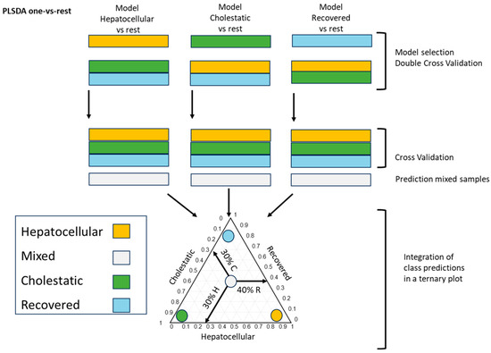
Drug-induced liver injury (DILI) is a serious adverse hepatic event presenting diagnostic and prognostic challenges. The clinical categorization of DILI into hepatocellular, cholestatic, or mixed phenotype is based on serum alanine aminotransferase (ALT) and alkaline phosphatase (ALP) values; however, this classification may not capture the full spectrum of DILI subtypes. With this aim, we explored the utility of assessing changes in the plasma metabolomic profiles of 79 DILI patients assessed by the RUCAM (Roussel Uclaf Causality Assessment Method) score to better characterize this condition and compare results obtained with the standard clinical characterization. Through the identification of various metabolites in the plasma (including free and conjugated bile acids and glycerophospholipids), and the integration of this information into predictive models, we were able to evaluate the extent of the hepatocellular or cholestatic phenotype and to assign a numeric value with the contribution of each specific DILI sub-phenotype into the patient’s general condition. Additionally, our results showed that metabolomic analysis enabled the monitoring of DILI variability responses to the same drug, the transitions between sub-phenotypes during disease progression, and identified a spectrum of residual DILI metabolic features, which can be overlooked using standard clinical diagnosis during patient follow-up. Full article
Show Figures

Figure 1

18 pages, 4393 KiB  
Article
Investigation of Gastrointestinal Toxicities Associated with Concurrent Abdominal Radiation Therapy and the Tyrosine Kinase Inhibitor Sunitinib in a Mouse Model
by Amber R. Prebble, Bailey Latka, Braden Burdekin, Del Leary, Mac Harris, Daniel Regan and Mary-Keara Boss
Int. J. Mol. Sci. 2024, 25(3), 1838; https://doi.org/10.3390/ijms25031838 - 2 Feb 2024
Viewed by 1397
Abstract
Tyrosine kinase inhibitors (TKIs) may be combined with radiation therapy (RT) to enhance tumor control; however, increased incidences of gastrointestinal (GI) toxicity have been reported with this combination. We hypothesize that toxicity is due to compromised intestinal healing caused by inhibition of vascular [...] Read more.
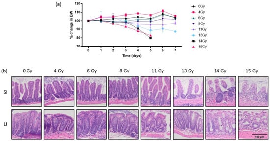
Tyrosine kinase inhibitors (TKIs) may be combined with radiation therapy (RT) to enhance tumor control; however, increased incidences of gastrointestinal (GI) toxicity have been reported with this combination. We hypothesize that toxicity is due to compromised intestinal healing caused by inhibition of vascular repair and proliferation pathways. This study explores underlying tissue toxicity associated with abdominal RT and concurrent sunitinib in a mouse model. Four groups of CD-1 mice were treated with 12 Gy abdominal RT, oral sunitinib, abdominal RT + sunitinib, or sham treatment. Mice received oral sunitinib or the vehicle via gavage for 14 days. On day 7, mice were irradiated with 12 Gy abdominal RT or sham treated. Mice were euthanized on day 14 and intestinal tract was harvested for semiquantitative histopathologic evaluation and immunohistochemical quantification of proliferation (Ki67) and vascular density (CD31). Non-irradiated groups had stable weights while abdominal irradiation resulted in weight loss, with mice receiving RT + SUN having greater weight loss than mice receiving RT alone. Semiquantitative analysis showed significant increases in inflammation in irradiated groups. The difference in the density of CD31+ cells was significantly increased in RT alone compared to SUN alone. Ki67+ density was not significant. In summary, we identify a lack of angiogenic response in irradiated GI tissues when abdominal RT is combined with a TKI, which may correlate with clinical toxicities seen in canine and human patients receiving combined treatment. Full article
Show Figures

Figure 1

17 pages, 5877 KiB  
Article
Effects of Waterpipe Smoke Exposure on Experimentally Induced Chronic Kidney Disease in Mice
by Sumaya Beegam, Suhail Al-Salam, Nur Elena Zaaba, Ozaz Elzaki, Badreldin H. Ali and Abderrahim Nemmar
Int. J. Mol. Sci. 2024, 25(1), 585; https://doi.org/10.3390/ijms25010585 - 2 Jan 2024
Viewed by 1493
Abstract
Tobacco smoking is an independent risk factor in the onset of kidney disease. To date, there have been no reports on the influence of waterpipe smoke (WPS) in experimentally induced chronic kidney disease (CKD) models. We studied the effects and mechanisms of actions [...] Read more.
Tobacco smoking is an independent risk factor in the onset of kidney disease. To date, there have been no reports on the influence of waterpipe smoke (WPS) in experimentally induced chronic kidney disease (CKD) models. We studied the effects and mechanisms of actions of WPS on a mouse model of adenine-induced CKD. Mice fed either a normal diet, or an adenine-added diet and were exposed to either air or WPS (30 min/day and 5 days/week) for four consecutive weeks. Plasma creatinine, urea and indoxyl sulfate increased and creatinine clearance decreased in adenine + WPS versus either WPS or adenine + saline groups. The urinary concentrations of kidney injury molecule-1 and adiponectin and the activities of neutrophil gelatinase-associated lipocalin and N-acetyl-β-D-glucosaminidase were augmented in adenine + WPS compared with either adenine + air or WPS groups. In the kidney tissue, several markers of oxidative stress and inflammation were higher in adenine + WPS than in either adenine + air or WPS groups. Compared with the controls, WPS inhalation in mice with CKD increased DNA damage, and urinary concentration of 8-hydroxy-2-deoxyguanosine. Furthermore, the expressions of nuclear factor κB (NF-κB) and mitogen-activated protein kinases (MAPKs) (ERK and p38) were elevated in the kidneys of adenine + WPS group, compared with the controls. Likewise, the kidneys of adenine + WPS group revealed more marked histological tubular injury, chronic inflammation and interstitial fibrosis. In conclusion, WPS inhalation aggravates kidney injury, oxidative stress, inflammation, DNA damage and fibrosis in mice with adenine-induced CKD, indicating that WPS exposure intensifies CKD. These effects were associated with a mechanism involving NF-κB, ERK and p38 activations. Full article
Show Figures

Figure 1

2023

Jump to: 2024, 2022, 2021, 2019

23 pages, 416 KiB  
Review
The Role of TGF-β3 in Radiation Response
by Ingunn Hanson, Kathinka E. Pitman and Nina F. J. Edin
Int. J. Mol. Sci. 2023, 24(8), 7614; https://doi.org/10.3390/ijms24087614 - 20 Apr 2023
Cited by 7 | Viewed by 2071
Abstract
Transforming growth factor-beta 3 (TGF-β3) is a ubiquitously expressed multifunctional cytokine involved in a range of physiological and pathological conditions, including embryogenesis, cell cycle regulation, immunoregulation, and fibrogenesis. The cytotoxic effects of ionizing radiation are employed in cancer radiotherapy, but its actions also [...] Read more.
Transforming growth factor-beta 3 (TGF-β3) is a ubiquitously expressed multifunctional cytokine involved in a range of physiological and pathological conditions, including embryogenesis, cell cycle regulation, immunoregulation, and fibrogenesis. The cytotoxic effects of ionizing radiation are employed in cancer radiotherapy, but its actions also influence cellular signaling pathways, including that of TGF-β3. Furthermore, the cell cycle regulating and anti-fibrotic effects of TGF-β3 have identified it as a potential mitigator of radiation- and chemotherapy-induced toxicity in healthy tissue. This review discusses the radiobiology of TGF-β3, its induction in tissue by ionizing radiation, and its potential radioprotective and anti-fibrotic effects. Full article
13 pages, 2596 KiB  
Article
The FomYjeF Protein Influences the Sporulation and Virulence of Fusarium oxysporum f. sp. momordicae
by Chenxing Wei, Caiyi Wen, Yuanyuan Zhang, Hongyan Du, Rongrong Zhong, Zhengzhe Guan, Mengjiao Wang, Yanhong Qin, Fei Wang, Luyang Song and Ying Zhao
Int. J. Mol. Sci. 2023, 24(8), 7260; https://doi.org/10.3390/ijms24087260 - 14 Apr 2023
Cited by 7 | Viewed by 2707
Abstract
Fusarium oxysporum causes vascular wilt in more than 100 plant species, resulting in massive economic losses. A deep understanding of the mechanisms of pathogenicity and symptom induction by this fungus is necessary to control crop wilt. The YjeF protein has been proven to [...] Read more.
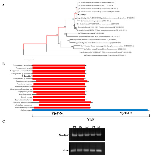
Fusarium oxysporum causes vascular wilt in more than 100 plant species, resulting in massive economic losses. A deep understanding of the mechanisms of pathogenicity and symptom induction by this fungus is necessary to control crop wilt. The YjeF protein has been proven to function in cellular metabolism damage-repair in Escherichia coli and to play an important role in Edc3 (enhancer of the mRNA decapping 3) function in Candida albicans, but no studies have been reported on related functions in plant pathogenic fungi. In this work, we report how the FomYjeF gene in F. oxysporum f. sp. momordicae contributes to conidia production and virulence. The deletion of the FomYjeF gene displayed a highly improved capacity for macroconidia production, and it was shown to be involved in carbendazim’s associated stress pathway. Meanwhile, this gene caused a significant increase in virulence in bitter gourd plants with a higher disease severity index and enhanced the accumulation of glutathione peroxidase and the ability to degrade hydrogen peroxide in F. oxysporum. These findings reveal that FomYjeF affects virulence by influencing the amount of spore formation and the ROS (reactive oxygen species) pathway of F. oxysporum f. sp. momordicae. Taken together, our study shows that the FomYjeF gene affects sporulation, mycelial growth, pathogenicity, and ROS accumulation in F. oxysporum. The results of this study provide a novel insight into the function of FomYjeF participation in the pathogenicity of F. oxysporum f. sp. momordicae. Full article
Show Figures

Figure 1

19 pages, 1345 KiB  
Review
Non-Protein Thiol Compounds and Antioxidant Responses Involved in Bryophyte Heavy-Metal Tolerance
by Giovanna Salbitani, Viviana Maresca, Piergiorgio Cianciullo, Rosanna Bossa, Simona Carfagna and Adriana Basile
Int. J. Mol. Sci. 2023, 24(6), 5302; https://doi.org/10.3390/ijms24065302 - 10 Mar 2023
Cited by 16 | Viewed by 3042
Abstract
Heavy-metal pollution represents a problem which has been widely discussed in recent years. The biological effects of heavy metals have been studied in both animals and plants, ranging from oxidative stress to genotoxicity. Plants, above all metal-tolerant species, have evolved a wide spectrum [...] Read more.
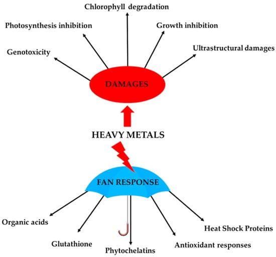
Heavy-metal pollution represents a problem which has been widely discussed in recent years. The biological effects of heavy metals have been studied in both animals and plants, ranging from oxidative stress to genotoxicity. Plants, above all metal-tolerant species, have evolved a wide spectrum of strategies to counteract exposure to toxic metal concentrations. Among these strategies, the chelation and vacuolar sequestration of heavy metals are, after cell-wall immobilization, the first line of defence that prevent heavy metals from interacting with cell components. Furthermore, bryophytes activate a series of antioxidant non-enzymatic and enzymatic responses to counteract the effects of heavy metal in the cellular compartments. In this review, the role of non-protein thiol compounds and antioxidant molecules in bryophytes will be discussed. Full article
Show Figures

Figure 1

15 pages, 6117 KiB  
Article
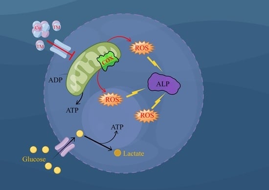
Tetrathiomolybdate Decreases the Expression of Alkaline Phosphatase in Dermal Papilla Cells by Increasing Mitochondrial ROS Production
by Fan Li, Hongli Liu, Xiaojing Wu, Zhicheng Song, Haojia Tang, Maohua Gong, Lei Liu and Fuchang Li
Int. J. Mol. Sci. 2023, 24(4), 3123; https://doi.org/10.3390/ijms24043123 - 4 Feb 2023
Cited by 2 | Viewed by 2379
Abstract
Dermal papilla cells (DPCs) play important roles in hair growth regulation. However, strategies to regrow hair are lacking. Here, global proteomic profiling identified the tetrathiomolybdate (TM)-mediated inactivation of copper (Cu) depletion-dependent mitochondrial cytochrome c oxidase (COX) as the primary metabolic defect in DPCs, [...] Read more.
Dermal papilla cells (DPCs) play important roles in hair growth regulation. However, strategies to regrow hair are lacking. Here, global proteomic profiling identified the tetrathiomolybdate (TM)-mediated inactivation of copper (Cu) depletion-dependent mitochondrial cytochrome c oxidase (COX) as the primary metabolic defect in DPCs, leading to decreased Adenosine Triphosphate (ATP) production, mitochondrial membrane potential depolarization, increased total cellular reactive oxygen species (ROS) levels, and reduced expression of the key marker of hair growth in DPCs. By using several known mitochondrial inhibitors, we found that excessive ROS production was responsible for the impairment of DPC function. We therefore subsequently showed that two ROS scavengers, N-acetyl cysteine (NAC) and ascorbic acid (AA), partially prevented the TM- and ROS-mediated inhibition of alkaline phosphatase (ALP). Overall, these findings established a direct link between Cu and the key marker of DPCs, whereby copper depletion strongly impaired the key marker of hair growth in the DPCs by increasing excessive ROS production. Full article
Show Figures

Graphical abstract

14 pages, 2382 KiB  
Article
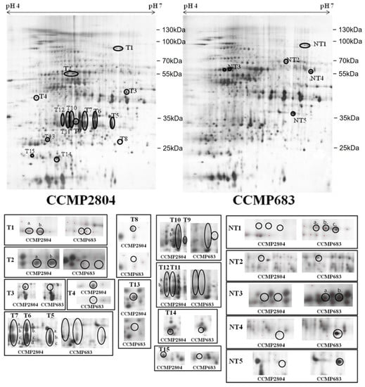
Upregulation of Peridinin-Chlorophyll A-Binding Protein in a Toxic Strain of Prorocentrum hoffmannianum under Normal and Phosphate-Depleted Conditions
by Thomas Chun-Hung Lee, Kaze King-Yip Lai, Steven Jing-Liang Xu and Fred Wang-Fat Lee
Int. J. Mol. Sci. 2023, 24(2), 1735; https://doi.org/10.3390/ijms24021735 - 15 Jan 2023
Cited by 2 | Viewed by 2318
Abstract
Some strains of the dinoflagellate species Prorocentrum hoffmannianum show contrasting ability to produce diarrhetic shellfish poisoning (DSP) toxins. We previously compared the okadaic acid (OA) production level between a highly toxic strain (CCMP2804) and a non-toxic strain (CCMP683) of P. hoffmannianum and revealed [...] Read more.
Some strains of the dinoflagellate species Prorocentrum hoffmannianum show contrasting ability to produce diarrhetic shellfish poisoning (DSP) toxins. We previously compared the okadaic acid (OA) production level between a highly toxic strain (CCMP2804) and a non-toxic strain (CCMP683) of P. hoffmannianum and revealed that the cellular concentration of OA in CCMP2804 would increase significantly under the depletion of phosphate. To understand the molecular mechanisms, here, we compared and analyzed the proteome changes of both strains growing under normal condition and at phosphate depletion using two-dimensional gel electrophoresis (2-DE). There were 41 and 33 differential protein spots observed under normal condition and phosphate depletion, respectively, of which most were upregulated in CCMP2804 and 22 were common to both conditions. Due to the lack of matched peptide mass fingerprints in the database, de novo peptide sequencing was applied to identify the differentially expressed proteins. Of those upregulated spots in CCMP2804, nearly 60% were identified as peridinin-chlorophyll a-binding protein (PCP), an important light-harvesting protein for photosynthesis in dinoflagellates. We postulated that the high expression of PCP encourages the production of DSP toxins by enhancing the yields of raw materials such as acetate, glycolate and glycine. Other possible mechanisms of toxicity related to PCP might be through triggering the transcription of non-ribosomal peptide synthetase/polyketide synthase genes and the transportation of dinophysistoxin-4 from chloroplast to vacuoles. Full article
Show Figures

Figure 1

23 pages, 4624 KiB  
Article
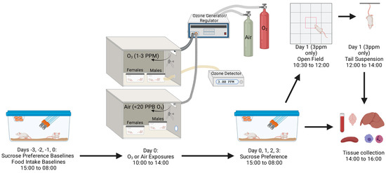
Effects of Ozone on Sickness and Depressive-like Behavioral and Biochemical Phenotypes and Their Regulation by Serum Amyloid A in Mice
by Kristen K. Baumann, W. Sandy Liang, Daniel V. Quaranta, Miranda L. Wilson, Helina S. Asrat, Jarl A. Thysell, Angelo V. Sarchi, William A. Banks and Michelle A. Erickson
Int. J. Mol. Sci. 2023, 24(2), 1612; https://doi.org/10.3390/ijms24021612 - 13 Jan 2023
Cited by 3 | Viewed by 2562
Abstract
Ozone (O3) is an air pollutant that primarily damages the lungs, but growing evidence supports the idea that O3 also harms the brain; acute exposure to O3 has been linked to central nervous system (CNS) symptoms such as depressed [...] Read more.
Ozone (O3) is an air pollutant that primarily damages the lungs, but growing evidence supports the idea that O3 also harms the brain; acute exposure to O3 has been linked to central nervous system (CNS) symptoms such as depressed mood and sickness behaviors. However, the mechanisms by which O3 inhalation causes neurobehavioral changes are limited. One hypothesis is that factors in the circulation bridge communication between the lungs and brain following O3 exposure. In this study, our goals were to characterize neurobehavioral endpoints of O3 exposure as they relate to markers of systemic and pulmonary inflammation, with a particular focus on serum amyloid A (SAA) and kynurenine as candidate mediators of O3 behavioral effects. We evaluated O3-induced dose-, time- and sex-dependent changes in pulmonary inflammation, circulating SAA and kynurenine and its metabolic enzymes, and sickness and depressive-like behaviors in Balb/c and CD-1 mice. We found that 3 parts per million (ppm) O3, but not 2 or 1 ppm O3, increased circulating SAA and lung inflammation, which were resolved by 48 h and was worse in females. We also found that indoleamine 2,3-dioxygenase (Ido1) mRNA expression was increased in the brain and spleen 24 h after 3 ppm O3 and that kynurenine was increased in blood. Sickness and depressive-like behaviors were observed at all O3 doses (1–3 ppm), suggesting that behavioral responses to O3 can occur independently of increased SAA or neutrophils in the lungs. Using SAA knockout mice, we found that SAA did not contribute to O3-induced pulmonary damage or inflammation, systemic increases in kynurenine post-O3, or depressive-like behavior but did contribute to weight loss. Together, these findings indicate that acute O3 exposure induces transient symptoms of sickness and depressive-like behaviors that may occur in the presence or absence of overt pulmonary neutrophilia and systemic increases of SAA. SAA does not appear to contribute to pulmonary inflammation induced by O3, although it may contribute to other aspects of sickness behavior, as reflected by a modest effect on weight loss. Full article
Show Figures

Figure 1

2022

Jump to: 2024, 2023, 2021, 2019

17 pages, 2935 KiB  
Article
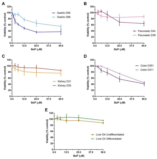
Metabolic Activation of Benzo[a]pyrene by Human Tissue Organoid Cultures
by Angela L. Caipa Garcia, Jill E. Kucab, Halh Al-Serori, Rebekah S. S. Beck, Franziska Fischer, Matthias Hufnagel, Andrea Hartwig, Andrew Floeder, Silvia Balbo, Hayley Francies, Mathew Garnett, Meritxell Huch, Jarno Drost, Matthias Zilbauer, Volker M. Arlt and David H. Phillips
Int. J. Mol. Sci. 2023, 24(1), 606; https://doi.org/10.3390/ijms24010606 - 29 Dec 2022
Cited by 6 | Viewed by 3470
Abstract
Organoids are 3D cultures that to some extent reproduce the structure, composition and function of the mammalian tissues from which they derive, thereby creating in vitro systems with more in vivo-like characteristics than 2D monocultures. Here, the ability of human organoids derived from [...] Read more.
Organoids are 3D cultures that to some extent reproduce the structure, composition and function of the mammalian tissues from which they derive, thereby creating in vitro systems with more in vivo-like characteristics than 2D monocultures. Here, the ability of human organoids derived from normal gastric, pancreas, liver, colon and kidney tissues to metabolise the environmental carcinogen benzo[a]pyrene (BaP) was investigated. While organoids from the different tissues showed varied cytotoxic responses to BaP, with gastric and colon organoids being the most susceptible, the xenobiotic-metabolising enzyme (XME) genes, CYP1A1 and NQO1, were highly upregulated in all organoid types, with kidney organoids having the highest levels. Furthermore, the presence of two key metabolites, BaP-t-7,8-dihydrodiol and BaP-tetrol-l-1, was detected in all organoid types, confirming their ability to metabolise BaP. BaP bioactivation was confirmed both by the activation of the DNA damage response pathway (induction of p-p53, pCHK2, p21 and γ-H2AX) and by DNA adduct formation. Overall, pancreatic and undifferentiated liver organoids formed the highest levels of DNA adducts. Colon organoids had the lowest responses in DNA adduct and metabolite formation, as well as XME expression. Additionally, high-throughput RT-qPCR explored differences in gene expression between organoid types after BaP treatment. The results demonstrate the potential usefulness of organoids for studying environmental carcinogenesis and genetic toxicology. Full article
Show Figures

Figure 1

21 pages, 3285 KiB  
Article
VANL-100 Attenuates Beta-Amyloid-Induced Toxicity in SH-SY5Y Cells
by Andrila E. Collins, Tarek M. Saleh and Bettina E. Kalisch
Int. J. Mol. Sci. 2023, 24(1), 442; https://doi.org/10.3390/ijms24010442 - 27 Dec 2022
Cited by 4 | Viewed by 2733
Abstract
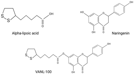
Antioxidants are being explored as novel therapeutics for the treatment of neurodegenerative diseases such as Alzheimer’s disease (AD) through strategies such as chemically linking antioxidants to synthesize novel co-drugs. The main objective of this study was to assess the cytoprotective effects of the [...] Read more.
Antioxidants are being explored as novel therapeutics for the treatment of neurodegenerative diseases such as Alzheimer’s disease (AD) through strategies such as chemically linking antioxidants to synthesize novel co-drugs. The main objective of this study was to assess the cytoprotective effects of the novel antioxidant compound VANL-100 in a cellular model of beta-amyloid (Aβ)-induced toxicity. The cytotoxic effects of Aβ in the presence and absence of all antioxidant compounds were measured using the 3-(4,5-dimethylthiazol-2-yl)2-5-diphenyl-2H-tetrazolium bromide (MTT) assay in SH-SY5Y cells in both pre-treatment and co-treatment experiments. In pre-treatment experiments, VANL-100, or one of its parent compounds, naringenin (NAR), alpha-lipoic acid (ALA), or naringenin + alpha-lipoic acid (NAR + ALA), was administrated 24 h prior to an additional 24-h incubation with 20 μM non-fibril or fibril Aβ25–35. Co-treatment experiments consisted of simultaneous treatment with Aβ and antioxidants. Pre-treatment and co-treatment with VANL-100 significantly attenuated Aβ-induced cell death. There were no significant differences between the protective effects of VANL-100, NAR, ALA, and NAR + ALA with either form of Aβ, or in the effect of VANL-100 between 24-h pre-treatment and co-treatment. These results demonstrate that the novel co-drug VANL-100 is capable of eliciting cytoprotective effects against Aβ-induced toxicity. Full article
Show Figures

Figure 1

10 pages, 2194 KiB  
Article
The Difference in Zinc Concentrations Required for Induction among Metallothionein Isoforms Can Be Explained by the Different MTF1 Affinities to MREs in Its Promoter
by Shoko Ogushi and Tomoki Kimura
Int. J. Mol. Sci. 2023, 24(1), 283; https://doi.org/10.3390/ijms24010283 - 23 Dec 2022
Cited by 2 | Viewed by 1925
Abstract
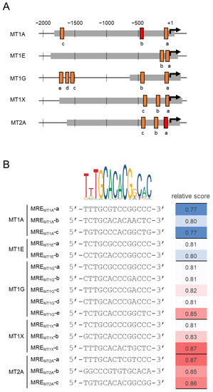
Metallothioneins (MTs) are cysteine-rich low-molecular-weight proteins that protect cells from heavy metal toxicity. MT1 and MT2 are considered ubiquitously expressed among the MT isoforms ranging from 1 to 4. These MT1 and MT2 transcriptions are regulated by metal regulatory transcription factor 1 (MTF1) [...] Read more.
Metallothioneins (MTs) are cysteine-rich low-molecular-weight proteins that protect cells from heavy metal toxicity. MT1 and MT2 are considered ubiquitously expressed among the MT isoforms ranging from 1 to 4. These MT1 and MT2 transcriptions are regulated by metal regulatory transcription factor 1 (MTF1) binding to the metal response element (MRE) of the promoter, which is upregulated in response to zinc. The functional MT isoforms are MT1A, MT1B, MT1E, MT1F, MT1G, MT1H, MT1M, MT1X, and MT2A in humans, but these expressions were differently regulated. Here, MT1A was shown to be significantly less upregulated by zinc than MT1E, MT1G, MT1X, and MT2A. The poor responsiveness of the MT1A zinc was suggested to be due to the MRE sequence in the MT1A promoter region having a lower MTF1 binding affinity compared to the other isoforms. MT1A may be induced via pathways other than the MTF1–MRE binding pathway. These findings may help elucidate the differential regulation of MT isoform expression. Full article
Show Figures

Figure 1

11 pages, 2082 KiB  
Article
Effects of Streamer Discharge on PM2.5 Containing Endotoxins and Polyaromatic Hydrocarbons and Their Biological Responses In Vitro
by Akiko Honda, Ken-ichiro Inoue, Shin Tamura, Michitaka Tanaka, Zaoshi Wang, Toshio Tanaka, Seitarou Hirai, Tomoaki Okuda, Kayo Ueda and Hirohisa Takano
Int. J. Mol. Sci. 2022, 23(24), 15891; https://doi.org/10.3390/ijms232415891 - 14 Dec 2022
Cited by 2 | Viewed by 1908
Abstract
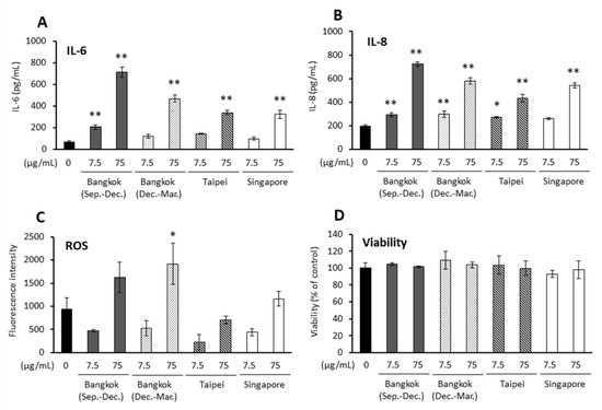
Experimental and epidemiological studies have demonstrated that fine particulate matter with a diameter of <2.5 μm (PM2.5) affects both the respiratory and immune systems. However, effective approaches to reduce PM2.5-induced hazardous effects have not been discovered yet. Streamer discharge is a category of [...] Read more.
Experimental and epidemiological studies have demonstrated that fine particulate matter with a diameter of <2.5 μm (PM2.5) affects both the respiratory and immune systems. However, effective approaches to reduce PM2.5-induced hazardous effects have not been discovered yet. Streamer discharge is a category of plasma discharge in which high-speed electrons collide with oxygen and nitrogen molecules. Although streamer discharge can reportedly eliminate bacteria, molds, chemical substances, and allergens, its ability to decontaminate PM2.5 has not been previously demonstrated. The present study explored whether streamer discharge treatment could reduce PM2.5-induced inflammatory responses by employing an in vitro system. PM2.5 was collected under four conditions (Bangkok (Sep.–Dec.), Bangkok (Dec.–Mar.), Singapore, and Taipei). Airway epithelial cells and antigen-presenting cells exposed to non-treated PM2.5 in several conditions resulted in inflammatory responses. Streamer-discharged PM2.5 (Bangkok (Sep.–Dec.)) decreased the expression of interleukin (IL)-6 and IL-8 compared to non-treated PM2.5. Moreover, composition analysis demonstrated that streamer discharge reduced some compounds, such as endotoxins and polycyclic aromatic hydrocarbons, included in PM2.5 that can elicit inflammatory responses. Streamer discharge treatment can reduce endotoxins, polycyclic aromatic hydrocarbons, and the subsequent inflammatory responses induced by PM2.5 in vitro. Full article
Show Figures

Figure 1

16 pages, 2212 KiB  
Article
Extensive Collection of Psychotropic Mushrooms with Determination of Their Tryptamine Alkaloids
by Klára Gotvaldová, Jan Borovička, Kateřina Hájková, Petra Cihlářová, Alan Rockefeller and Martin Kuchař
Int. J. Mol. Sci. 2022, 23(22), 14068; https://doi.org/10.3390/ijms232214068 - 15 Nov 2022
Cited by 20 | Viewed by 25244
Abstract
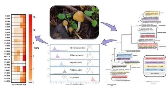
Since not only psilocybin (PSB) but also PSB-containing mushrooms are used for psychedelic therapy and microdosing, it is necessary to know their concentration variability in wild-grown mushrooms. This article aimed to determine the PSB, psilocin (PS), baeocystin (BA), norbaeocystin (NB), and aeruginascin (AE) [...] Read more.
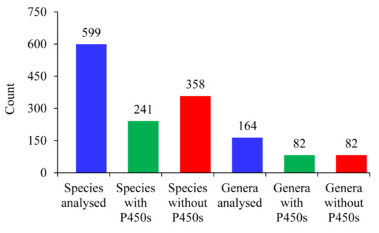
Since not only psilocybin (PSB) but also PSB-containing mushrooms are used for psychedelic therapy and microdosing, it is necessary to know their concentration variability in wild-grown mushrooms. This article aimed to determine the PSB, psilocin (PS), baeocystin (BA), norbaeocystin (NB), and aeruginascin (AE) concentrations in a large sample set of mushrooms belonging to genera previously reported to contain psychotropic tryptamines. Ultra-high performance liquid chromatography coupled with tandem mass spectrometry was used to quantify tryptamine alkaloids in the mushroom samples. Most mushroom collections were documented by fungarium specimens and/or ITS rDNA/LSU/EF1-α sequencing. Concentrations of five tryptamine alkaloids were determined in a large sample set of 226 fruiting bodies of 82 individual collections from seven mushroom genera. For many mushroom species, concentrations of BA, NB, and AE are reported for the first time. The highest PSB/PS concentrations were found in Psilocybe species, but no tryptamines were detected in the P. fuscofulva and P. fimetaria collections. The tryptamine concentrations in mushrooms are extremely variable, representing a problem for mushroom consumers due to the apparent risk of overdose. The varied cocktail of tryptamines in wild mushrooms could influence the medicinal effect compared to therapy with chemically pure PSB, posing a serious problem for data interpretation. Full article
Show Figures

Graphical abstract

24 pages, 4114 KiB  
Article
Filamentous Hemagglutinin of Bordetella pertussis Does Not Interact with the β2 Integrin CD11b/CD18
by Maryam Golshani, Waheed Ur Rahman, Adriana Osickova, Jana Holubova, Jinery Lora, Nataliya Balashova, Peter Sebo and Radim Osicka
Int. J. Mol. Sci. 2022, 23(20), 12598; https://doi.org/10.3390/ijms232012598 - 20 Oct 2022
Cited by 2 | Viewed by 2611
Abstract
The pertussis agent Bordetella pertussis produces a number of virulence factors, of which the filamentous hemagglutinin (FhaB) plays a role in B. pertussis adhesion to epithelial and phagocytic cells. Moreover, FhaB was recently found to play a crucial role in nasal cavity infection [...] Read more.
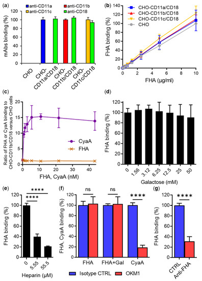
The pertussis agent Bordetella pertussis produces a number of virulence factors, of which the filamentous hemagglutinin (FhaB) plays a role in B. pertussis adhesion to epithelial and phagocytic cells. Moreover, FhaB was recently found to play a crucial role in nasal cavity infection and B. pertussis transmission to new hosts. The 367 kDa FhaB protein translocates through an FhaC pore to the outer bacterial surface and is eventually processed to a ~220 kDa N-terminal FHA fragment by the SphB1 protease. A fraction of the mature FHA then remains associated with bacterial cell surface, while most of FHA is shed into the bacterial environment. Previously reported indirect evidence suggested that FHA, or its precursor FhaB, may bind the β2 integrin CD11b/CD18 of human macrophages. Therefore, we assessed FHA binding to various cells producing or lacking the integrin and show that purified mature FHA does not bind CD11b/CD18. Further results then revealed that the adhesion of B. pertussis to cells does not involve an interaction between the bacterial surface-associated FhaB and/or mature FHA and the β2 integrin CD11b/CD18. In contrast, FHA binding was strongly inhibited at micromolar concentrations of heparin, corroborating that the cell binding of FHA is ruled by the interaction of its heparin-binding domain with sulfated glycosaminoglycans on the cell surface. Full article
Show Figures

Figure 1

25 pages, 5604 KiB  
Article
Chlorhexidine Promotes Psl Expression in Pseudomonas aeruginosa That Enhances Cell Aggregation with Preserved Pathogenicity Demonstrates an Adaptation against Antiseptic
by Uthaibhorn Singkham-In, Pornpimol Phuengmaung, Jiradej Makjaroen, Wilasinee Saisorn, Thansita Bhunyakarnjanarat, Tanittha Chatsuwan, Chintana Chirathaworn, Wiwat Chancharoenthana and Asada Leelahavanichkul
Int. J. Mol. Sci. 2022, 23(15), 8308; https://doi.org/10.3390/ijms23158308 - 27 Jul 2022
Cited by 10 | Viewed by 3031
Abstract
Because Pseudomonas aeruginosa is frequently in contact with Chlorhexidine (a regular antiseptic), bacterial adaptations are possible. In comparison with the parent strain, the Chlorhexidine-adapted strain formed smaller colonies with metabolic downregulation (proteomic analysis) with the cross-resistance against colistin (an antibiotic for several antibiotic-resistant [...] Read more.
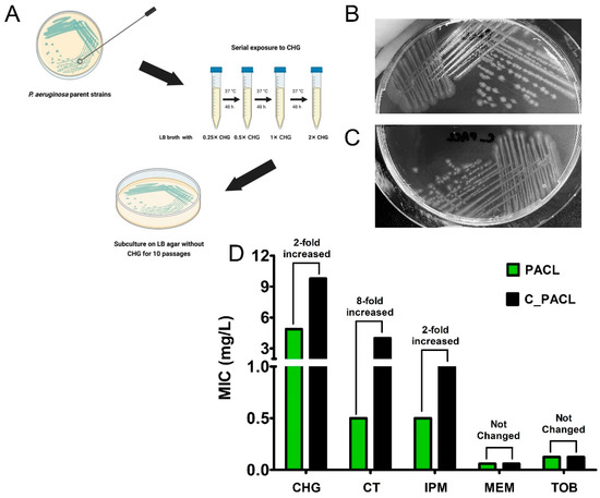
Because Pseudomonas aeruginosa is frequently in contact with Chlorhexidine (a regular antiseptic), bacterial adaptations are possible. In comparison with the parent strain, the Chlorhexidine-adapted strain formed smaller colonies with metabolic downregulation (proteomic analysis) with the cross-resistance against colistin (an antibiotic for several antibiotic-resistant bacteria), partly through the modification of L-Ara4N in the lipopolysaccharide at the outer membrane. Chlorhexidine-adapted strain formed dense liquid–solid interface biofilms with enhanced cell aggregation partly due to the Chlorhexidine-induced overexpression of psl (exopolysaccharide-encoded gene) through the LadS/GacSA pathway (c-di-GMP-independence) in 12 h biofilms and maintained the aggregation with SiaD-mediated c-di-GMP dependence in 24 h biofilms as evaluated by polymerase chain reaction (PCR). The addition of Ca2+ in the Chlorhexidine-adapted strain facilitated several Psl-associated genes, indicating an impact of Ca2+ in Psl production. The activation by Chlorhexidine-treated sessile bacteria demonstrated a lower expression of IL-6 and IL-8 on fibroblasts and macrophages than the activation by the parent strain, indicating the less inflammatory reactions from Chlorhexidine-exposed bacteria. However, the 14-day severity of the wounds in mouse caused by Chlorhexidine-treated bacteria versus the parent strain was similar, as indicated by wound diameters and bacterial burdens. In conclusion, Chlorhexidine induced psl over-expression and colistin cross-resistance that might be clinically important. Full article
Show Figures

Figure 1

25 pages, 962 KiB  
Review
Sexual Dimorphism in Adipose-Hypothalamic Crosstalk and the Contribution of Aryl Hydrocarbon Receptor to Regulate Energy Homeostasis
by Nazmul Haque and Shelley A. Tischkau
Int. J. Mol. Sci. 2022, 23(14), 7679; https://doi.org/10.3390/ijms23147679 - 12 Jul 2022
Cited by 7 | Viewed by 3905
Abstract
There are fundamental sex differences in the regulation of energy homeostasis. Better understanding of the underlying mechanisms of energy balance that account for this asymmetry will assist in developing sex-specific therapies for sexually dimorphic diseases such as obesity. Multiple organs, including the hypothalamus [...] Read more.
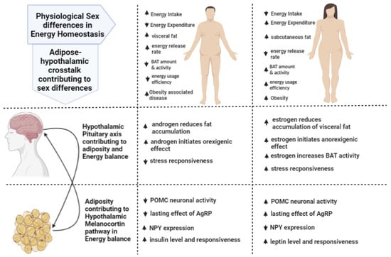
There are fundamental sex differences in the regulation of energy homeostasis. Better understanding of the underlying mechanisms of energy balance that account for this asymmetry will assist in developing sex-specific therapies for sexually dimorphic diseases such as obesity. Multiple organs, including the hypothalamus and adipose tissue, play vital roles in the regulation of energy homeostasis, which are regulated differently in males and females. Various neuronal populations, particularly within the hypothalamus, such as arcuate nucleus (ARC), can sense nutrient content of the body by the help of peripheral hormones such leptin, derived from adipocytes, to regulate energy homeostasis. This review summarizes how adipose tissue crosstalk with homeostatic network control systems in the brain, which includes energy regulatory regions and the hypothalamic–pituitary axis, contribute to energy regulation in a sex-specific manner. Moreover, development of obesity is contingent upon diet and environmental factors. Substances from diet and environmental contaminants can exert insidious effects on energy metabolism, acting peripherally through the aryl hydrocarbon receptor (AhR). Developmental AhR activation can impart permanent alterations of neuronal development that can manifest a number of sex-specific physiological changes, which sometimes become evident only in adulthood. AhR is currently being investigated as a potential target for treating obesity. The consensus is that impaired function of the receptor protects from obesity in mice. AhR also modulates sex steroid receptors, and hence, one of the objectives of this review is to explain why investigating sex differences while examining this receptor is crucial. Overall, this review summarizes sex differences in the regulation of energy homeostasis imparted by the adipose–hypothalamic axis and examines how this axis can be affected by xenobiotics that signal through AhR. Full article
Show Figures

Figure 1

20 pages, 1057 KiB  
Review
Neurotoxicity Associated with Treatment of Acute Lymphoblastic Leukemia Chemotherapy and Immunotherapy
by Patrycja Śliwa-Tytko, Agnieszka Kaczmarska, Monika Lejman and Joanna Zawitkowska
Int. J. Mol. Sci. 2022, 23(10), 5515; https://doi.org/10.3390/ijms23105515 - 15 May 2022
Cited by 11 | Viewed by 7716
Abstract
Immunotherapy is a milestone in the treatment of poor-prognosis pediatric acute lymphoblastic leukemia (ALL) and is expected to improve treatment outcomes and reduce doses of conventional chemotherapy without compromising the effectiveness of the therapy. However, both chemotherapy and immunotherapy cause side effects, including [...] Read more.
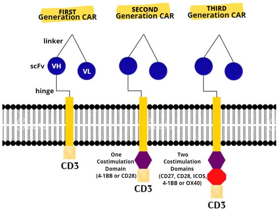
Immunotherapy is a milestone in the treatment of poor-prognosis pediatric acute lymphoblastic leukemia (ALL) and is expected to improve treatment outcomes and reduce doses of conventional chemotherapy without compromising the effectiveness of the therapy. However, both chemotherapy and immunotherapy cause side effects, including neurological ones. Acute neurological complications occur in 3.6–11% of children treated for ALL. The most neurotoxical chemotherapeutics are L-asparaginase (L-ASP), methotrexate (MTX), vincristine (VCR), and nelarabine (Ara-G). Neurotoxicity associated with methotrexate (MTX-NT) occurs in 3–7% of children treated for ALL and is characterized by seizures, stroke-like symptoms, speech disturbances, and encephalopathy. Recent studies indicate that specific polymorphisms in genes related to neurogenesis may have a predisposition to MTX toxicity. One of the most common complications associated with CAR T-cell therapy is immune effector cell-associated neurotoxicity syndrome (ICANS). Mechanisms of neurotoxicity in CAR T-cell therapy are still unknown and may be due to disruption of the blood–brain barrier and the effects of elevated cytokine levels on the central nervous system (CNS). In this review, we present an analysis of the current knowledge on the mechanisms of neurotoxicity of standard chemotherapy and the targeted therapy in children with ALL. Full article
Show Figures

Figure 1

14 pages, 2129 KiB  
Article
Direct T-2 Toxicity on Human Skin—Fibroblast Hs68 Cell Line—In Vitro Study
by Edyta Janik-Karpinska, Michal Ceremuga, Magdalena Wieckowska, Monika Szyposzynska, Marcin Niemcewicz, Ewelina Synowiec, Tomasz Sliwinski and Michal Bijak
Int. J. Mol. Sci. 2022, 23(9), 4929; https://doi.org/10.3390/ijms23094929 - 29 Apr 2022
Cited by 6 | Viewed by 2866
Abstract
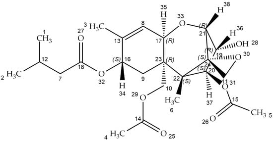
T-2 toxin is produced by different Fusarium species, and it can infect crops such as wheat, barley, and corn. It is known that the T-2 toxin induces various forms of toxicity such as hepatotoxicity, nephrotoxicity, immunotoxicity, and neurotoxicity. In addition, T-2 toxin possesses [...] Read more.
T-2 toxin is produced by different Fusarium species, and it can infect crops such as wheat, barley, and corn. It is known that the T-2 toxin induces various forms of toxicity such as hepatotoxicity, nephrotoxicity, immunotoxicity, and neurotoxicity. In addition, T-2 toxin possesses a strong dermal irritation effect and can be absorbed even through intact skin. As a dermal irritant agent, it is estimated to be 400 times more toxic than sulfur mustard. Toxic effects can include redness, blistering, and necrosis, but the molecular mechanism of these effects still remains unknown. This in vitro study focused on the direct toxicity of T-2 toxin on human skin—fibroblast Hs68 cell line. As a result, the level of toxicity of T-2 toxin and its cytotoxic mechanism of action was determined. In cytotoxicity assays, the dose and time-dependent cytotoxic effect of T-2 on a cell line was observed. Bioluminometry results showed that relative levels of ATP in treated cells were decreased. Further analysis of the toxin’s impact on the induction of apoptosis and necrosis processes showed the significant predominance of PI-stained cells, lack of caspase 3/7 activity, and increased concentration of released Human Cytokeratin 18 in treated cells, which indicates the necrosis process. In conclusion, the results of an in vitro human skin fibroblast model revealed for the first time that the T-2 toxin induces necrosis as a toxicity effect. These results provide new insight into the toxic T-2 mechanism on the skin. Full article
Show Figures

Figure 1

26 pages, 7705 KiB  
Article
CD44+ and CD133+ Non-Small Cell Lung Cancer Cells Exhibit DNA Damage Response Pathways and Dormant Polyploid Giant Cancer Cell Enrichment Relating to Their p53 Status
by Margarita Pustovalova, Taisia Blokhina, Lina Alhaddad, Anna Chigasova, Roman Chuprov-Netochin, Alexander Veviorskiy, Gleb Filkov, Andreyan N. Osipov and Sergey Leonov
Int. J. Mol. Sci. 2022, 23(9), 4922; https://doi.org/10.3390/ijms23094922 - 28 Apr 2022
Cited by 22 | Viewed by 4528
Abstract
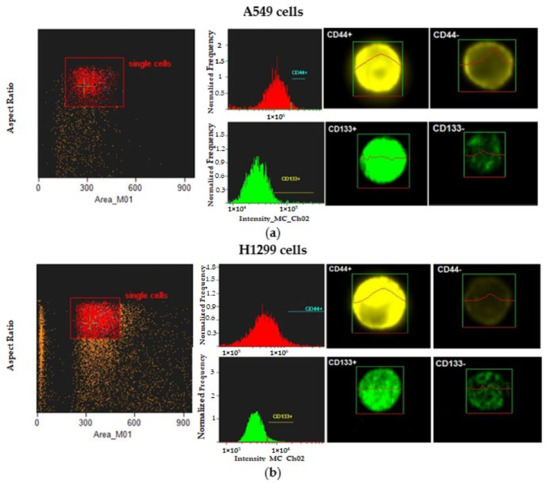
Cancer stem cells (CSCs) play a critical role in the initiation, progression and therapy relapse of many cancers including non-small cell lung cancer (NSCLC). Here, we aimed to address the question of whether the FACS-sorted CSC-like (CD44 + &CD133 +) vs. non-CSC (CD44−/CD133− [...] Read more.
Cancer stem cells (CSCs) play a critical role in the initiation, progression and therapy relapse of many cancers including non-small cell lung cancer (NSCLC). Here, we aimed to address the question of whether the FACS-sorted CSC-like (CD44 + &CD133 +) vs. non-CSC (CD44−/CD133− isogenic subpopulations of p53wt A549 and p53null H1299 cells differ in terms of DNA-damage signaling and the appearance of “dormant” features, including polyploidy, which are early markers (predictors) of their sensitivity to genotoxic stress. X-ray irradiation (IR) at 5 Gy provoked significantly higher levels of the ATR-Chk1/Chk2-pathway activity in CD44−/CD133− and CD133+ subpopulations of H1299 cells compared to the respective subpopulations of A549 cells, which only excited ATR-Chk2 activation as demonstrated by the Multiplex DNA-Damage/Genotoxicity profiling. The CD44+ subpopulations did not demonstrate IR-induced activation of ATR, while significantly augmenting only Chk2 and Chk1/2 in the A549- and H1299-derived cells, respectively. Compared to the A549 cells, all the subpopulations of H1299 cells established an increased IR-induced expression of the γH2AX DNA-repair protein. The CD44−/CD133− and CD133+ subpopulations of the A549 cells revealed IR-induced activation of ATR-p53-p21 cell dormancy signaling-mediated pathway, while none of the CD44+ subpopulations of either cell line possessed any signs of such activity. Our data indicated, for the first time, the transcription factor MITF–FAM3C axis operative in p53-deficient H1299 cells, specifically their CD44+ and CD133+ populations, in response to IR, which warrants further investigation. The p21-mediated quiescence is likely the predominant surviving pathway in CD44−/CD133− and CD133+ populations of A549 cells as indicated by single-cell high-content imaging and analysis of Ki67- and EdU-coupled fluorescence after IR stress. SA-beta-galhistology revealed that cellular-stress-induced premature senescence (SIPS) likely has a significant influence on the temporary dormant state of H1299 cells. For the first time, we demonstrated polyploid giant and/or multinucleated cancer-cell (PGCC/MGCC) fractions mainly featuring the progressively augmenting Ki67low phenotype in CD44+ and CD133+ A549 cells at 24–48 h after IR. In contrast, the Ki67high phenotype enrichment in the same fractions of all the sorted H1299 cells suggested an increase in their cycling/heterochromatin reorganization activity after IR stress. Our results proposed that entering the “quiescence” state rather than p21-mediated SIPS may play a significant role in the survival of p53wt CSC-like NSCLC cells after IR. The results obtained are important for the selection of therapeutic schemes for the treatment of patients with NSCLC, depending on the functioning of the p53 system in tumor cells. Full article
Show Figures

Figure 1

19 pages, 6357 KiB  
Article
1,2,3,4,6-Penta-O-galloyl-d-glucose Interrupts the Early Adipocyte Lifecycle and Attenuates Adiposity and Hepatic Steatosis in Mice with Diet-Induced Obesity
by Ashish Rao Sathyanarayana, Chung-Kuang Lu, Chih-Chuang Liaw, Chia-Chuan Chang, Hsin-Ying Han, Brian D. Green, Wei-Jan Huang, Cheng Huang, Wen-Di He, Lin-Chien Lee and Hui-Kang Liu
Int. J. Mol. Sci. 2022, 23(7), 4052; https://doi.org/10.3390/ijms23074052 - 6 Apr 2022
Cited by 7 | Viewed by 3120
Abstract
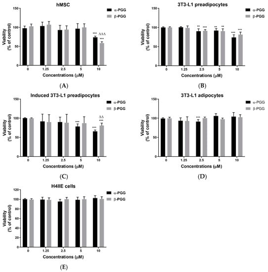
Phytochemicals that interrupt adipocyte lifecycle can provide anti-obesity effects. 1,2,3,4,6-penta-O-galloyl-d-glucose (PGG) is a tannin with two isomers that occurs widely in plants and exhibits various pharmacological activities. The aim of the investigation is to comprehensively examine effects of PGG isomer(s) on [...] Read more.
Phytochemicals that interrupt adipocyte lifecycle can provide anti-obesity effects. 1,2,3,4,6-penta-O-galloyl-d-glucose (PGG) is a tannin with two isomers that occurs widely in plants and exhibits various pharmacological activities. The aim of the investigation is to comprehensively examine effects of PGG isomer(s) on adipocyte lifecycle and diet-induced obesity. Human mesenchymal stem cells (hMSC), 3T3-L1 fibroblasts, and H4IIE hepatoma cells were used to determine the effects of PGG isomers on cell viability and adipogenesis. Mice with diet-induced obesity were generated from male C57/BL6 mice fed with a 45% high fat diet. Oral administration of β-PGG (0.1 and 5 mg/kg) lasted for 14 weeks. Viability was reduced by repeated PGG treatment in hMSC, preadipocytes, and cells under differentiation. PGG mainly induces apoptosis, and this effect is independent of its insulin mimetic action. In vivo, administration of β-PGG attenuated shortening of the colon, hyperlipidaemia, fat cells and islet hypertrophy in DIO mice. Hepatic steatosis and related gene expression were improved along with glucose intolerance. Increased serum adiponectin, leptin, and glucagon-like peptide-1 levels were also observed. In conclusion, repeated PGG treatment interrupts the adipocyte lifecycle. PGG administration reduces adiposity and fatty liver development in DIO mice, and therefore, PGG could aid in clinical management of obesity. Full article
Show Figures

Figure 1

13 pages, 3381 KiB  
Article
Protective Effects of Mitochondrial Uncoupling Protein 2 against Aristolochic Acid I-Induced Toxicity in HK-2 Cells
by Chen Feng, Etienne Empweb Anger, Xiong Zhang, Shengdi Su, Chenlin Su, Shuxin Zhao, Feng Yu and Ji Li
Int. J. Mol. Sci. 2022, 23(7), 3674; https://doi.org/10.3390/ijms23073674 - 27 Mar 2022
Cited by 6 | Viewed by 2689
Abstract
Aristolochic acid I (AA I) is one of the most abundant and toxic aristolochic acids that is reported to cause Aristolochic acid nephropathy (AAN). This paper was designed to assess whether mitochondrial Uncoupling Protein 2 (UCP2), which plays an antioxidative and antiapoptotic role, [...] Read more.
Aristolochic acid I (AA I) is one of the most abundant and toxic aristolochic acids that is reported to cause Aristolochic acid nephropathy (AAN). This paper was designed to assess whether mitochondrial Uncoupling Protein 2 (UCP2), which plays an antioxidative and antiapoptotic role, could protect human renal proximal tubular epithelial (HK-2) cells from toxicity induced by AA I. In this study, HK-2 cells were treated with different concentrations of AA I with or without UCP2 inhibitor (genipin). To upregulate the expression of UCP2 in HK-2 cells, UCP2-DNA transfection was performed. The cell viability was evaluated by colorimetric method using MTT. A series of related biological events such as Reactive Oxygen Species (ROS), Glutathione peroxidase (GSH-Px), and Malondialdehyde (MDA) were evaluated. The results showed that the cytotoxicity of AA I with genipin group was much higher than that of AA I alone. Genipin dramatically boosted oxidative stress and exacerbated AA I-induced apoptosis. Furthermore, the increased expression of UCP2 can reduce the toxicity of AA I on HK-2 cells and upregulation of UCP2 expression can reduce AA I-induced oxidative stress and apoptosis. In conclusion, UCP2 might be a potential target for alleviating AA I-induced nephrotoxicity. Full article
Show Figures

Figure 1

17 pages, 4085 KiB  
Article
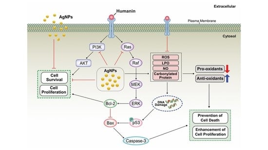
Activation of Autophagy by Low-Dose Silica Nanoparticles Enhances Testosterone Secretion in Leydig Cells
by Jinlong Zhang, Rongrong Ye, Jason William Grunberger, Jiaqi Jin, Qianru Zhang, Raziye Mohammadpour, Nitish Khurana, Xianyu Xu, Hamidreza Ghandehari and Fenglei Chen
Int. J. Mol. Sci. 2022, 23(6), 3104; https://doi.org/10.3390/ijms23063104 - 13 Mar 2022
Cited by 11 | Viewed by 3142
Abstract
Silica nanoparticles (SNPs) can cause abnormal spermatogenesis in male reproductive toxicity. However, the toxicity and toxicological mechanisms of SNPs in testosterone synthesis and secretion in Leydig cells are not well known. Therefore, this study aimed to determine the effect and molecular mechanism of [...] Read more.
Silica nanoparticles (SNPs) can cause abnormal spermatogenesis in male reproductive toxicity. However, the toxicity and toxicological mechanisms of SNPs in testosterone synthesis and secretion in Leydig cells are not well known. Therefore, this study aimed to determine the effect and molecular mechanism of low doses of SNPs in testosterone production in Leydig cells. For this, mouse primary Leydig cells (PLCs) were exposed to 100 nm Stöber nonporous spherical SNPs. We observed significant accumulation of SNPs in the cytoplasm of PLCs via transmission electron microscopy (TEM). CCK-8 and flow cytometry assays confirmed that low doses (50 and 100 μg/mL) of SNPs had no significant effect on cell viability and apoptosis, whereas high doses (more than 200 μg/mL) decreased cell viability and increased cell apoptosis in PLCs. Monodansylcadaverine (MDC) staining showed that SNPs caused the significant accumulation of autophagosomes in the cytoplasm of PLCs. SNPs activated autophagy by upregulating microtubule-associated protein light chain 3 (LC3-II) and BCL-2-interacting protein (BECLIN-1) levels, in addition to downregulating sequestosome 1 (SQSTM1/P62) level at low doses. In addition, low doses of SNPs enhanced testosterone secretion and increased steroidogenic acute regulatory protein (StAR) expression. SNPs combined with rapamycin (RAP), an autophagy activator, enhanced testosterone production and increased StAR expression, whereas SNPs combined with 3-methyladenine (3-MA) and chloroquine (CQ), autophagy inhibitors, had an opposite effect. Furthermore, BECLIN-1 depletion inhibited testosterone production and StAR expression. Altogether, our results demonstrate that low doses of SNPs enhanced testosterone secretion via the activation of autophagy in PLCs. Full article
Show Figures

Figure 1

22 pages, 1810 KiB  
Review
Blood Thiol Redox State in Chronic Kidney Disease
by Maria Lisa Garavaglia, Daniela Giustarini, Graziano Colombo, Francesco Reggiani, Silvia Finazzi, Marta Calatroni, Lucia Landoni, Nicola Marcello Portinaro, Aldo Milzani, Salvatore Badalamenti, Ranieri Rossi and Isabella Dalle-Donne
Int. J. Mol. Sci. 2022, 23(5), 2853; https://doi.org/10.3390/ijms23052853 - 5 Mar 2022
Cited by 8 | Viewed by 2784
Abstract
Thiols (sulfhydryl groups) are effective antioxidants that can preserve the correct structure of proteins, and can protect cells and tissues from damage induced by oxidative stress. Abnormal levels of thiols have been measured in the blood of patients with moderate-to-severe chronic kidney disease [...] Read more.
Thiols (sulfhydryl groups) are effective antioxidants that can preserve the correct structure of proteins, and can protect cells and tissues from damage induced by oxidative stress. Abnormal levels of thiols have been measured in the blood of patients with moderate-to-severe chronic kidney disease (CKD) compared to healthy subjects, as well as in end-stage renal disease (ESRD) patients on haemodialysis or peritoneal dialysis. The levels of protein thiols (a measure of the endogenous antioxidant capacity inversely related to protein oxidation) and S-thiolated proteins (mixed disulphides of protein thiols and low molecular mass thiols), and the protein thiolation index (the molar ratio of the S-thiolated proteins to free protein thiols in plasma) have been investigated in the plasma or red blood cells of CKD and ESRD patients as possible biomarkers of oxidative stress. This type of minimally invasive analysis provides valuable information on the redox status of the less-easily accessible tissues and organs, and of the whole organism. This review provides an overview of reversible modifications in protein thiols in the setting of CKD and renal replacement therapy. The evidence suggests that protein thiols, S-thiolated proteins, and the protein thiolation index are promising biomarkers of reversible oxidative stress that could be included in the routine monitoring of CKD and ESRD patients. Full article
Show Figures

Figure 1

10 pages, 4774 KiB  
Article
Underestimated Properties of Nanosized Amorphous Titanium Dioxide
by Marek Wiśniewski and Katarzyna Roszek
Int. J. Mol. Sci. 2022, 23(5), 2460; https://doi.org/10.3390/ijms23052460 - 23 Feb 2022
Cited by 5 | Viewed by 2456
Abstract
Titanium dioxide is one of the best described photosensitive materials used in photocatalysis, solar cells, self-cleaning coatings, and sunscreens. The scientific and industrial attention has been focused on the highly photoactive crystalline phase of titanium dioxide (TiO2). It is commonly accepted [...] Read more.
Titanium dioxide is one of the best described photosensitive materials used in photocatalysis, solar cells, self-cleaning coatings, and sunscreens. The scientific and industrial attention has been focused on the highly photoactive crystalline phase of titanium dioxide (TiO2). It is commonly accepted that the smaller TiO2 particles, the higher photoactivity they present. Therefore, titanium dioxide nanoparticles are massively produced and widely used in everyday products. The amorphous phase of titanium dioxide has been treated with neglect, as the lack of its photocatalytic properties is assumed in advance. In this work, the complex experimental proof of the UV-protective properties of the nano-sized amorphous TiO2 phase is reported. Amorphous n-TiO2 is characterized by photocatalytic inactivity and, as a consequence, low cytotoxicity to fibroblast cells. When exposed to UV radiation, cells with amorphous TiO2 better survive under stress conditions. Thus, we postulate that amorphous n-TiO2 will be more beneficial and completely safe for cosmetic applications. Moreover, the results from in situ FTIR studies let us correlate the low toxicity of amorphous samples with low ability to form hydroperoxo surface species. Full article
Show Figures

Graphical abstract

28 pages, 4098 KiB  
Article
The Effects of a Meldonium Pre-Treatment on the Course of the LPS-Induced Sepsis in Rats
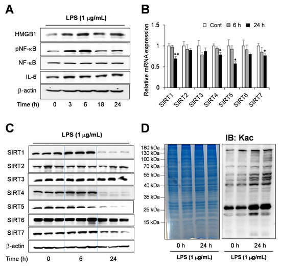
by Siniša Đurašević, Aleksandra Ružičić, Iva Lakić, Tomislav Tosti, Saša Đurović, Sofija Glumac, Snežana Pejić, Ana Todorović, Dunja Drakulić, Sanja Stanković, Nebojša Jasnić, Jelena Đorđević and Zoran Todorović
Int. J. Mol. Sci. 2022, 23(4), 2395; https://doi.org/10.3390/ijms23042395 - 21 Feb 2022
Cited by 9 | Viewed by 3736
Abstract
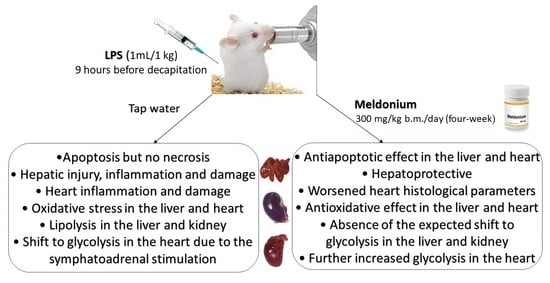
A dysregulated and overwhelming response to an infection accompanied by the exaggerated pro-inflammatory state and metabolism disturbance leads to the fatal outcome in sepsis. Previously we showed that meldonium, an anti-ischemic drug clinically used to treat myocardial and cerebral ischemia, strongly increases mortality [...] Read more.
A dysregulated and overwhelming response to an infection accompanied by the exaggerated pro-inflammatory state and metabolism disturbance leads to the fatal outcome in sepsis. Previously we showed that meldonium, an anti-ischemic drug clinically used to treat myocardial and cerebral ischemia, strongly increases mortality in faecal-induced peritonitis (FIP) in rats. We postulated that the same mechanism that is responsible for the otherwise strong anti-inflammatory effects of meldonium could be the culprit of the increased mortality. In the present study, we applied the LPS-induced model of sepsis to explore the presence of any differences from and/or similarities to the FIP model. When it comes to energy production, despite some shared similarities, it is evident that LPS and FIP models of sepsis differ greatly. A different profile of sympathoadrenal activation may account for this observation, as it was lacking in the FIP model, whereas in the LPS model it was strong enough to overcome the effects of meldonium. Therefore, choosing the appropriate model of sepsis induction is of great importance, especially if energy homeostasis is the main focus of the study. Even when differences in the experimental design of the two models are acknowledged, the role of different patterns of energy production cannot be excluded. On that account, our results draw attention to the importance of uninterrupted energy production in sepsis but also call for much-needed revisions of the current recommendations for its treatment. Full article
Show Figures

Graphical abstract

11 pages, 1271 KiB  
Article
Chemosensory Proteins Are Associated with Thiamethoxam and Spirotetramat Tolerance in Aphis gossypii Glover
by Hongfei Xu, Kunpeng Yan, Yaping Ding, Yuntong Lv, Jianyi Li, Fengting Yang, Xuewei Chen, Xiwu Gao, Yiou Pan and Qingli Shang
Int. J. Mol. Sci. 2022, 23(4), 2356; https://doi.org/10.3390/ijms23042356 - 21 Feb 2022
Cited by 12 | Viewed by 2543
Abstract
Chemosensory proteins (CSPs) are a class of transporters in arthropods. Deeper research on CSPs showed that CSPs may be involved in some physiological processes beyond chemoreception, such as insect resistance to pesticides. We identified two upregulated CSPs in two resistant strains of Aphis [...] Read more.
Chemosensory proteins (CSPs) are a class of transporters in arthropods. Deeper research on CSPs showed that CSPs may be involved in some physiological processes beyond chemoreception, such as insect resistance to pesticides. We identified two upregulated CSPs in two resistant strains of Aphis gossypii Glover. To understand their role in the resistance of aphids to pesticides, we performed the functional verification of CSP1 and CSP4 in vivo and in vitro. Results showed that the sensitivity of the thiamethoxam-resistant strain to thiamethoxam increased significantly with the silencing of CSP1 and CSP4 by RNAi (RNA interference), and the sensitivity of the spirotetramat-resistant strain to spirotetramat increased significantly with the silencing of CSP4. Transgenic Drosophila melanogaster expressing CSPs exhibited stronger resistance to thiamethoxam, spirotetramat, and alpha-cypermethrin than the control did. In the bioassay of transgenic Drosophila, CSPs showed different tolerance mechanisms for different pesticides, and the overexpressed CSPs may play a role in processes other than resistance to pesticides. In brief, the present results prove that CSPs are related to the resistance of cotton aphids to insecticides. Full article
Show Figures

Figure 1

19 pages, 2654 KiB  
Article
A Mixture of Endocrine Disrupting Chemicals Associated with Lower Birth Weight in Children Induces Adipogenesis and DNA Methylation Changes in Human Mesenchymal Stem Cells
by Polina Lizunkova, Elin Engdahl, Gábor Borbély, Chris Gennings, Christian Lindh, Carl-Gustaf Bornehag and Joëlle Rüegg
Int. J. Mol. Sci. 2022, 23(4), 2320; https://doi.org/10.3390/ijms23042320 - 19 Feb 2022
Cited by 10 | Viewed by 4210
Abstract
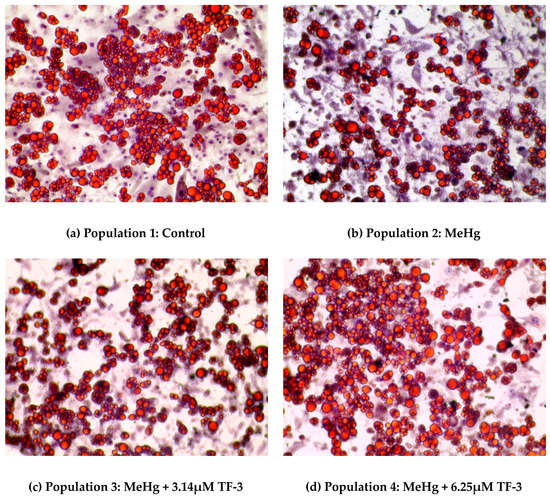
Endocrine Disrupting Chemicals (EDCs) are man-made compounds that alter functions of the endocrine system. Environmental mixtures of EDCs might have adverse effects on human health, even though their individual concentrations are below regulatory levels of concerns. However, studies identifying and experimentally testing adverse [...] Read more.
Endocrine Disrupting Chemicals (EDCs) are man-made compounds that alter functions of the endocrine system. Environmental mixtures of EDCs might have adverse effects on human health, even though their individual concentrations are below regulatory levels of concerns. However, studies identifying and experimentally testing adverse effects of real-life mixtures are scarce. In this study, we aimed at evaluating an epidemiologically identified EDC mixture in an experimental setting to delineate its cellular and epigenetic effects. The mixture was established using data from the Swedish Environmental Longitudinal Mother and child Asthma and allergy (SELMA) study where it was associated with lower birth weight, an early marker for prenatal metabolic programming. This mixture was then tested for its ability to change metabolic programming of human mesenchymal stem cells. In these cells, we assessed if the mixture induced adipogenesis and genome-wide DNA methylation changes. The mixture increased lipid droplet accumulation already at concentrations corresponding to levels measured in the pregnant women of the SELMA study. Furthermore, we identified differentially methylated regions in genes important for adipogenesis and thermogenesis. This study shows that a mixture reflecting human real-life exposure can induce molecular and cellular changes during development that could underlie adverse outcomes. Full article
Show Figures

Figure 1

20 pages, 2072 KiB  
Review
Metalloprotein-Specific or Critical Amino Acid Residues: Perspectives on Plant-Precise Detoxification and Recognition Mechanisms under Cadmium Stress
by Dandan Li, Tengbing He, Muhammad Saleem and Guandi He
Int. J. Mol. Sci. 2022, 23(3), 1734; https://doi.org/10.3390/ijms23031734 - 3 Feb 2022
Cited by 27 | Viewed by 3888
Abstract
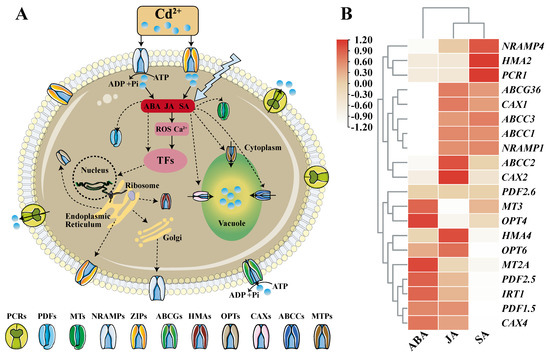
Cadmium (Cd) pollution in cultivated land is caused by irresistible geological factors and human activities; intense diffusion and migration have seriously affected the safety of food crops. Plants have evolved mechanisms to control excessive influx of Cd in the environment, such as directional [...] Read more.
Cadmium (Cd) pollution in cultivated land is caused by irresistible geological factors and human activities; intense diffusion and migration have seriously affected the safety of food crops. Plants have evolved mechanisms to control excessive influx of Cd in the environment, such as directional transport, chelation and detoxification. This is done by some specific metalloproteins, whose key amino acid motifs have been investigated by scientists one by one. The application of powerful cell biology, crystal structure science, and molecular probe targeted labeling technology has identified a series of protein families involved in the influx, transport and detoxification of the heavy metal Cd. This review summarizes them as influx proteins (NRAMP, ZIP), chelating proteins (MT, PDF), vacuolar proteins (CAX, ABCC, MTP), long-distance transport proteins (OPT, HMA) and efflux proteins (PCR, ABCG). We selected representative proteins from each family, and compared their amino acid sequence, motif structure, subcellular location, tissue specific distribution and other characteristics of differences and common points, so as to summarize the key residues of the Cd binding target. Then, we explain its special mechanism of action from the molecular structure. In conclusion, this review is expected to provide a reference for the exploration of key amino acid targets of Cd, and lay a foundation for the intelligent design and breeding of crops with high/low Cd accumulation. Full article
Show Figures

Figure 1

20 pages, 1746 KiB  
Review
The Aryl Hydrocarbon Receptor (AHR): A Novel Therapeutic Target for Pulmonary Diseases?
by Binoy Shivanna, Chun Chu and Bhagavatula Moorthy
Int. J. Mol. Sci. 2022, 23(3), 1516; https://doi.org/10.3390/ijms23031516 - 28 Jan 2022
Cited by 22 | Viewed by 5832
Abstract
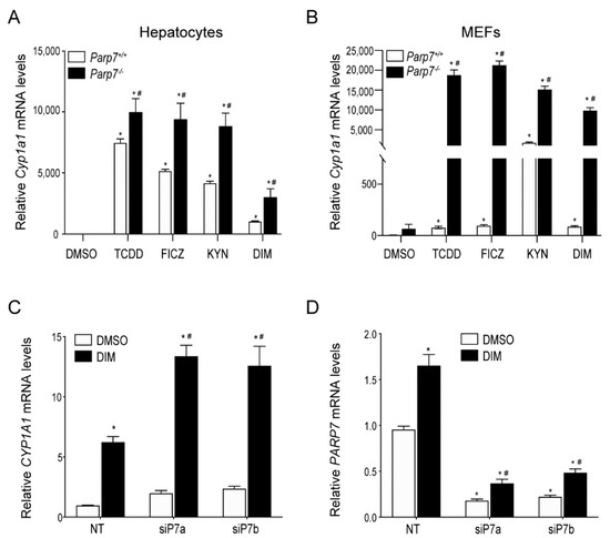
The aryl hydrocarbon receptor (AHR) is a cytoplasmic transcription factor that is well-known for regulating xenobiotic metabolism. Studies in knockout and transgenic mice indicate that the AHR plays a vital role in the development of liver and regulation of reproductive, cardiovascular, hematopoietic, and [...] Read more.
The aryl hydrocarbon receptor (AHR) is a cytoplasmic transcription factor that is well-known for regulating xenobiotic metabolism. Studies in knockout and transgenic mice indicate that the AHR plays a vital role in the development of liver and regulation of reproductive, cardiovascular, hematopoietic, and immune homeostasis. In this focused review on lung diseases associated with acute injury and alveolar development, we reviewed and summarized the current literature on the mechanistic role(s) and therapeutic potential of the AHR in acute lung injury, chronic obstructive pulmonary disease, and bronchopulmonary dysplasia (BPD). Pre-clinical studies indicate that endogenous AHR activation is necessary to protect neonatal and adult lungs against hyperoxia- and cigarette smoke-induced injury. Our goal is to provide insight into the high translational potential of the AHR in the meaningful management of infants and adults with these lung disorders that lack curative therapies. Full article
Show Figures

Figure 1

21 pages, 4956 KiB  
Article
Metabolites of Cannabis Induce Cardiac Toxicity and Morphological Alterations in Cardiac Myocytes
by Ayse Orme Merve, Pola Sobiecka, Vytautas Remeškevičius, Luke Taylor, Lili Saskoy, Scott Lawton, Ben P. Jones, Ahmed Elwakeel, Francesca E. Mackenzie, Elena Polycarpou, Jason Bennett and Brian Rooney
Int. J. Mol. Sci. 2022, 23(3), 1401; https://doi.org/10.3390/ijms23031401 - 26 Jan 2022
Cited by 6 | Viewed by 5165
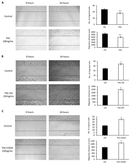
Abstract
Cannabis is one of the most commonly used recreational drugs worldwide. Rrecent epidemiology studies have linked increased cardiac complications to cannabis use. However, this literature is predominantly based on case incidents and post-mortem investigations. This study elucidates the molecular mechanism of Δ9-tetrahydrocannabinol (THC), [...] Read more.
Cannabis is one of the most commonly used recreational drugs worldwide. Rrecent epidemiology studies have linked increased cardiac complications to cannabis use. However, this literature is predominantly based on case incidents and post-mortem investigations. This study elucidates the molecular mechanism of Δ9-tetrahydrocannabinol (THC), and its primary metabolites 11-Hydroxy-Δ9-THC (THC-OH) and 11-nor-9-carboxy-Δ⁹-tetrahydrocannabinol (THC-COOH). Treatment of cardiac myocytes with THC-OH and THC-COOH increased cell migration and proliferation (p < 0.05), with no effect on cell adhesion, with higher doses (250–100 ng/mL) resulting in increased cell death and significant deterioration in cellular architecture. Conversely, no changes in cell morphology or viability were observed in response to THC. Expression of key ECM proteins α-SMA and collagen were up-regulated in response to THC-OH and THC-COOH treatments with concomitant modulation of PI3K and MAPK signalling. Investigations in the planarian animal model Polycelis nigra demonstrated that treatments with cannabinoid metabolites resulted in increased protein deposition at transection sites while higher doses resulted in significant lethality and decline in regeneration. These results highlight that the key metabolites of cannabis elicit toxic effects independent of the parent and psychoactive compound, with implications for cardiotoxicity relating to hypertrophy and fibrogenesis. Full article
Show Figures

Figure 1

15 pages, 2832 KiB  
Article
Reduced Colonic Mucosal Injury in 2,3,7,8-Tetrachlorodibenzo-p-Dioxin Poly ADP-Ribose Polymerase (TIPARP/PARP7)-Deficient Mice
by David Hutin, Karoline Alvik Hagen, Peng Shao, Kim Sugamori, Denis M. Grant and Jason Matthews
Int. J. Mol. Sci. 2022, 23(2), 920; https://doi.org/10.3390/ijms23020920 - 15 Jan 2022
Cited by 5 | Viewed by 2639
Abstract
Poly-ADP-ribose polymerases (PARPs) are important regulators of the immune system, including TCDD-inducible poly-ADP-ribose polymerase (TIPARP), also known as poly-ADP-ribose polymerase 7 (PARP7). PARP7 negatively regulates aryl hydrocarbon receptor (AHR) and type I interferon (IFN-I) signaling, both of which have been implicated in intestinal [...] Read more.
Poly-ADP-ribose polymerases (PARPs) are important regulators of the immune system, including TCDD-inducible poly-ADP-ribose polymerase (TIPARP), also known as poly-ADP-ribose polymerase 7 (PARP7). PARP7 negatively regulates aryl hydrocarbon receptor (AHR) and type I interferon (IFN-I) signaling, both of which have been implicated in intestinal homeostasis and immunity. Since the loss of PARP7 expression increases AHR and IFN-I signaling, we used a murine dextran sulfate sodium (DSS)-induced colitis model to investigate the effect of PARP7 loss on DSS-induced intestinal inflammation. DSS-exposed Parp7−/− mice had less body weight loss, lower disease index scores, and reduced expression of several inflammation genes, including interleukin IL-6, C-x-c motif chemokine ligand 1 (Cxcl1), and lipocalin-2, when compared with wild-type mice. However, no significant difference was observed between genotypes in the colonic expression of the AHR target gene cytochrome P450 1A1 (Cyp1a1). Moreover, no significant differences in microbial composition were observed between the genotypes. Our findings demonstrate that the absence of PARP7 protein results in an impaired immune response to colonic inflammation and suggests that PARP7 may participate in the recruitment of immune cells to the inflammation site, which may be due to its role in IFN-I signaling rather than AHR signaling. Full article
Show Figures

Figure 1

20 pages, 5171 KiB  
Article
Pharmacological Dissection of the Crosstalk between NaV and CaV Channels in GH3b6 Cells
by Léa Réthoré, Joohee Park, Jérôme Montnach, Sébastien Nicolas, Joseph Khoury, Elodie Le Seac’h, Kamel Mabrouk, Harold De Pomyers, Hélène Tricoire-Leignel, César Mattei, Daniel Henrion, Ziad Fajloun, Michel De Waard, Claire Legendre and Christian Legros
Int. J. Mol. Sci. 2022, 23(2), 827; https://doi.org/10.3390/ijms23020827 - 13 Jan 2022
Cited by 4 | Viewed by 3090
Abstract
Thanks to the crosstalk between Na+ and Ca2+ channels, Na+ and Ca2+ homeostasis interplay in so-called excitable cells enables the generation of action potential in response to electrical stimulation. Here, we investigated the impact of persistent activation of voltage-gated [...] Read more.
Thanks to the crosstalk between Na+ and Ca2+ channels, Na+ and Ca2+ homeostasis interplay in so-called excitable cells enables the generation of action potential in response to electrical stimulation. Here, we investigated the impact of persistent activation of voltage-gated Na+ (NaV) channels by neurotoxins, such as veratridine (VTD), on intracellular Ca2+ concentration ([Ca2+]i) in a model of excitable cells, the rat pituitary GH3b6 cells, in order to identify the molecular actors involved in Na+-Ca2+ homeostasis crosstalk. By combining RT-qPCR, immunoblotting, immunocytochemistry, and patch-clamp techniques, we showed that GH3b6 cells predominantly express the NaV1.3 channel subtype, which likely endorses their voltage-activated Na+ currents. Notably, these Na+ currents were blocked by ICA-121431 and activated by the β-scorpion toxin Tf2, two selective NaV1.3 channel ligands. Using Fura-2, we showed that VTD induced a [Ca2+]i increase. This effect was suppressed by the selective NaV channel blocker tetrodotoxin, as well by the selective L-type CaV channel (LTCC) blocker nifedipine. We also evidenced that crobenetine, a NaV channel blocker, abolished VTD-induced [Ca2+]i elevation, while it had no effects on LTCC. Altogether, our findings highlight a crosstalk between NaV and LTCC in GH3b6 cells, providing a new insight into the mode of action of neurotoxins. Full article
Show Figures

Figure 1

2021

Jump to: 2024, 2023, 2022, 2019

24 pages, 6007 KiB  
Article
Dopamine and Methamphetamine Differentially Affect Electron Transport Chain Complexes and Parkin in Rat Striatum: New Insight into Methamphetamine Neurotoxicity
by Viktoriia Bazylianska, Akhil Sharma, Heli Chauhan, Bernard Schneider and Anna Moszczynska
Int. J. Mol. Sci. 2022, 23(1), 363; https://doi.org/10.3390/ijms23010363 - 29 Dec 2021
Cited by 14 | Viewed by 4844
Abstract
Methamphetamine (METH) is a highly abused psychostimulant that is neurotoxic to dopaminergic (DAergic) nerve terminals in the striatum and increases the risk of developing Parkinson’s disease (PD). In vivo, METH-mediated DA release, followed by DA-mediated oxidative stress and mitochondrial dysfunction in pre- and [...] Read more.
Methamphetamine (METH) is a highly abused psychostimulant that is neurotoxic to dopaminergic (DAergic) nerve terminals in the striatum and increases the risk of developing Parkinson’s disease (PD). In vivo, METH-mediated DA release, followed by DA-mediated oxidative stress and mitochondrial dysfunction in pre- and postsynaptic neurons, mediates METH neurotoxicity. METH-triggered oxidative stress damages parkin, a neuroprotective protein involved in PD etiology via its involvement in the maintenance of mitochondria. It is not known whether METH itself contributes to mitochondrial dysfunction and whether parkin regulates complex I, an enzymatic complex downregulated in PD. To determine this, we separately assessed the effects of METH or DA alone on electron transport chain (ETC) complexes and the protein parkin in isolated striatal mitochondria. We show that METH decreases the levels of selected complex I, II, and III subunits (NDUFS3, SDHA, and UQCRC2, respectively), whereas DA decreases the levels only of the NDUFS3 subunit in our preparations. We also show that the selected subunits are not decreased in synaptosomal mitochondria under similar experimental conditions. Finally, we found that parkin overexpression does not influence the levels of the NDUFS3 subunit in rat striatum. The presented results indicate that METH itself is a factor promoting dysfunction of striatal mitochondria; therefore, it is a potential drug target against METH neurotoxicity. The observed decreases in ETC complex subunits suggest that DA and METH decrease activities of the ETC complexes via oxidative damage to their subunits and that synaptosomal mitochondria may be somewhat “resistant” to DA- and METH-induced disruption in mitochondrial ETC complexes than perikaryal mitochondria. The results also suggest that parkin does not regulate NDUFS3 turnover in rat striatum. Full article
Show Figures

Figure 1

22 pages, 3457 KiB  
Article
A Genus-Wide Bioactivity Analysis of Daboia (Viperinae: Viperidae) Viper Venoms Reveals Widespread Variation in Haemotoxic Properties
by Bianca op den Brouw, Francisco C. P. Coimbra, Nicholas R. Casewell, Syed Abid Ali, Freek J. Vonk and Bryan G. Fry
Int. J. Mol. Sci. 2021, 22(24), 13486; https://doi.org/10.3390/ijms222413486 - 15 Dec 2021
Cited by 8 | Viewed by 2828
Abstract
The snake genus Daboia (Viperidae: Viperinae; Oppel, 1811) contains five species: D. deserti, D. mauritanica, and D. palaestinae, found in Afro-Arabia, and the Russell’s vipers D. russelii and D. siamensis, found in Asia. Russell’s vipers are responsible for a [...] Read more.
The snake genus Daboia (Viperidae: Viperinae; Oppel, 1811) contains five species: D. deserti, D. mauritanica, and D. palaestinae, found in Afro-Arabia, and the Russell’s vipers D. russelii and D. siamensis, found in Asia. Russell’s vipers are responsible for a major proportion of the medically important snakebites that occur in the regions they inhabit, and their venoms are notorious for their coagulopathic effects. While widely documented, the extent of venom variation within the Russell’s vipers is poorly characterised, as is the venom activity of other species within the genus. In this study we investigated variation in the haemotoxic activity of Daboia using twelve venoms from all five species, including multiple variants of D. russelii, D. siamensis, and D. palaestinae. We tested the venoms on human plasma using thromboelastography, dose-response coagulometry analyses, and calibrated automated thrombography, and on human fibrinogen by thromboelastography and fibrinogen gels. We assessed activation of blood factors X and prothrombin by the venoms using fluorometry. Variation in venom activity was evident in all experiments. The Asian species D. russelii and D. siamensis and the African species D. mauritanica possessed procoagulant venom, while D. deserti and D. palaestinae were net-anticoagulant. Of the Russell’s vipers, the venom of D. siamensis from Myanmar was most toxic and D. russelli of Sri Lanka the least. Activation of both factor X and prothrombin was evident by all venoms, though at differential levels. Fibrinogenolytic activity varied extensively throughout the genus and followed no phylogenetic trends. This venom variability underpins one of the many challenges facing treatment of Daboia snakebite envenoming. Comprehensive analyses of available antivenoms in neutralising these variable venom activities are therefore of utmost importance. Full article
Show Figures

Figure 1

24 pages, 9170 KiB  
Article
A Complex Evaluation of the In-Vivo Biocompatibility and Degradation of an Extruded ZnMgSr Absorbable Alloy Implanted into Rabbit Bones for 360 Days
by Karel Klíma, Dan Ulmann, Martin Bartoš, Michal Španko, Jaroslava Dušková, Radka Vrbová, Jan Pinc, Jiří Kubásek, Marek Vlk, Tereza Ulmannová, René Foltán, Eitan Brizman, Milan Drahoš, Michal Beňo, Vladimír Machoň and Jaroslav Čapek
Int. J. Mol. Sci. 2021, 22(24), 13444; https://doi.org/10.3390/ijms222413444 - 14 Dec 2021
Cited by 7 | Viewed by 3119
Abstract
The increasing incidence of trauma in medicine brings with it new demands on the materials used for the surgical treatment of bone fractures. Titanium, its alloys, and steel are used worldwide in the treatment of skeletal injuries. These metallic materials, although inert, are [...] Read more.
The increasing incidence of trauma in medicine brings with it new demands on the materials used for the surgical treatment of bone fractures. Titanium, its alloys, and steel are used worldwide in the treatment of skeletal injuries. These metallic materials, although inert, are often removed after the injured bone has healed. The second-stage procedure—the removal of the plates and screws—can overwhelm patients and overload healthcare systems. The development of suitable absorbable metallic materials would help us to overcome these issues. In this experimental study, we analyzed an extruded Zn-0.8Mg-0.2Sr (wt.%) alloy on a rabbit model. From this alloy we developed screws which were implanted into the rabbit tibia. After 120, 240, and 360 days, we tested the toxicity at the site of implantation and also within the vital organs: the liver, kidneys, and brain. The results were compared with a control group, implanted with a Ti-based screw and sacrificed after 360 days. The samples were analyzed using X-ray, micro-CT, and a scanning electron microscope. Chemical analysis revealed only small concentrations of zinc, strontium, and magnesium in the liver, kidneys, and brain. Histologically, the alloy was verified to possess very good biocompatibility after 360 days, without any signs of toxicity at the site of implantation. We did not observe raised levels of Sr, Zn, or Mg in any of the vital organs when compared with the Ti group at 360 days. The material was found to slowly degrade in vivo, forming solid corrosion products on its surface. Full article
Show Figures

Figure 1

24 pages, 2395 KiB  
Article
The Influence of an Adrenergic Antagonist Guanethidine (GUA) on the Distribution Pattern and Chemical Coding of Dorsal Root Ganglia (DRG) Neurons Supplying the Porcine Urinary Bladder
by Paweł Janikiewicz, Barbara Wasilewska, Urszula Mazur, Amelia Franke-Radowiecka, Mariusz Majewski and Agnieszka Bossowska
Int. J. Mol. Sci. 2021, 22(24), 13399; https://doi.org/10.3390/ijms222413399 - 13 Dec 2021
Cited by 3 | Viewed by 2604
Abstract
Although guanethidine (GUA) was used in the past as a drug to suppress hyperactivity of the sympathetic nerve fibers, there are no available data concerning the possible action of this substance on the sensory component of the peripheral nervous system supplying the urinary [...] Read more.
Although guanethidine (GUA) was used in the past as a drug to suppress hyperactivity of the sympathetic nerve fibers, there are no available data concerning the possible action of this substance on the sensory component of the peripheral nervous system supplying the urinary bladder. Thus, the present study was aimed at disclosing the influence of intravesically instilled GUA on the distribution, relative frequency, and chemical coding of dorsal root ganglion neurons associated with the porcine urinary bladder. The investigated sensory neurons were visualized with a retrograde tracing method using Fast Blue (FB), while their chemical profile was disclosed with single-labeling immunohistochemistry using antibodies against substance P (SP), calcitonin gene-related peptide (CGRP), pituitary adenylate cyclase activating polypeptide (PACAP), galanin (GAL), neuronal nitric oxide synthase (nNOS), somatostatin (SOM), and calbindin (CB). After GUA treatment, a slight decrease in the number of FB+ neurons containing SP was observed when compared with untreated animals (34.6 ± 6.5% vs. 45.6 ± 1.3%), while the number of retrogradely traced cells immunolabeled for GAL, nNOS, and CB distinctly increased (12.3 ± 1.0% vs. 7.4 ± 0.6%, 11.9 ± 0.6% vs. 5.4 ± 0.5% and 8.6 ± 0.5% vs. 2.7 ± 0.4%, respectively). However, administration of GUA did not change the number of FB+ neurons containing CGRP, PACAP, or SOM. The present study provides evidence that GUA significantly modifies the sensory innervation of the porcine urinary bladder wall and thus may be considered a potential tool for studying the plasticity of this subdivision of the bladder innervation. Full article
Show Figures

Figure 1

25 pages, 3099 KiB  
Article
Juvenile African Clawed Frogs (Xenopus laevis) Express Growth, Metamorphosis, Mortality, Gene Expression, and Metabolic Changes When Exposed to Thiamethoxam and Clothianidin
by Jill A. Jenkins, Katherine R. Hartop, Ghadeer Bukhari, Debra E. Howton, Kelly L. Smalling, Scott V. Mize, Michelle L. Hladik, Darren Johnson, Rassa O. Draugelis-Dale and Bonnie L. Brown
Int. J. Mol. Sci. 2021, 22(24), 13291; https://doi.org/10.3390/ijms222413291 - 10 Dec 2021
Cited by 6 | Viewed by 3745
Abstract
Neonicotinoids (NEO) represent the main class of insecticides currently in use, with thiamethoxam (THX) and clothianidin (CLO) primarily applied agriculturally. With few comprehensive studies having been performed with non-target amphibians, the aim was to investigate potential biomarker responses along an adverse outcome pathway [...] Read more.
Neonicotinoids (NEO) represent the main class of insecticides currently in use, with thiamethoxam (THX) and clothianidin (CLO) primarily applied agriculturally. With few comprehensive studies having been performed with non-target amphibians, the aim was to investigate potential biomarker responses along an adverse outcome pathway of NEO exposure, whereby data were collected on multiple biological hierarchies. Juvenile African clawed frogs, Xenopus laevis, were exposed to commercial formulations of THX and CLO at high (100 ppm) and low (20 ppm) concentrations of the active ingredient. Mortality, growth, development, liver metabolic enzyme activity, and gene expression endpoints were quantified. Tadpoles (n > 1000) from NF 47 through tail resorption stage (NF 66) were exposed to NEO or to NEO-free media treatments. Liver cell reductase activity and cytotoxicity were quantified by flow cytometry. Compared to control reference gene expressions, levels of expression for NEO receptor subunits, cell structure, function, and decontamination processes were measured by RT-qPCR by using liver and brain. Mortality in THX high was 21.5% compared to the control (9.1%); the metabolic conversion of THX to CLO may explain these results. The NF 57 control tadpoles were heavier, longer, and more developed than the others. The progression of development from NF 57–66 was reduced by THX low, and weight gain was impaired. Liver reductases were highest in the control (84.1%), with low NEO exhibiting the greatest reductions; the greatest cytotoxicity was seen with THX high. More transcriptional activity was noted in brains than in livers. Results affirm the utility of a study approach that considers multiple complexities in ecotoxicological studies with non-target amphibians, underscoring the need for simultaneously considering NEO concentration-response relationships with both whole-organism and biomarker endpoints. Full article
Show Figures

Figure 1

18 pages, 4093 KiB  
Article
Prenatal Exposure to Triclocarban Impairs ESR1 Signaling and Disrupts Epigenetic Status in Sex-Specific Ways as Well as Dysregulates the Expression of Neurogenesis- and Neurotransmitter-Related Genes in the Postnatal Mouse Brain
by Agnieszka Wnuk, Joanna Rzemieniec, Karolina Przepiórska, Bernadeta Angelika Pietrzak, Marzena Maćkowiak and Małgorzata Kajta
Int. J. Mol. Sci. 2021, 22(23), 13121; https://doi.org/10.3390/ijms222313121 - 4 Dec 2021
Cited by 9 | Viewed by 2483
Abstract
Triclocarban is a highly effective and broadly used antimicrobial agent. Humans are continually exposed to triclocarban, but the safety of prenatal exposure to triclocarban in the context of neurodevelopment remains unknown. In this study, we demonstrated for the first time that mice that [...] Read more.
Triclocarban is a highly effective and broadly used antimicrobial agent. Humans are continually exposed to triclocarban, but the safety of prenatal exposure to triclocarban in the context of neurodevelopment remains unknown. In this study, we demonstrated for the first time that mice that had been prenatally exposed to environmentally relevant doses of triclocarban had impaired estrogen receptor 1 (ESR1) signaling in the brain. These mice displayed decreased mRNA and protein expression levels of ESR1 as well as hypermethylation of the Esr1 gene in the cerebral cortex. Prenatal exposure to triclocarban also diminished the mRNA expression of Esr2, Gper1, Ahr, Arnt, Cyp19a1, Cyp1a1, and Atg7, and the protein levels of CAR, ARNT, and MAP1LC3AB in female brains and decreased the protein levels of BCL2, ARNT, and MAP1LC3AB in male brains. In addition, exposure to triclocarban caused sex-specific alterations in the methylation levels of global DNA and estrogen receptor genes. Microarray and enrichment analyses showed that, in males, triclocarban dysregulated mainly neurogenesis-related genes, whereas, in females, the compound dysregulated mainly neurotransmitter-related genes. In conclusion, our data identified triclocarban as a neurodevelopmental risk factor that particularly targets ESR1, affects apoptosis and autophagy, and in sex-specific ways disrupts the epigenetic status of brain tissue and dysregulates the postnatal expression of neurogenesis- and neurotransmitter-related genes. Full article
Show Figures

Graphical abstract

13 pages, 3505 KiB  
Article
Intracellular Exposure Dose-Associated Susceptibility of Steatotic Hepatocytes to Metallic Nanoparticles
by Xiaoli Zhang, Yongyi Wei, Chengjun Li, Weiyu Wang, Rui Zhang, Jianbo Jia and Bing Yan
Int. J. Mol. Sci. 2021, 22(23), 12643; https://doi.org/10.3390/ijms222312643 - 23 Nov 2021
Cited by 5 | Viewed by 2366
Abstract
Non-alcoholic fatty liver disease (NAFLD), mainly characterized by the accumulation of excess fat in hepatocytes, is the most prevalent liver disorder afflicting ~25% of adults worldwide. In vivo studies have shown that adult rodents with NAFLD were more sensitive to metallic nanoparticles (MNPs) [...] Read more.
Non-alcoholic fatty liver disease (NAFLD), mainly characterized by the accumulation of excess fat in hepatocytes, is the most prevalent liver disorder afflicting ~25% of adults worldwide. In vivo studies have shown that adult rodents with NAFLD were more sensitive to metallic nanoparticles (MNPs) than healthy MNPs. However, due to the complex interactions between various cell types in a fatty liver, it has become a major challenge to reveal the toxic effects of MNPs to specific types of liver cells such as steatotic hepatocytes. In this study, we reported the susceptibility of steatotic hepatocytes in cytotoxicity and the induction of oxidative stress to direct exposures to MNPs with different components (silver, ZrO2, and TiO2 NPs) and sizes (20–30 nm and 125 nm) in an oleic acid (OA) -induced steatotic HepG2 (sHepG2) cell model. Furthermore, the inhibitory potential of MNPs against the process of fatty acid oxidation (FAO) were obvious in sHepG2 cells, even at extremely low doses of 2 or 4 μg/mL, which was not observed in non-steatotic HepG2 (nHepG2) cells. Further experiments on the differential cell uptake of MNPs in nHepG2 and sHepG2 cells demonstrated that the susceptibility of steatotic hepatocytes to MNP exposures was in association with the higher cellular accumulation of MNPs. Overall, our study demonstrated that it is necessary and urgent to take the intracellular exposure dose into consideration when assessing the potential toxicity of environmentally exposed MNPs. Full article
Show Figures

Graphical abstract

17 pages, 4714 KiB  
Article
Enhancing Paraoxon Binding to Organophosphorus Hydrolase Active Site
by Léa El Khoury, David L. Mobley, Dongmei Ye and Susan B. Rempe
Int. J. Mol. Sci. 2021, 22(23), 12624; https://doi.org/10.3390/ijms222312624 - 23 Nov 2021
Cited by 3 | Viewed by 2377
Abstract
Organophosphorus hydrolase (OPH) is a metalloenzyme that can hydrolyze organophosphorus agents resulting in products that are generally of reduced toxicity. The best OPH substrate found to date is diethyl p-nitrophenyl phosphate (paraoxon). Most structural and kinetic studies assume that the binding orientation of [...] Read more.
Organophosphorus hydrolase (OPH) is a metalloenzyme that can hydrolyze organophosphorus agents resulting in products that are generally of reduced toxicity. The best OPH substrate found to date is diethyl p-nitrophenyl phosphate (paraoxon). Most structural and kinetic studies assume that the binding orientation of paraoxon is identical to that of diethyl 4-methylbenzylphosphonate, which is the only substrate analog co-crystallized with OPH. In the current work, we used a combined docking and molecular dynamics (MD) approach to predict the likely binding mode of paraoxon. Then, we used the predicted binding mode to run MD simulations on the wild type (WT) OPH complexed with paraoxon, and OPH mutants complexed with paraoxon. Additionally, we identified three hot-spot residues (D253, H254, and I255) involved in the stability of the OPH active site. We then experimentally assayed single and double mutants involving these residues for paraoxon binding affinity. The binding free energy calculations and the experimental kinetics of the reactions between each OPH mutant and paraoxon show that mutated forms D253E, D253E-H254R, and D253E-I255G exhibit enhanced substrate binding affinity over WT OPH. Interestingly, our experimental results show that the substrate binding affinity of the double mutant D253E-H254R increased by 19-fold compared to WT OPH. Full article
Show Figures

Figure 1

19 pages, 2862 KiB  
Article
MicroRNA Expression Profiling in Porcine Liver, Jejunum and Serum upon Dietary DON Exposure Reveals Candidate Toxicity Biomarkers
by Maia Segura-Wang, Bertrand Grenier, Suzana Ilic, Ursula Ruczizka, Maximiliane Dippel, Moritz Bünger, Matthias Hackl and Veronika Nagl
Int. J. Mol. Sci. 2021, 22(21), 12043; https://doi.org/10.3390/ijms222112043 - 7 Nov 2021
Cited by 4 | Viewed by 2378
Abstract
Deoxynivalenol (DON), a frequent mycotoxin worldwide, impairs human and animal health. The response of microRNAs, small non-coding RNAs, to DON has been scarcely investigated, but holds remarkable potential for biomarker applications. Hence, we aimed to investigate DON-induced changes in the microRNA expression in [...] Read more.
Deoxynivalenol (DON), a frequent mycotoxin worldwide, impairs human and animal health. The response of microRNAs, small non-coding RNAs, to DON has been scarcely investigated, but holds remarkable potential for biomarker applications. Hence, we aimed to investigate DON-induced changes in the microRNA expression in porcine liver, jejunum and serum by combining targeted and untargeted analyses. Piglets received uncontaminated feed or feed containing 900 µg/kg and 2500 µg/kg DON for four weeks, followed by a wash-out period. In tissue, only slight changes in microRNA expression were detected, with ssc-miR-10b being downregulated in liver of DON-exposed piglets. In serum, several microRNAs were differentially expressed upon DON exposure, four of which were validated by qPCR (ssc-miR-16, ssc-miR-128, ssc-miR-451, ssc-miR-205). The serum microRNA response to DON increased over time and declined after removal of contaminated diets. Receiver operating curve analyses for individual microRNAs were significant, and a combination of the four microRNAs increased the predictive capacity for DON exposure. Predicted microRNA target genes showed enrichment of several pathways including PIK3-AKT, Wnt/β-catenin, and adherens junctions. This study gives, for the first time, a comprehensive view of the porcine microRNA response to DON, providing a basis for future research on microRNAs as biomarkers for mycotoxins. Full article
Show Figures

Figure 1

13 pages, 3995 KiB  
Article
Experimental Evaluation of Food-Grade Semi-Refined Carrageenan Toxicity
by Denys Pogozhykh, Yevgen Posokhov, Valeriy Myasoedov, Galina Gubina-Vakulyck, Tetyana Chumachenko, Oleksandr Knigavko, Hanna Polikarpova, Yuliia Kalashnyk-Vakulenko, Ketino Sharashydze, Oksana Nakonechna, Volodymyr Prokopyuk, Anatolii Onishchenko and Anton Tkachenko
Int. J. Mol. Sci. 2021, 22(20), 11178; https://doi.org/10.3390/ijms222011178 - 16 Oct 2021
Cited by 12 | Viewed by 3469
Abstract
The safety of food additives E407 and E407a has raised concerns in the scientific community. Thus, this study aims to assess the local and systemic toxic effects of the common food additive E407a in rats orally exposed to it for two weeks. Complex [...] Read more.
The safety of food additives E407 and E407a has raised concerns in the scientific community. Thus, this study aims to assess the local and systemic toxic effects of the common food additive E407a in rats orally exposed to it for two weeks. Complex evaluations of the effects of semi-refined carrageenan (E407a) on rats upon oral exposure were performed. Local effects of E407a on the intestine were analyzed using routine histological stains and CD68 immunostaining. Furthermore, circulating levels of inflammatory markers were assessed. A fluorescent probe O1O (2- (2′-OH-phenyl)-5-phenyl-1,3-oxazole) was used for evaluating the state of leukocyte cell membranes. Cell death modes of leukocytes were analyzed by flow cytometry using Annexin V and 7-aminoactinomycin D staining. Oral administration of the common food additive E407a was found to be associated with altered small and large intestinal morphology, infiltration of the lamina propria in the small intestine with macrophages (CD68+ cells), high systemic levels of inflammation markers, and changes in the lipid order of the phospholipid bilayer in the cell membranes of leukocytes, alongside the activation of their apoptosis. Our findings suggest that oral exposure to E407a through rats results in the development of intestinal inflammation. Full article
Show Figures

Figure 1

14 pages, 2818 KiB  
Article
Porphyrin Based 2D-MOF Structures as Dual-Kinetic Sorafenib Nanocarriers for Hepatoma Treatment
by Adam Bieniek, Marek Wiśniewski, Joanna Czarnecka, Jędrzej Wierzbicki, Marcin Ziętek, Maciej Nowacki, Dariusz Grzanka, Tomasz Kloskowski and Katarzyna Roszek
Int. J. Mol. Sci. 2021, 22(20), 11161; https://doi.org/10.3390/ijms222011161 - 16 Oct 2021
Cited by 10 | Viewed by 2879
Abstract
The existing clinical protocols of hepatoma treatment require improvement of drug efficacy that can be achieved by harnessing nanomedicine. Porphyrin-based, paddle-wheel framework (PPF) structures were obtained and tested as dual-kinetic Sorafenib (SOR) nanocarriers against hepatoma. We experimentally proved that sloughing of PPF structures [...] Read more.
The existing clinical protocols of hepatoma treatment require improvement of drug efficacy that can be achieved by harnessing nanomedicine. Porphyrin-based, paddle-wheel framework (PPF) structures were obtained and tested as dual-kinetic Sorafenib (SOR) nanocarriers against hepatoma. We experimentally proved that sloughing of PPF structures combined with gradual dissolving are effective mechanisms for releasing the drug from the nanocarrier. By controlling the PPF degradation and size of adsorbed SOR deposits, we were able to augment SOR anticancer effects, both in vitro and in vivo, due to the dual kinetic behavior of SOR@PPF. Obtained drug delivery systems with slow and fast release of SOR influenced effectively, although in a different way, the cancer cells proliferation (reflected with EC50 and ERK 1/2 phosphorylation level). The in vivo studies proved that fast-released SOR@PPF reduces the tumor size considerably, while the slow-released SOR@PPF much better prevents from lymph nodes involvement and distant metastases. Full article
Show Figures

Graphical abstract

17 pages, 1852 KiB  
Article
Elucidating Carfilzomib’s Induced Cardiotoxicity in an In Vivo Model of Aging: Prophylactic Potential of Metformin
by Panagiotis Efentakis, Garyfalia Psarakou, Aimilia Varela, Eleni Dimitra Papanagnou, Michail Chatzistefanou, Panagiota-Efstathia Nikolaou, Costantinos H. Davos, Maria Gavriatopoulou, Ioannis P. Trougakos, Meletios Athanasios Dimopoulos, Ioanna Andreadou and Evangelos Terpos
Int. J. Mol. Sci. 2021, 22(20), 10956; https://doi.org/10.3390/ijms222010956 - 11 Oct 2021
Cited by 11 | Viewed by 2641
Abstract
Background: Carfilzomib is a first-line proteasome inhibitor indicated for relapsed/refractory multiple myeloma (MM), with its clinical use being hampered by cardiotoxic phenomena. We have previously established a translational model of carfilzomib cardiotoxicity in young adult mice, in which metformin emerged as a prophylactic [...] Read more.
Background: Carfilzomib is a first-line proteasome inhibitor indicated for relapsed/refractory multiple myeloma (MM), with its clinical use being hampered by cardiotoxic phenomena. We have previously established a translational model of carfilzomib cardiotoxicity in young adult mice, in which metformin emerged as a prophylactic therapy. Considering that MM is an elderly disease and that age is an independent risk factor for cardiotoxicity, herein, we sought to validate carfilzomib’s cardiotoxicity in an in vivo model of aging. Methods: Aged mice underwent the translational two- and four-dose protocols without and with metformin. Mice underwent echocardiography and were subsequently sacrificed for molecular analyses in the blood and cardiac tissue. Results: Carfilzomib decreased proteasomal activity both in PBMCs and myocardium in both protocols. Carfilzomib induced mild cardiotoxicity after two doses and more pronounced cardiomyopathy in the four-dose protocol, while metformin maintained cardiac function. Carfilzomib led to an increased Bip expression and decreased AMPKα phosphorylation, while metformin coadministration partially decreased Bip expression and induced AMPKα phosphorylation, leading to enhanced myocardial LC3B-dependent autophagy. Conclusion: Carfilzomib induced cardiotoxicity in aged mice, an effect significantly reversed by metformin. The latter possesses translational importance as it further supports the clinical use of metformin as a potent prophylactic therapy. Full article
Show Figures

Graphical abstract

43 pages, 5860 KiB  
Review
Metabolic Toxification of 1,2-Unsaturated Pyrrolizidine Alkaloids Causes Human Hepatic Sinusoidal Obstruction Syndrome: The Update
by Rolf Teschke, Noudeng Vongdala, Nguyen Van Quan, Tran Ngoc Quy and Tran Dang Xuan
Int. J. Mol. Sci. 2021, 22(19), 10419; https://doi.org/10.3390/ijms221910419 - 27 Sep 2021
Cited by 32 | Viewed by 4179
Abstract
Saturated and unsaturated pyrrolizidine alkaloids (PAs) are present in more than 6000 plant species growing in countries all over the world. They have a typical heterocyclic structure in common, but differ in their potential toxicity, depending on the presence or absence of a [...] Read more.
Saturated and unsaturated pyrrolizidine alkaloids (PAs) are present in more than 6000 plant species growing in countries all over the world. They have a typical heterocyclic structure in common, but differ in their potential toxicity, depending on the presence or absence of a double bond between C1 and C2. Fortunately, most plants contain saturated PAs without this double bond and are therefore not toxic for consumption by humans or animals. In a minority of plants, however, PAs with this double bond between C1 and C2 exhibit strong hepatotoxic, genotoxic, cytotoxic, neurotoxic, and tumorigenic potentials. If consumed in error and in large emouns, plants with 1,2-unsaturated PAs induce metabolic breaking-off of the double bonds of the unsaturated PAs, generating PA radicals that may trigger severe liver injury through a process involving microsomal P450 (CYP), with preference of its isoforms CYP 2A6, CYP 3A4, and CYP 3A5. This toxifying CYP-dependent conversion occurs primarily in the endoplasmic reticulum of the hepatocytes equivalent to the microsomal fraction. Toxified PAs injure the protein membranes of hepatocytes, and after passing their plasma membranes, more so the liver sinusoidal endothelial cells (LSECs), leading to life-threatening hepatic sinusoidal obstruction syndrome (HSOS). This injury is easily diagnosed by blood pyrrolizidine protein adducts, which are perfect diagnostic biomarkers, supporting causality evaluation using the updated RUCAM (Roussel Uclaf Causality Assessment Method). HSOS is clinically characterized by weight gain due to fluid accumulation (ascites, pleural effusion, and edema), and may lead to acute liver failure, liver transplantation, or death. In conclusion, plant-derived PAs with a double bond between C1 and C2 are potentially hepatotoxic after metabolic removal of the double bond, and may cause PA-HSOS with a potential lethal outcome, even if PA consumption is stopped. Full article
Show Figures

Figure 1

14 pages, 3784 KiB  
Article
Self-Assembly Nanoparticles of Natural Bioactive Abietane Diterpenes
by Epole Ntungwe, Eva María Domínguez-Martín, Gabrielle Bangay, Catarina Garcia, Iris Guerreiro, Eleonora Colombo, Lucilia Saraiva, Ana María Díaz-Lanza, Andreia Rosatella, Marta M. Alves, Catarina Pinto Reis, Daniele Passarella and Patricia Rijo
Int. J. Mol. Sci. 2021, 22(19), 10210; https://doi.org/10.3390/ijms221910210 - 22 Sep 2021
Cited by 5 | Viewed by 3627
Abstract
Different approaches have been reported to enhance penetration of small drugs through physiological barriers; among them is the self-assembly drug conjugates preparation that shows to be a promising approach to improve activity and penetration, as well as to reduce side effects. In recent [...] Read more.
Different approaches have been reported to enhance penetration of small drugs through physiological barriers; among them is the self-assembly drug conjugates preparation that shows to be a promising approach to improve activity and penetration, as well as to reduce side effects. In recent years, the use of drug-conjugates, usually obtained by covalent coupling of a drug with biocompatible lipid moieties to form nanoparticles, has gained considerable attention. Natural products isolated from plants have been a successful source of potential drug leads with unique structural diversity. In the present work three molecules derived from natural products were employed as lead molecules for the synthesis of self-assembled nanoparticles. The first molecule is the cytotoxic royleanone 7α-acetoxy-6β-hydroxyroyleanone (Roy, 1) that has been isolated from hairy coleus (Plectranthus hadiensis (Forssk.) Schweinf). ex Sprenger leaves in a large amount. This royleanone, its hemisynthetic derivative 7α-acetoxy-6β-hydroxy-12-benzoyloxyroyleanone (12BzRoy, 2) and 6,7-dehydroroyleanone (DHR, 3), isolated from the essential oil of thicket coleus (P. madagascariensis (Pers.) Benth.) were employed in this study. The royleanones were conjugated with squalene (sq), oleic acid (OA), and/or 1-bromododecane (BD) self-assembly inducers. Roy-OA, DHR-sq, and 12BzRoy-sq conjugates were successfully synthesized and characterized. The cytotoxic effect of DHR-sq was previously assessed on three human cell lines: NCI-H460 (IC50 74.0 ± 2.2 µM), NCI-H460/R (IC50 147.3 ± 3.7 µM), and MRC-5 (IC50 127.3 ± 7.3 µM), and in this work Roy-OA NPs was assayed against Vero-E6 cells at different concentrations (0.05, 0.1, and 0.2 mg/mL). The cytotoxicity of DHR-sq NPs was lower when compared with DHR alone in these cell lines: NCI-H460 (IC50 10.3 ± 0.5 µM), NCI-H460/R (IC50 10.6 ± 0.4 µM), and MRC-5 (IC5016.9 ± 0.5 µM). The same results were observed with Roy-OA NPs against Vero-E6 cells as was found to be less cytotoxic than Roy alone in all the concentrations tested. From the obtained DLS results, 12BzRoy-sq assemblies were not in the nano range, although Roy-OA NP assemblies show a promising size (509.33 nm), Pdl (0.249), zeta potential (−46.2 mV), and spherical morphology from SEM. In addition, these NPs had a low release of Roy at physiological pH 7.4 after 24 h. These results suggest the nano assemblies can act as prodrugs for the release of cytotoxic lead molecules. Full article
Show Figures

Figure 1

12 pages, 3113 KiB  
Article
Microcystin-LR (MC-LR) Triggers Inflammatory Responses in Macrophages
by Robin C. Su, Joshua D. Breidenbach, Khaled Alganem, Fatimah K. Khalaf, Benjamin W. French, Prabhatchandra Dube, Deepak Malhotra, Robert McCullumsmith, John B. Presloid, R. Mark Wooten, David J. Kennedy and Steven T. Haller
Int. J. Mol. Sci. 2021, 22(18), 9939; https://doi.org/10.3390/ijms22189939 - 14 Sep 2021
Cited by 11 | Viewed by 3096
Abstract
We were the first to previously report that microcystin-LR (MC-LR) has limited effects within the colons of healthy mice but has toxic effects within colons of mice with pre-existing inflammatory bowel disease. In the current investigation, we aimed to elucidate the mechanism by [...] Read more.
We were the first to previously report that microcystin-LR (MC-LR) has limited effects within the colons of healthy mice but has toxic effects within colons of mice with pre-existing inflammatory bowel disease. In the current investigation, we aimed to elucidate the mechanism by which MC-LR exacerbates colitis and to identify effective therapeutic targets. Through our current investigation, we report that there is a significantly greater recruitment of macrophages into colonic tissue with pre-existing colitis in the presence of MC-LR than in the absence of MC-LR. This is seen quantitatively through IHC staining and the enumeration of F4/80-positive macrophages and through gene expression analysis for Cd68, Cd11b, and Cd163. Exposure of isolated macrophages to MC-LR was found to directly upregulate macrophage activation markers Tnf and Il1b. Through a high-throughput, unbiased kinase activity profiling strategy, MC-LR-induced phosphorylation events were compared with potential inhibitors, and doramapimod was found to effectively prevent MC-LR-induced inflammatory responses in macrophages. Full article
Show Figures

Figure 1

18 pages, 5596 KiB  
Article
The In Vitro Anti-Pseudomonal Activity of Cu2+, Strawberry Furanone, Gentamicin, and Lytic Phages Alone and in Combination: Pros and Cons
by Agata Dorotkiewicz-Jach, Pawel Markwitz and Zuzanna Drulis-Kawa
Int. J. Mol. Sci. 2021, 22(18), 9830; https://doi.org/10.3390/ijms22189830 - 11 Sep 2021
Cited by 3 | Viewed by 2236
Abstract
In this study, we investigated the anti-pseudomonal activity of cupric ions (Cu2+), strawberry furanone (HDMF), gentamicin (GE), and three lytic Pseudomonas aeruginosa bacteriophages (KT28, KTN4, LUZ19), separately and in combination. HDMF showed an anti-virulent effect but only when applied with Cu [...] Read more.
In this study, we investigated the anti-pseudomonal activity of cupric ions (Cu2+), strawberry furanone (HDMF), gentamicin (GE), and three lytic Pseudomonas aeruginosa bacteriophages (KT28, KTN4, LUZ19), separately and in combination. HDMF showed an anti-virulent effect but only when applied with Cu2+ or GE. GE, at a sub-minimal inhibitory concentration, slowed down phage progeny production due to protein synthesis inhibition. Cu2+ significantly reduced both the bacterial cell count and the number of infective phage particles, likely due to its genotoxicity or protein inactivation and cell membrane disruption effects. Furthermore, Cu2+‘s probable sequestration by phage particles led to the reduction of free toxic metal ions available in the solution. An additive antibacterial effect was only observed for the combination of GE and Cu2+, potentially due to enhanced ROS production or to outer membrane permeabilization. This study indicates that possible interference between antibacterial agents needs to be carefully investigated for the preparation of effective therapeutic cocktails. Full article
Show Figures

Figure 1

14 pages, 2038 KiB  
Article
Probiotic Administration Mitigates Bisphenol A Reproductive Toxicity in Zebrafish
by Christian Giommi, Hamid R. Habibi, Michela Candelma, Oliana Carnevali and Francesca Maradonna
Int. J. Mol. Sci. 2021, 22(17), 9314; https://doi.org/10.3390/ijms22179314 - 27 Aug 2021
Cited by 20 | Viewed by 5210
Abstract
Although the use of bisphenol A (BPA) has been banned in a number of countries, its presence in the environment still creates health issues both for humans and wildlife. So far, BPA toxicity has been largely investigated on different biological processes, from reproduction [...] Read more.
Although the use of bisphenol A (BPA) has been banned in a number of countries, its presence in the environment still creates health issues both for humans and wildlife. So far, BPA toxicity has been largely investigated on different biological processes, from reproduction to development, immune system, and metabolism. In zebrafish, Danio rerio, previous studies revealed the ability of environmentally relevant concentrations of this contaminant to significantly impair fertility via epigenetic modification. In addition, several studies demonstrated the ability of different probiotic strains to improve organism health. This study provides information on the role of the probiotic mixture SLAb51 to counteract adverse BPA effects on reproduction. A 28-day trial was set up with different experimental groups: BPA, exposed to 10 µg/L BPA; P, receiving a dietary supplementation of SLAb51 at a final concentration of 109 CFU/g; BPA+P exposed to 10 µg/L BPA and receiving SLAb51 at a final concentration of 109 CFU/g and a C group. Since oocyte growth and maturation represent key aspects for fertility in females, studies were performed on isolated class III (vitellogenic) and IV (in maturation) follicles and liver, with emphasis on the modulation of the different vitellogenin isoforms. In males, key signals regulating spermatogenesis were investigated. Results demonstrated that in fish exposed to the combination of BPA and probiotic, most of the transcripts were closer to C or P levels, supporting the hypothesis of SLAb51 to antagonize BPA toxicity. This study represents the first evidence related to the use of SLAb51 to improve reproduction and open new fields of investigation regarding its use to reduce endocrine disrupting compound impacts on health. Full article
Show Figures

Graphical abstract

18 pages, 2108 KiB  
Article
Doxorubicin Paradoxically Ameliorates Tumor-Induced Inflammation in Young Mice
by Ibrahim Y. Abdelgawad, Marianne K. O. Grant, Flavia E. Popescu, David A. Largaespada and Beshay N. Zordoky
Int. J. Mol. Sci. 2021, 22(16), 9023; https://doi.org/10.3390/ijms22169023 - 21 Aug 2021
Cited by 7 | Viewed by 4233
Abstract
Doxorubicin (DOX) is one of the most widely used chemo-therapeutic agents in pediatric oncology. DOX elicits an inflammatory response in multiple organs, which contributes to DOX-induced adverse effects. Cancer itself causes inflammation leading to multiple pathologic conditions. The current study investigated the inflammatory [...] Read more.
Doxorubicin (DOX) is one of the most widely used chemo-therapeutic agents in pediatric oncology. DOX elicits an inflammatory response in multiple organs, which contributes to DOX-induced adverse effects. Cancer itself causes inflammation leading to multiple pathologic conditions. The current study investigated the inflammatory response to DOX and tumors using an EL4-lymphoma, immunocompetent, juvenile mouse model. Four-week old male C57BL/6N mice were injected subcutaneously with EL4 lymphoma cells (5 × 10⁴ cells/mouse) in the flank region, while tumor-free mice were injected with vehicle. Three days following tumor implantation, both tumor-free and tumor-bearing mice were injected intraperitoneally with either DOX (4 mg/kg/week) or saline for 3 weeks. One week after the last DOX injection, the mice were euthanized and the hearts, livers, kidneys, and serum were harvested. Gene expression and serum concentration of inflammatory markers were quantified using real-time PCR and ELISA, respectively. DOX treatment significantly suppressed tumor growth in tumor-bearing mice and caused significant cardiac atrophy in tumor-free and tumor-bearing mice. EL4 tumors elicited a strong inflammatory response in the heart, liver, and kidney. Strikingly, DOX treatment ameliorated tumor-induced inflammation paradoxical to the effect of DOX in tumor-free mice, demonstrating a widely divergent effect of DOX treatment in tumor-free versus tumor-bearing mice. Full article
Show Figures

Figure 1

14 pages, 831 KiB  
Review
Quantitative Structure-Activity Relationship (QSAR) Studies on the Toxic Effects of Nitroaromatic Compounds (NACs): A Systematic Review
by Tao Huang, Guohui Sun, Lijiao Zhao, Na Zhang, Rugang Zhong and Yongzhen Peng
Int. J. Mol. Sci. 2021, 22(16), 8557; https://doi.org/10.3390/ijms22168557 - 9 Aug 2021
Cited by 52 | Viewed by 4953
Abstract
Nitroaromatic compounds (NACs) are ubiquitous in the environment due to their extensive industrial applications. The recalcitrance of NACs causes their arduous degradation, subsequently bringing about potential threats to human health and environmental safety. The problem of how to effectively predict the toxicity of [...] Read more.
Nitroaromatic compounds (NACs) are ubiquitous in the environment due to their extensive industrial applications. The recalcitrance of NACs causes their arduous degradation, subsequently bringing about potential threats to human health and environmental safety. The problem of how to effectively predict the toxicity of NACs has drawn public concern over time. Quantitative structure–activity relationship (QSAR) is introduced as a cost-effective tool to quantitatively predict the toxicity of toxicants. Both OECD (Organization for Economic Co-operation and Development) and REACH (Registration, Evaluation and Authorization of Chemicals) legislation have promoted the use of QSAR as it can significantly reduce living animal testing. Although numerous QSAR studies have been conducted to evaluate the toxicity of NACs, systematic reviews related to the QSAR modeling of NACs toxicity are less reported. The purpose of this review is to provide a thorough summary of recent QSAR studies on the toxic effects of NACs according to the corresponding classes of toxic response endpoints. Full article
Show Figures

Figure 1

42 pages, 2399 KiB  
Review
Single- and Two-Electron Reduction of Nitroaromatic Compounds by Flavoenzymes: Mechanisms and Implications for Cytotoxicity
by Narimantas Čėnas, Aušra Nemeikaitė-Čėnienė and Lidija Kosychova
Int. J. Mol. Sci. 2021, 22(16), 8534; https://doi.org/10.3390/ijms22168534 - 8 Aug 2021
Cited by 18 | Viewed by 3630
Abstract
Nitroaromatic compounds (ArNO2) maintain their importance in relation to industrial processes, environmental pollution, and pharmaceutical application. The manifestation of toxicity/therapeutic action of nitroaromatics may involve their single- or two-electron reduction performed by various flavoenzymes and/or their physiological redox partners, metalloproteins. The [...] Read more.
Nitroaromatic compounds (ArNO2) maintain their importance in relation to industrial processes, environmental pollution, and pharmaceutical application. The manifestation of toxicity/therapeutic action of nitroaromatics may involve their single- or two-electron reduction performed by various flavoenzymes and/or their physiological redox partners, metalloproteins. The pivotal and still incompletely resolved questions in this area are the identification and characterization of the specific enzymes that are involved in the bioreduction of ArNO2 and the establishment of their contribution to cytotoxic/therapeutic action of nitroaromatics. This review addresses the following topics: (i) the intrinsic redox properties of ArNO2, in particular, the energetics of their single- and two-electron reduction in aqueous medium; (ii) the mechanisms and structure-activity relationships of reduction in ArNO2 by flavoenzymes of different groups, dehydrogenases-electrontransferases (NADPH:cytochrome P-450 reductase, ferredoxin:NADP(H) oxidoreductase and their analogs), mammalian NAD(P)H:quinone oxidoreductase, bacterial nitroreductases, and disulfide reductases of different origin (glutathione, trypanothione, and thioredoxin reductases, lipoamide dehydrogenase), and (iii) the relationships between the enzymatic reactivity of compounds and their activity in mammalian cells, bacteria, and parasites. Full article
Show Figures

Figure 1

15 pages, 2538 KiB  
Article
Global Lysine Acetylome Analysis of LPS-Stimulated HepG2 Cells Identified Hyperacetylation of PKM2 as a Metabolic Regulator in Sepsis
by Ann-Yae Na, Sanjita Paudel, Soyoung Choi, Jun Hyung Lee, Min-Sik Kim, Jong-Sup Bae and Sangkyu Lee
Int. J. Mol. Sci. 2021, 22(16), 8529; https://doi.org/10.3390/ijms22168529 - 8 Aug 2021
Cited by 7 | Viewed by 3023
Abstract
Sepsis-induced liver dysfunction (SILD) is a common event and is strongly associated with mortality. Establishing a causative link between protein post-translational modification and diseases is challenging. We studied the relationship among lysine acetylation (Kac), sirtuin (SIRTs), and the factors involved in SILD, which [...] Read more.
Sepsis-induced liver dysfunction (SILD) is a common event and is strongly associated with mortality. Establishing a causative link between protein post-translational modification and diseases is challenging. We studied the relationship among lysine acetylation (Kac), sirtuin (SIRTs), and the factors involved in SILD, which was induced in LPS-stimulated HepG2 cells. Protein hyperacetylation was observed according to SIRTs reduction after LPS treatment for 24 h. We identified 1449 Kac sites based on comparative acetylome analysis and quantified 1086 Kac sites on 410 proteins for acetylation. Interestingly, the upregulated Kac proteins are enriched in glycolysis/gluconeogenesis pathways in the Kyoto Encyclopedia of Genes and Genomes (KEGG) category. Among the proteins in the glycolysis pathway, hyperacetylation, a key regulator of lactate level in sepsis, was observed at three pyruvate kinase M2 (PKM2) sites. Hyperacetylation of PKM2 induced an increase in its activity, consequently increasing the lactate concentration. In conclusion, this study is the first to conduct global profiling of Kac, suggesting that the Kac mechanism of PKM2 in glycolysis is associated with sepsis. Moreover, it helps to further understand the systematic information regarding hyperacetylation during the sepsis process. Full article
Show Figures

Figure 1

14 pages, 2479 KiB  
Article
Protein Corona Hinders N-CQDs Oxidative Potential and Favors Their Application as Nanobiocatalytic System
by Joanna Czarnecka, Mateusz Kwiatkowski, Marek Wiśniewski and Katarzyna Roszek
Int. J. Mol. Sci. 2021, 22(15), 8136; https://doi.org/10.3390/ijms22158136 - 29 Jul 2021
Cited by 7 | Viewed by 2302
Abstract
The oxidative properties of nanomaterials arouse legitimate concerns about oxidative damage in biological systems. On the other hand, the undisputable benefits of nanomaterials promote them for biomedical applications; thus, the strategies to reduce oxidative potential are urgently needed. We aimed at analysis of [...] Read more.
The oxidative properties of nanomaterials arouse legitimate concerns about oxidative damage in biological systems. On the other hand, the undisputable benefits of nanomaterials promote them for biomedical applications; thus, the strategies to reduce oxidative potential are urgently needed. We aimed at analysis of nitrogen-containing carbon quantum dots (N-CQDs) in terms of their biocompatibility and internalization by different cells. Surprisingly, N-CQD uptake does not contribute to the increased oxidative stress inside cells and lacks cytotoxic influence even at high concentrations, primarily through protein corona formation. We proved experimentally that the protein coating effectively limits the oxidative capacity of N-CQDs. Thus, N-CQDs served as an immobilization support for three different enzymes with the potential to be used as therapeutics. Various kinetic parameters of immobilized enzymes were analyzed. Regardless of the enzyme structure and type of reaction catalyzed, adsorption on the nanocarrier resulted in increased catalytic efficiency. The enzymatic-protein-to-nanomaterial ratio is the pivotal factor determining the course of kinetic parameter changes that can be tailored for enzyme application. We conclude that the above properties of N-CQDs make them an ideal support for enzymatic drugs required for multiple biomedical applications, including personalized medical therapies. Full article
Show Figures

Figure 1

31 pages, 5681 KiB  
Review
Metal Oxide Nanoparticles: Evidence of Adverse Effects on the Male Reproductive System
by Mariana Vassal, Sandra Rebelo and Maria de Lourdes Pereira
Int. J. Mol. Sci. 2021, 22(15), 8061; https://doi.org/10.3390/ijms22158061 - 28 Jul 2021
Cited by 34 | Viewed by 4003
Abstract
Metal oxide nanoparticles (MONPs) are inorganic materials that have become a valuable tool for many industrial sectors, especially in healthcare, due to their versatility, unique intrinsic properties, and relatively inexpensive production cost. As a consequence of their wide applications, human exposure to MONPs [...] Read more.
Metal oxide nanoparticles (MONPs) are inorganic materials that have become a valuable tool for many industrial sectors, especially in healthcare, due to their versatility, unique intrinsic properties, and relatively inexpensive production cost. As a consequence of their wide applications, human exposure to MONPs has increased dramatically. More recently, their use has become somehow controversial. On one hand, MONPs can interact with cellular macromolecules, which makes them useful platforms for diagnostic and therapeutic interventions. On the other hand, research suggests that these MONPs can cross the blood–testis barrier and accumulate in the testis. Although it has been demonstrated that some MONPs have protective effects on male germ cells, contradictory reports suggest that these nanoparticles compromise male fertility by interfering with spermatogenesis. In fact, in vitro and in vivo studies indicate that exposure to MONPs could induce the overproduction of reactive oxygen species, resulting in oxidative stress, which is the main suggested molecular mechanism that leads to germ cells’ toxicity. The latter results in subsequent damage to proteins, cell membranes, and DNA, which ultimately may lead to the impairment of the male reproductive system. The present manuscript overviews the therapeutic potential of MONPs and their biomedical applications, followed by a critical view of their potential risks in mammalian male fertility, as suggested by recent scientific literature. Full article
Show Figures

Figure 1

23 pages, 1173 KiB  
Review
How to Improve the Biocompatibility of Peritoneal Dialysis Solutions (without Jeopardizing the Patient’s Health)
by Mario Bonomini, Valentina Masola, Giuseppe Procino, Victor Zammit, José C. Divino-Filho, Arduino Arduini and Giovanni Gambaro
Int. J. Mol. Sci. 2021, 22(15), 7955; https://doi.org/10.3390/ijms22157955 - 26 Jul 2021
Cited by 18 | Viewed by 6845
Abstract
Peritoneal dialysis (PD) is an important, if underprescribed, modality for the treatment of patients with end-stage kidney disease. Among the barriers to its wider use are the deleterious effects of currently commercially available glucose-based PD solutions on the morphological integrity and function of [...] Read more.
Peritoneal dialysis (PD) is an important, if underprescribed, modality for the treatment of patients with end-stage kidney disease. Among the barriers to its wider use are the deleterious effects of currently commercially available glucose-based PD solutions on the morphological integrity and function of the peritoneal membrane due to fibrosis. This is primarily driven by hyperglycaemia due to its effects, through multiple cytokine and transcription factor signalling—and their metabolic sequelae—on the synthesis of collagen and other extracellular membrane components. In this review, we outline these interactions and explore how novel PD solution formulations are aimed at utilizing this knowledge to minimise the complications associated with fibrosis, while maintaining adequate rates of ultrafiltration across the peritoneal membrane and preservation of patient urinary volumes. We discuss the development of a new generation of reduced-glucose PD solutions that employ a variety of osmotically active constituents and highlight the biochemical rationale underlying optimization of oxidative metabolism within the peritoneal membrane. They are aimed at achieving optimal clinical outcomes and improving the whole-body metabolic profile of patients, particularly those who are glucose-intolerant, insulin-resistant, or diabetic, and for whom daily exposure to high doses of glucose is contraindicated. Full article
Show Figures

Figure 1

10 pages, 727 KiB  
Review
Microbial Toxins in Insect and Nematode Pest Biocontrol
by Subbaiah Chalivendra
Int. J. Mol. Sci. 2021, 22(14), 7657; https://doi.org/10.3390/ijms22147657 - 17 Jul 2021
Cited by 20 | Viewed by 4258
Abstract
Invertebrate pests, such as insects and nematodes, not only cause or transmit human and livestock diseases but also impose serious crop losses by direct injury as well as vectoring pathogenic microbes. The damage is global but greater in developing countries, where human health [...] Read more.
Invertebrate pests, such as insects and nematodes, not only cause or transmit human and livestock diseases but also impose serious crop losses by direct injury as well as vectoring pathogenic microbes. The damage is global but greater in developing countries, where human health and food security are more at risk. Although synthetic pesticides have been in use, biological control measures offer advantages via their biodegradability, environmental safety and precise targeting. This is amply demonstrated by the successful and widespread use of Bacillusthuringiensis to control mosquitos and many plant pests, the latter by the transgenic expression of insecticidal proteins from B. thuringiensis in crop plants. Here, I discuss the prospects of using bacterial and fungal toxins for pest control, including the molecular basis of their biocidal activity. Full article
Show Figures

Figure 1

16 pages, 2531 KiB  
Article
Integrated Analysis of miR-430 on Steroidogenesis-Related Gene Expression of Larval Rice Field Eel Monopterus albus
by Lihan Zhang, Qiushi Yang, Weitong Xu, Zhaojun Wu and Dapeng Li
Int. J. Mol. Sci. 2021, 22(13), 6994; https://doi.org/10.3390/ijms22136994 - 29 Jun 2021
Cited by 5 | Viewed by 2705
Abstract
The present study aims to reveal the mechanism by which miR-430s regulate steroidogenesis in larval rice field eel Monopterus albus. To this end, M. albus embryos were respectively microinjected with miRNA-overexpressing mimics (agomir430a, agomir430b, and agomir430c) or miRNA-knockdown inhibitors (antagomir430a, antagomir430b, and [...] Read more.
The present study aims to reveal the mechanism by which miR-430s regulate steroidogenesis in larval rice field eel Monopterus albus. To this end, M. albus embryos were respectively microinjected with miRNA-overexpressing mimics (agomir430a, agomir430b, and agomir430c) or miRNA-knockdown inhibitors (antagomir430a, antagomir430b, and antagomir430c). Transcriptome profiling of the larvae indicated that a total of more than 149 differentially expressed genes (DEGs) were identified among the eight treatments. Specifically, DEGs related to steroidogenesis, the GnRH signaling pathway, the erbB signaling pathway, the Wnt signaling pathway, and other pathways were characterized in the transcriptome. We found that steroidogenesis-related genes (hydroxysteroid 17-beta dehydrogenase 3 (17β-hsdb3), hydroxysteroid 17-beta dehydrogenase 7 (17β-hsdb7), hydroxysteroid 17-beta dehydrogenase 12 (17β-hsdb12), and cytochrome P450 family 19 subfamily a (cyp19a1b)) were significantly downregulated in miR-430 knockdown groups. The differential expressions of miR-430 in three gonads indicated different roles of three miR-430 (a, b, and c) isoforms in regulating steroidogenesis and sex differentiation. Mutation of the miR-430 sites reversed the downregulation of cytochrome P450 family 17 (cyp17), cyp19a1b, and forkhead box L2 (foxl2) reporter activities by miR-430, indicating that miR-430 directly interacted with cyp17, cyp19a1b, and foxl2 genes to inhibit their expressions. Combining these findings, we concluded that miR-430 regulated the steroidogenesis and the biosynthesis of steroid hormones by targeting cyp19a1b in larval M. albus. Our results provide a novel insight into steroidogenesis at the early stage of fish at the molecular level. Full article
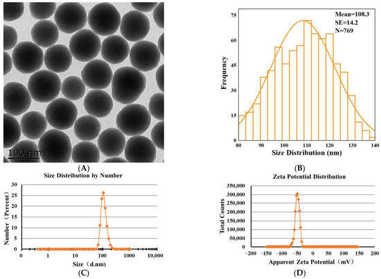
Show Figures

Figure 1

17 pages, 2732 KiB  
Article
Pharmacological Characterisation of Pseudocerastes and Eristicophis Viper Venoms Reveal Anticancer (Melanoma) Properties and a Potentially Novel Mode of Fibrinogenolysis
by Bianca op den Brouw, Parviz Ghezellou, Nicholas R. Casewell, Syed Abid Ali, Behzad Fathinia, Bryan G. Fry, Mettine H.A. Bos and Maria P. Ikonomopoulou
Int. J. Mol. Sci. 2021, 22(13), 6896; https://doi.org/10.3390/ijms22136896 - 27 Jun 2021
Cited by 10 | Viewed by 2667
Abstract
Venoms are a rich source of potential lead compounds for drug discovery, and descriptive studies of venom form the first phase of the biodiscovery process. In this study, we investigated the pharmacological potential of crude Pseudocerastes and Eristicophis snake venoms in haematological disorders [...] Read more.
Venoms are a rich source of potential lead compounds for drug discovery, and descriptive studies of venom form the first phase of the biodiscovery process. In this study, we investigated the pharmacological potential of crude Pseudocerastes and Eristicophis snake venoms in haematological disorders and cancer treatment. We assessed their antithrombotic potential using fibrinogen thromboelastography, fibrinogen gels with and without protease inhibitors, and colourimetric fibrinolysis assays. These assays indicated that the anticoagulant properties of the venoms are likely induced by the hydrolysis of phospholipids and by selective fibrinogenolysis. Furthermore, while most fibrinogenolysis occurred by the direct activity of snake venom metalloproteases and serine proteases, modest evidence indicated that fibrinogenolytic activity may also be mediated by selective venom phospholipases and an inhibitory venom-derived serine protease. We also found that the Pseudocerastes venoms significantly reduced the viability of human melanoma (MM96L) cells by more than 80%, while it had almost no effect on the healthy neonatal foreskin fibroblasts (NFF) as determined by viability assays. The bioactive properties of these venoms suggest that they contain a number of toxins suitable for downstream pharmacological development as candidates for antithrombotic or anticancer agents. Full article
Show Figures

Figure 1

16 pages, 3959 KiB  
Article
Combinatory Effects of Cerium Dioxide Nanoparticles and Acetaminophen on the Liver—A Case Study of Low-Dose Interactions in Human HuH-7 Cells
by Benjamin C. Krause, Fabian L. Kriegel, Victoria Tartz, Harald Jungnickel, Philipp Reichardt, Ajay Vikram Singh, Peter Laux, Mohamed Shemis and Andreas Luch
Int. J. Mol. Sci. 2021, 22(13), 6866; https://doi.org/10.3390/ijms22136866 - 25 Jun 2021
Cited by 8 | Viewed by 3274
Abstract
The interactions between pharmaceuticals and nanomaterials and its potentially resulting toxicological effects in living systems are only insufficiently investigated. In this study, two model compounds, acetaminophen, a pharmaceutical, and cerium dioxide, a manufactured nanomaterial, were investigated in combination and individually. Upon inhalation, cerium [...] Read more.
The interactions between pharmaceuticals and nanomaterials and its potentially resulting toxicological effects in living systems are only insufficiently investigated. In this study, two model compounds, acetaminophen, a pharmaceutical, and cerium dioxide, a manufactured nanomaterial, were investigated in combination and individually. Upon inhalation, cerium dioxide nanomaterials were shown to systemically translocate into other organs, such as the liver. Therefore we picked the human liver cell line HuH-7 cells as an in vitro system to investigate liver toxicity. Possible synergistic or antagonistic metabolic changes after co-exposure scenarios were investigated. Toxicological data of the water soluble tetrazolium (WST-1) assay for cell proliferation and genotoxicity assessment using the Comet assay were combined with an untargeted as well as a targeted lipidomics approach. We found an attenuated cytotoxicity and an altered metabolic profile in co-exposure experiments with cerium dioxide, indicating an interaction of both compounds at these endpoints. Single exposure against cerium dioxide showed a genotoxic effect in the Comet assay. Conversely, acetaminophen exhibited no genotoxic effect. Comet assay data do not indicate an enhancement of genotoxicity after co-exposure. The results obtained in this study highlight the advantage of investigating co-exposure scenarios, especially for bioactive substances. Full article
Show Figures

Figure 1

17 pages, 2900 KiB  
Article
SIRT3 Overexpression Ameliorates Asbestos-Induced Pulmonary Fibrosis, mt-DNA Damage, and Lung Fibrogenic Monocyte Recruitment
by Paul Cheresh, Seok-Jo Kim, Renea Jablonski, Satoshi Watanabe, Ziyan Lu, Monica Chi, Kathryn A. Helmin, David Gius, G. R. Scott Budinger and David W. Kamp
Int. J. Mol. Sci. 2021, 22(13), 6856; https://doi.org/10.3390/ijms22136856 - 25 Jun 2021
Cited by 31 | Viewed by 3858
Abstract
Alveolar epithelial cell (AEC) mitochondrial (mt) DNA damage and fibrotic monocyte-derived alveolar macrophages (Mo-AMs) are implicated in the pathobiology of pulmonary fibrosis. We showed that sirtuin 3 (SIRT3), a mitochondrial protein regulating cell fate and aging, is deficient in the AECs of idiopathic [...] Read more.
Alveolar epithelial cell (AEC) mitochondrial (mt) DNA damage and fibrotic monocyte-derived alveolar macrophages (Mo-AMs) are implicated in the pathobiology of pulmonary fibrosis. We showed that sirtuin 3 (SIRT3), a mitochondrial protein regulating cell fate and aging, is deficient in the AECs of idiopathic pulmonary fibrosis (IPF) patients and that asbestos- and bleomycin-induced lung fibrosis is augmented in Sirt3 knockout (Sirt3−/−) mice associated with AEC mtDNA damage and intrinsic apoptosis. We determined whether whole body transgenic SIRT3 overexpression (Sirt3Tg) protects mice from asbestos-induced pulmonary fibrosis by mitigating lung mtDNA damage and Mo-AM recruitment. Crocidolite asbestos (100 µg/50 µL) or control was instilled intratracheally in C57Bl6 (Wild-Type) mice or Sirt3Tg mice, and at 21 d lung fibrosis (histology, fibrosis score, Sircol assay) and lung Mo-AMs (flow cytometry) were assessed. Compared to controls, Sirt3Tg mice were protected from asbestos-induced pulmonary fibrosis and had diminished lung mtDNA damage and Mo-AM recruitment. Further, pharmacologic SIRT3 inducers (i.e., resveratrol, viniferin, and honokiol) each diminish oxidant-induced AEC mtDNA damage in vitro and, in the case of honokiol, protection occurs in a SIRT3-dependent manner. We reason that SIRT3 preservation of AEC mtDNA is a novel therapeutic focus for managing patients with IPF and other types of pulmonary fibrosis. Full article
Show Figures

Figure 1

17 pages, 2707 KiB  
Article
Sub-Chronic Effects of Slight PAH- and PCB-Contaminated Mesocosms in Paracentrotus lividus Lmk: A Multi-Endpoint Approach and De Novo Transcriptomic
by Luisa Albarano, Valerio Zupo, Davide Caramiello, Maria Toscanesi, Marco Trifuoggi, Marco Guida, Giovanni Libralato and Maria Costantini
Int. J. Mol. Sci. 2021, 22(13), 6674; https://doi.org/10.3390/ijms22136674 - 22 Jun 2021
Cited by 9 | Viewed by 2415
Abstract
Sediment pollution is a major issue in coastal areas, potentially endangering human health and the marine environments. We investigated the short-term sublethal effects of sediments contaminated with polycyclic aromatic hydrocarbons (PAHs) and polychlorinated biphenyls (PCBs) on the sea urchin Paracentrotus lividus for two [...] Read more.
Sediment pollution is a major issue in coastal areas, potentially endangering human health and the marine environments. We investigated the short-term sublethal effects of sediments contaminated with polycyclic aromatic hydrocarbons (PAHs) and polychlorinated biphenyls (PCBs) on the sea urchin Paracentrotus lividus for two months. Spiking occurred at concentrations below threshold limit values permitted by the law (TLVPAHs = 900 µg/L, TLVPCBs = 8 µg/L, Legislative Italian Decree 173/2016). A multi-endpoint approach was adopted, considering both adults (mortality, bioaccumulation and gonadal index) and embryos (embryotoxicity, genotoxicity and de novo transcriptome assembly). The slight concentrations of PAHs and PCBs added to the mesocosms were observed to readily compartmentalize in adults, resulting below the detection limits just one week after their addition. Reconstructed sediment and seawater, as negative controls, did not affect sea urchins. PAH- and PCB-spiked mesocosms were observed to impair P. lividus at various endpoints, including bioaccumulation and embryo development (mainly PAHs) and genotoxicity (PAHs and PCBs). In particular, genotoxicity tests revealed that PAHs and PCBs affected the development of P. lividus embryos deriving from exposed adults. Negative effects were also detected by generating a de novo transcriptome assembly and its annotation, as well as by real-time qPCR performed to identify genes differentially expressed in adults exposed to the two contaminants. The effects on sea urchins (both adults and embryos) at background concentrations of PAHs and PCBs below TLV suggest a need for further investigations on the impact of slight concentrations of such contaminants on marine biota. Full article
Show Figures

Figure 1

15 pages, 2217 KiB  
Article
In Vitro Evaluation of the Individual and Combined Cytotoxic and Estrogenic Effects of Zearalenone, Its Reduced Metabolites, Alternariol, and Genistein
by Adrienn Balázs, Zelma Faisal, Rita Csepregi, Tamás Kőszegi, Balázs Kriszt, István Szabó and Miklós Poór
Int. J. Mol. Sci. 2021, 22(12), 6281; https://doi.org/10.3390/ijms22126281 - 11 Jun 2021
Cited by 22 | Viewed by 2971
Abstract
Mycotoxins are toxic metabolites of filamentous fungi. Previous studies demonstrated the co-occurrence of Fusarium and Alternaria toxins, including zearalenone (ZEN), ZEN metabolites, and alternariol (AOH). These xenoestrogenic mycotoxins appear in soy-based meals and dietary supplements, resulting in the co-exposure to ZEN and AOH [...] Read more.
Mycotoxins are toxic metabolites of filamentous fungi. Previous studies demonstrated the co-occurrence of Fusarium and Alternaria toxins, including zearalenone (ZEN), ZEN metabolites, and alternariol (AOH). These xenoestrogenic mycotoxins appear in soy-based meals and dietary supplements, resulting in the co-exposure to ZEN and AOH with the phytoestrogen genistein (GEN). In this study, the cytotoxic and estrogenic effects of ZEN, reduced ZEN metabolites, AOH, and GEN are examined to evaluate their individual and combined impacts. Our results demonstrate that reduced ZEN metabolites, AOH, and GEN can aggravate ZEN-induced toxicity; in addition, the compounds tested exerted mostly synergism or additive combined effects regarding cytotoxicity and/or estrogenicity. Therefore, these observations underline the importance and the considerable risk of mycotoxin co-exposure and the combined effects of mycoestrogens with phytoestrogens. Full article
Show Figures

Figure 1

50 pages, 3119 KiB  
Review
Ionic Liquids—A Review of Their Toxicity to Living Organisms
by Ana R.P. Gonçalves, Xavier Paredes, A. F. Cristino, F. J.V. Santos and Carla S.G.P. Queirós
Int. J. Mol. Sci. 2021, 22(11), 5612; https://doi.org/10.3390/ijms22115612 - 25 May 2021
Cited by 119 | Viewed by 7097
Abstract
Ionic liquids (ILs) were initially hailed as a green alternative to traditional solvents because of their almost non-existent vapor pressure as ecological replacement of most common volatile solvents in industrial processes for their damaging effects on the environment. It is common knowledge that [...] Read more.
Ionic liquids (ILs) were initially hailed as a green alternative to traditional solvents because of their almost non-existent vapor pressure as ecological replacement of most common volatile solvents in industrial processes for their damaging effects on the environment. It is common knowledge that they are not as green as desired, and more thought must be put into the biological consequences of their industrial use. Still, compared to the amount of research studying their physicochemical properties and potential applications in different areas, there is a scarcity of scientific papers regarding how these substances interact with different organisms. The intent of this review was to compile the information published in this area since 2015 to allow the reader to better understand how, for example, bacteria, plants, fish, etc., react to the presence of this family of liquids. In general, lipophilicity is one of the main drivers of toxicity and thus the type of cation. The anion tends to play a minor (but not negligible) role, but more research is needed since, owing to the very nature of ILs, except for the most common ones (imidazolium and ammonium-based), many of them are subject to only one or two articles. Full article
Show Figures

Graphical abstract

23 pages, 4318 KiB  
Article
Ancient Bacterial Class Alphaproteobacteria Cytochrome P450 Monooxygenases Can Be Found in Other Bacterial Species
by Nomfundo Nzuza, Tiara Padayachee, Puleng Rosinah Syed, Justyna Dorota Kryś, Wanping Chen, Dominik Gront, David R. Nelson and Khajamohiddin Syed
Int. J. Mol. Sci. 2021, 22(11), 5542; https://doi.org/10.3390/ijms22115542 - 24 May 2021
Cited by 9 | Viewed by 2859
Abstract
Cytochrome P450 monooxygenases (CYPs/P450s), heme-thiolate proteins, are well-known players in the generation of chemicals valuable to humans and as a drug target against pathogens. Understanding the evolution of P450s in a bacterial population is gaining momentum. In this study, we report comprehensive analysis [...] Read more.
Cytochrome P450 monooxygenases (CYPs/P450s), heme-thiolate proteins, are well-known players in the generation of chemicals valuable to humans and as a drug target against pathogens. Understanding the evolution of P450s in a bacterial population is gaining momentum. In this study, we report comprehensive analysis of P450s in the ancient group of the bacterial class Alphaproteobacteria. Genome data mining and annotation of P450s in 599 alphaproteobacterial species belonging to 164 genera revealed the presence of P450s in only 241 species belonging to 82 genera that are grouped into 143 P450 families and 214 P450 subfamilies, including 77 new P450 families. Alphaproteobacterial species have the highest average number of P450s compared to Firmicutes species and cyanobacterial species. The lowest percentage of alphaproteobacterial species P450s (2.4%) was found to be part of secondary metabolite biosynthetic gene clusters (BGCs), compared other bacterial species, indicating that during evolution large numbers of P450s became part of BGCs in other bacterial species. Our study identified that some of the P450 families found in alphaproteobacterial species were passed to other bacterial species. This is the first study to report on the identification of CYP125 P450, cholesterol and cholest-4-en-3-one hydroxylase in alphaproteobacterial species (Phenylobacterium zucineum) and to predict cholesterol side-chain oxidation capability (based on homolog proteins) by P. zucineum. Full article
Show Figures

Figure 1

13 pages, 1838 KiB  
Article
Bisphenol A Inhibits the Transporter Function of the Blood-Brain Barrier by Directly Interacting with the ABC Transporter Breast Cancer Resistance Protein (BCRP)
by Elin Engdahl, Maarten D. M. van Schijndel, Dimitrios Voulgaris, Michela Di Criscio, Kerry A. Ramsbottom, Daniel J. Rigden, Anna Herland and Joëlle Rüegg
Int. J. Mol. Sci. 2021, 22(11), 5534; https://doi.org/10.3390/ijms22115534 - 24 May 2021
Cited by 19 | Viewed by 4710
Abstract
The breast cancer resistance protein (BCRP) is an important efflux transporter in the blood-brain barrier (BBB), protecting the brain from a wide range of substances. In this study, we investigated if BCRP function is affected by bisphenol A (BPA), a high production volume [...] Read more.
The breast cancer resistance protein (BCRP) is an important efflux transporter in the blood-brain barrier (BBB), protecting the brain from a wide range of substances. In this study, we investigated if BCRP function is affected by bisphenol A (BPA), a high production volume chemical used in common consumer products, as well as by bisphenol F (BPF) and bisphenol S (BPS), which are used to substitute BPA. We employed a transwell-based in vitro cell model of iPSC-derived brain microvascular endothelial cells, where BCRP function was assessed by measuring the intracellular accumulation of its substrate Hoechst 33342. Additionally, we used in silico modelling to predict if the bisphenols could directly interact with BCRP. Our results showed that BPA significantly inhibits the transport function of BCRP. Additionally, BPA was predicted to bind to the cavity that is targeted by known BCRP inhibitors. Taken together, our findings demonstrate that BPA inhibits BCRP function in vitro, probably by direct interaction with the transporter. This effect might contribute to BPA’s known impact on neurodevelopment. Full article
Show Figures

Figure 1

2019

Jump to: 2024, 2023, 2022, 2021

13 pages, 1383 KiB  
Article
Evaluation of the Major Steps in the Conventional Protocol for the Alkaline Comet Assay
by Mahsa Karbaschi, Yunhee Ji, Abdulhadi Mohammed S. Abdulwahed, Alhanoof Alohaly, Juan F. Bedoya, Shanna L. Burke, Thomas M. Boulos, Helen G. Tempest and Marcus S. Cooke
Int. J. Mol. Sci. 2019, 20(23), 6072; https://doi.org/10.3390/ijms20236072 - 2 Dec 2019
Cited by 24 | Viewed by 8030
Abstract
Single cell gel electrophoresis, also known as the comet assay, has become a widespread DNA damage assessment tool due to its sensitivity, adaptability, low cost, ease of use, and reliability. Despite these benefits, this assay has shortcomings, such as long assay running time, [...] Read more.
Single cell gel electrophoresis, also known as the comet assay, has become a widespread DNA damage assessment tool due to its sensitivity, adaptability, low cost, ease of use, and reliability. Despite these benefits, this assay has shortcomings, such as long assay running time, the manipulation of multiple slides, individually, through numerous process steps, the challenge of working in a darkened environment, and reportedly considerable inter- and intra-laboratory variation. All researchers typically perform the comet assay based upon a common core approach; however, it appears that some steps in this core have little proven basis, and may exist, partly, out of convenience, or dogma. The aim of this study was to critically re-evaluate key steps in the comet assay, using our laboratory’s protocol as a model, firstly to understand the scientific basis for why certain steps in the protocol are performed in a particular manner, and secondly to simplify the assay, and decrease the cost and run time. Here, the shelf life of the lysis and neutralization buffers, the effect of temperature and incubation period during the lysis step, the necessity for drying the slides between the electrophoresis and staining step, and the need to perform the sample workup and electrophoresis steps under subdued light were all evaluated. Full article
Show Figures

Graphical abstract

18 pages, 2121 KiB  
Article
Genome-Wide Adductomics Analysis Reveals Heterogeneity in the Induction and Loss of Cyclobutane Thymine Dimers across Both the Nuclear and Mitochondrial Genomes
by Alaa S. Alhegaili, Yunhee Ji, Nicolas Sylvius, Matthew J. Blades, Mahsa Karbaschi, Helen G. Tempest, George D. D. Jones and Marcus S. Cooke
Int. J. Mol. Sci. 2019, 20(20), 5112; https://doi.org/10.3390/ijms20205112 - 15 Oct 2019
Cited by 9 | Viewed by 3658
Abstract
The distribution of DNA damage and repair is considered to occur heterogeneously across the genome. However, commonly available techniques, such as the alkaline comet assay or HPLC-MS/MS, measure global genome levels of DNA damage, and do not reflect potentially significant events occurring at [...] Read more.
The distribution of DNA damage and repair is considered to occur heterogeneously across the genome. However, commonly available techniques, such as the alkaline comet assay or HPLC-MS/MS, measure global genome levels of DNA damage, and do not reflect potentially significant events occurring at the gene/sequence-specific level, in the nuclear or mitochondrial genomes. We developed a method, which comprises a combination of Damaged DNA Immunoprecipitation and next generation sequencing (DDIP-seq), to assess the induction and repair of DNA damage induced by 0.1 J/cm2 solar-simulated radiation at the sequence-specific level, across both the entire nuclear and mitochondrial genomes. DDIP-seq generated a genome-wide, high-resolution map of cyclobutane thymine dimer (T<>T) location and intensity. In addition to being a straightforward approach, our results demonstrated a clear differential distribution of T<>T induction and loss, across both the nuclear and mitochondrial genomes. For nuclear DNA, this differential distribution existed at both the sequence and chromosome level. Levels of T<>T were much higher in the mitochondrial DNA, compared to nuclear DNA, and decreased with time, confirmed by qPCR, despite no reported mechanisms for their repair in this organelle. These data indicate the existence of regions of sensitivity and resistance to damage formation, together with regions that are fully repaired, and those for which > 90% of damage remains, after 24 h. This approach offers a simple, yet more detailed approach to studying cellular DNA damage and repair, which will aid our understanding of the link between DNA damage and disease. Full article
Show Figures

Figure 1

29 pages, 4785 KiB  
Article
Mitochondrial Peptide Humanin Protects Silver Nanoparticles-Induced Neurotoxicity in Human Neuroblastoma Cancer Cells (SH-SY5Y)
by Sangiliyandi Gurunathan, Muniyandi Jeyaraj, Min-Hee Kang and Jin-Hoi Kim
Int. J. Mol. Sci. 2019, 20(18), 4439; https://doi.org/10.3390/ijms20184439 - 9 Sep 2019
Cited by 31 | Viewed by 4957
Abstract
The extensive usage of silver nanoparticles (AgNPs) as medical products such as antimicrobial and anticancer agents has raised concerns about their harmful effects on human beings. AgNPs can potentially induce oxidative stress and apoptosis in cells. However, humanin (HN) is a small secreted [...] Read more.
The extensive usage of silver nanoparticles (AgNPs) as medical products such as antimicrobial and anticancer agents has raised concerns about their harmful effects on human beings. AgNPs can potentially induce oxidative stress and apoptosis in cells. However, humanin (HN) is a small secreted peptide that has cytoprotective and neuroprotective cellular effects. The aim of this study was to assess the harmful effects of AgNPs on human neuroblastoma SH-SY5Y cells and also to investigate the protective effect of HN from AgNPs-induced cell death, mitochondrial dysfunctions, DNA damage, and apoptosis. AgNPs were prepared with an average size of 18 nm diameter to study their interaction with SH-SY5Y cells. AgNPs caused a dose-dependent decrease of cell viability and proliferation, induced loss of plasma-membrane integrity, oxidative stress, loss of mitochondrial membrane potential (MMP), and loss of ATP content, amongst other effects. Pretreatment or co-treatment of HN with AgNPs protected cells from several of these AgNPs induced adverse effects. Thus, this study demonstrated for the first time that HN protected neuroblastoma cells against AgNPs-induced neurotoxicity. The mechanisms of the HN-mediated protective effect on neuroblastoma cells may provide further insights for the development of novel therapeutic agents against neurodegenerative diseases. Full article
Show Figures

Graphical abstract

17 pages, 2260 KiB  
Article
Effects of Methylmercury and Theaflavin Digallate on Adipokines in Mature 3T3-L1 Adipocytes
by Shubhangi Chauhan, Kriya Dunlap and Lawrence K. Duffy
Int. J. Mol. Sci. 2019, 20(11), 2755; https://doi.org/10.3390/ijms20112755 - 5 Jun 2019
Cited by 15 | Viewed by 4314
Abstract
Diabetes is a contributor to morbidity across the globe and is often associated with obesity, metabolic syndrome and other inflammatory diseases associated with aging. In addition to genetic and lifestyle factors, environmental factors such as metals and persistent organic pollutants may increase the [...] Read more.
Diabetes is a contributor to morbidity across the globe and is often associated with obesity, metabolic syndrome and other inflammatory diseases associated with aging. In addition to genetic and lifestyle factors, environmental factors such as metals and persistent organic pollutants may increase the severity or lower the threshold of these conditions. In cell culture, methylmercury is toxic to adipocytes and may impact adipokine secretions. In this study, we determined the effects of different concentrations of theaflavin digallate on methylmercury exposed 3T3-L1 adipocytes in cell culture. Secretions of resistin, adiponectin and lipid peroxidation product, 4-hydroxynonenal (4-HNE) were monitored using ELISA assays. Cell morphology of methylmercury and theaflavin-3,3′-digallate treated adipocytes was assessed using Lipid (Oil Red O) staining. Exposure to methylmercury increased the levels of resistin and adiponectin as well as 4-HNE when compared to the control cells. Methylmercury treated cells resulted in smaller number of adipocytes and clumped lipid droplets. These results suggest that methylmercury induces reactive oxygen species leading to development of an inflammatory response. Theaflavin-3,3′-digallate reduced the impact of methylmercury by maintaining the adipocytes morphology and secretion patterns of adiponectin, resistin and 4-hydroxynonenal. With this experimental model system other anti-inflammatory and signaling agents could be tested at the biochemical level before eventually leading to studies in animal models. Full article
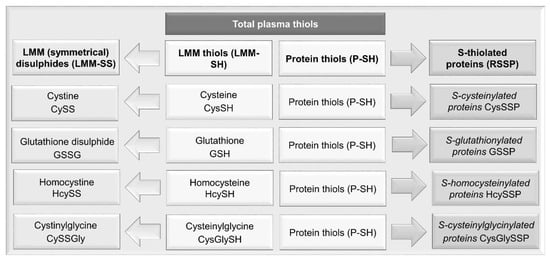
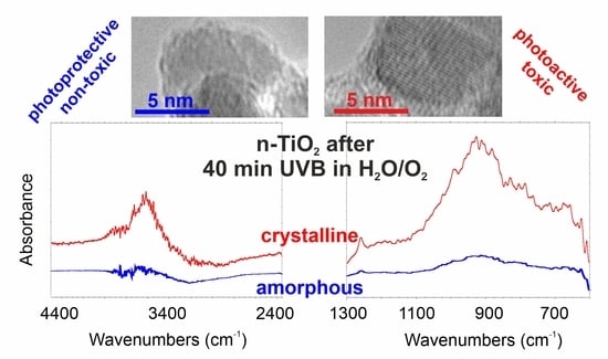
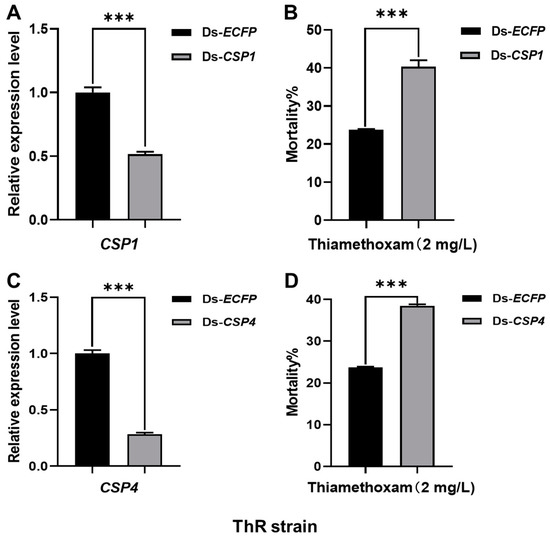
Show Figures

Figure 1

Back to TopTop